Understanding Vibrio and Related Organisms in Medical Bacteriology

Vibrio, Aeromonas, and Plesiomonas are gram-negative bacilli commonly found in water sources, causing gastrointestinal diseases. They exhibit similarities to Enterobacteriaceae but with distinct characteristics such as polar flagella and being oxidase-positive. Vibrios like V. cholerae are crucial human pathogens with unique growth requirements and identification methods. Antigenic structure and classifications further distinguish these organisms in medical bacteriology.

Download Presentation

Please find below an Image/Link to download the presentation.
The content on the website is provided AS IS for your information and personal use only. It may not be sold, licensed, or shared on other websites without obtaining consent from the author. Download presentation by click this link. If you encounter any issues during the download, it is possible that the publisher has removed the file from their server.
E N D
Presentation Transcript
Vibrio,and related organisms ( Aeromonas &plesiomonas) Prof. Dr. Lamyaa Kadhim Baqer Medical bacteriology Third year
General Characteristics of Vibrio, Aeromonas , and Plesiomonas Vibrio , Aeromonas , and Plesiomonas species are gram negative bacilli that are all widely distributed in nature. Primarily found in water sources. They cause gastrointestinal disease Similarities to Enterobacteriaceae .Gram-negative .Facultative anaerobes .Fermentative bacilli Differences from Enterobacteriaceae .Having polar flagella (in contrast to peritrichous flagella of the motile Enterobacteriaceae). .Oxidase positive
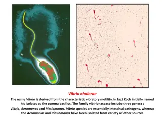
The Vibrios Morphology and physiology of Vibrios -They are comma-shaped (vibrioid) gram negative bacilli, are motile possessing a polar flagellum. They belong to the family Vibrionaceae .The most significant human Vibrios are : V. cholerae, V. parahaemolyticus, andV. vulnificus. --Broad temperature and pH range for growth on media: .18 -37 C .PH 7.0 9.0 (useful for enrichment). - For rapid isolation from faeces , Alkaline Peptone Water APW ( pH 8.6) is used (enrichment medium for V. cholerae ). -Vibrios can grow on a variety of simple media such as ,blood agar and MacConkey`s agar. The selective medium is TCBS agar (Thiosulfate Citrate Bile salts Sucrose agar ) - V. cholerae grow without salt. Most other Vibrios are halophilic ( salt-loving).
VIBRIO CHOLERAE Morphology and Identification Vibrio cholerae : - Gram-negative ,facultatively anaerobic, fermentative comma-shaped ,curved rods - 2-4 m long - Actively mobile by means of a polar flagellum
Culture Vibrio cholerae can grow on a variety of simple media like blood agar and MacConkey`s agar with a broad temperature range from (18 C to 37 C) . And tolerate a wide range of pH (7.0 to 9.0) but are susceptible to stomach acids. V.cholerae grows well on thiosulfate-citrate-bile-sucrose (TCBS) agar ,which is the selective medium for Vibrios.
On TCBS V. cholerae produces large (2 4 mm in diameter) yellow-colored colonies, slightly flattened with opaque centers and translucent peripheries. The yellow color is due to the fermentation of sucrose in TCBS medium. Non-sucrose-fermenting Vibrios such as V. parahaemolyticus produce green to blue-green colonies.
Antigenic structure and Biologic Classifications Many Vibrios share a single heat -labile flagellar H antigen which is nonspecific. -V.choleraehas O liposaccharides antigens that give serological specificity. Based on variable somatic O-antigen composition ,more than 200 O antigen groups of V. cholerae have been recognized 1- O1 serogroup V. cholerae strains of O1 and O139 serogroups cause classic epidemic cholera ( both are toxigenic) -These V. cholerae serogroups O1 antigen have extra determinants that make possible further typing: the serotypes are Ogawa, Inaba, and Hikogima . -Two biotypes of epidemicV. cholerae (different biochemical reactions) 1-Classic 2-El Tor Include other strains that are identical to O1 strains but do not agglutinate in O1 antiserum They are called: Non-cholera Vibrios (NCV) or Non-agglutinating (NAG) Vibrios (O139 serogroups) Occasionally non- O1/non-O139 V.cholerae causes cholera-like disease
Laboratory Diagnosis of V. cholerae A-Specimen B-Smears C-Transport media D-Culture E-Specific Tests A- Specimens: specimens for culture include watery stool and mucous flakes from patients or carriers. B- Smears: The microscopic appearance of smears made from stool samples is not reliable. Under dark- field microscopy ,the rapidly motile Vibrios might be seen (darting movement).
C- Transport media: The ideal transport medium is Cary Blair media , and if not available, Alkaline Peptone Water (APW) can be used. D- Culture: Culture is the gold standard in the isolation and diagnosis of the organism from the stool sample. Vibrio can grow on a variety of simple media like blood agar and MacConkey`s agar Selective / differential medium is thiosulfate-citrate-bile salts agar (TCBS) producing yellow colonies. Enrichment medium is Alkaline peptone broth (high pH 8.0-9.0 : vibrio can survive and replicate while other organisms can not)
E-Specific Tests 1-Biochemical tests 2-String test 3- Serological tests 4-Molecular techniques
Biochemical tests: -Ferments glucose, maltose, mannitol, and sucrose but not lactose and arabinose -Reduces nitrate -Oxidase positive :a key step in the preliminary identification of V.cholerae and other Vibrios. - Indole positive - String test positive. - Most vibrio species are halotolerant ,and NaCl often stimulates their growth. This differentiates Vibrios from Aeromonas since Vibrios grow on media containing 6% NaCl ,but Aeromonads does not. Differentiation between Classic and El Tor ( bio- typing): El Tor biotype produces a hemolysin, + Voges-Proskauer test, and is resistant to polymyxin B .
String Test Uses of String test : to separate Vibrio species from other bacterial species that share similarities, particularly Aeromonas species. All V. cholerae strains, as well as most other Vibrios, are positive, whereas Aeromonas strains are negative. Principle of String test The test is done by emulsifying a large colony in a small drop of 0.5% sodium deoxycholate in sterile distilled H2O. If the result is positive, the suspension will lose turbidity ,and DNA will be released from the lysed cells causing the mixture to become viscous. a mucoid string is formed when an inoculating loop is drawn slowly suspension. away from the
Serologic identification As mentioned before, there are more than 200 serogroups of V. cholerae , among which O1 and 0139 serogroups are the causative agent of most of the epidemics and pandemics. Serologic identification of V. cholera O1 and 0139 serogroup is the most rapid and specific method of identification. It is carried out by slide agglutination test with the specific antisera. Isolates of O1 serogroup is further divided into three serotypes Ogawa Inaba and Hikojima (rare).
Summary of Laboratory Diagnosis of V. cholerae Collect stool sample Place in transport medium and send to laboratory Streak on selective or non-selective medium Incubate 12-18 hours at 37 Identify colonies Perform a slide agglutination with polyvalent O1 group antiserum and type specific antisera OR Perform biochemical tests such as string test, oxidase test,
Virulence factors: 1-Endotoxin: has only a negligible significance as a virulence factor 2-Enterotoxin (Cholera toxin CT - ) : an exotoxin which is the main factor of pathogenity. It is antigenically and pharmacologically identical in all sero and biotypes of V. cholerae causing epidemic cholera. 3-Adherence factor providing ability to attach to the intestinal epithelial cells 4-Mucinase :causes extreme cellular desquamation 5-Vascular permeability factor
Vibrio cholera Enterotoxin :Mechanism of Action -The cholera toxin is a complex A-B toxin. The B subunits are for binding and the A subunit is the active. -The toxin binds ,through its (B) subunits to specific receptors on intestinal epithelial cells. The active portion of the A subunit is released and enters the intestinal epithelial cells. The active portion interacts with G proteins that control the enzyme adenylate cyclase ,leading to catabolic conversion of adenosine triphosphate (ATP) to cyclic adenosine monophosphate (cAMP) .The rise in cAMP results in hypersecretion of water and electrolytes (Na+ ,K+, CL-, and HCO3- ) into intestinal lumen causing diarrhea.
Pathogenesis of V. cholerae Incubation period : 2-3 days Source of infection: human carriers or patients with cholera and marine shellfish Predisposing factors: Poor sanitation , overcrowding , malnutrition. Mode of transmission : ingestion of contaminated food and water Infective dose: organisms are very sensitive to gastric acid ,so high infective dose >108 CFU is needed in normal person. Lowerinfective dose 103 -105 CFU in patients having reduced gastric acid secretion (achlorhydria or hypochlorhydria and patients using antacid treatment). Clinical Findings : sudden onset of nausea and vomiting and profuse diarrhea with abdominal cramps. Stools, which resemble ``rice water``, contain mucus, epithelial cells ,and large number of Vibrios. There is rapid loss of fluid and electrolytes, which leads to profound dehydration, circulatory collapse ,and anuria which if not replaced adequately may lead to death.
Pathogenesis of V. cholerae (Summary) Ingestion of contaminated food or water (in large number) The bacteria is sensitive to gastric acid so large dose needed to cause disease unless patient achlorhydric ( reduced gastric acid )or taking antacids Colonization of small intestine: -Bacterial motility (polar flagella) -Production of mucinase Attachment to specific receptors Massive loss of fluid and electrolytes (no damage to intestinal epithelial cells, no blood or leucocytes in stool) Toxin production
Treatment of Cholera 1- Fluid and electrolytes replacement. 2- Acid base balance adjustment. 3- Antibiotics such tetracycline.
Preventive measures 1-Education and improvement of sanitation 2-Pure water supply and clean food preparation 3-Proper sewage disposal 4-Isolation of patients 5-Detection of carriers 6-Vaccination Immunity An attack of cholera is followed by immunity to reinfection but the duration and degree of immunity are unknown. Re- attacks are not common for 6-12 months.
Other Vibrio Infections V. Cholerae serogroups O1 and O139 cause cholera in humans - Other Vibrios such as Vibrio cholera serogroups non-O1/ non-O139 and Vibrio parahaemolyticus may cause cholera-like diarrhea ,gastroenteritis and extraintestinal sepsis Vibrio parahaemolyticus: It is a halophilic (salt-loving) Vibrio associated with enteritis produced by thermostable direct hemolysin. Infection is acquired by ingestion of raw or improperly cooked sea foods. V. vulnificus: it is a halophilic vibrio, which ferments lactose associated with wound infections as well as fatal septicemias. Non-O1/0139 serogroups of V. cholerae: cause diarrhea
Other Vibrionaceae genera Vibrios-related organisms Aeromonas & Plesiomonas
Aeromonas -a gram negative facultative anaerobic rod that morphologically resembles members of the family Enterobacteriaceae -16 species have been recognized most of which are associated with human disease. The most important pathogens are : Aeromonas hydrophila , Aeromonas caviae, and Aeromonas veroni bivar sobria. -The organisms are present in fresh and brackish water throughout the world. Motile species have single polar flagellum. Non motile species apparently are not associated with human disease. -Infection is acquired by ingestion or exposure to contaminated water or food -The two major diseases associated with Aeromonas are gastroenteritis and wound infections (with or without bacteremia). It causes opportunistic systemic disease in immunocompromised person.
Plesiomonas Single species Plesiomonas shigelloides -The bacteria are oxidase positive and have multiple polar flagella (lophotrichous) -Found in soil and aquatic environment (fresh or estuarine water) , some cold blooded animals ( such as reptiles, fishes, amphibians ) are carriers - Infection is acquired by ingestion or exposure to contaminated water or sea food or through occupational exposure (fish handlers, zookeeper) - Primarily causes gastroenteritis or rarely extraintestinal infections