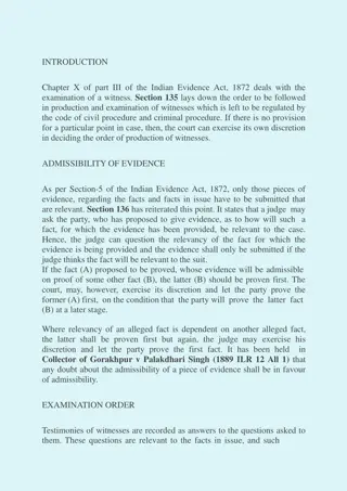
Diagnosis-Driven Physical Examination of the Shoulder Part 2: Key Considerations and Case Study
This resource delves into the diagnosis-driven physical examination of shoulder conditions, covering common conditions, key historical factors, and provocative maneuvers for differential diagnosis. It presents an organizational scheme for musculoskeletal assessments and details various shoulder conditions and their presentations. A case study illustrates the application of history taking and physical examination in diagnosing shoulder pain, including observations, palpation findings, range of motion assessments, strength evaluations, and specialty testing.
- Shoulder conditions
- Differential diagnosis
- Musculoskeletal evaluation
- Physical examination
- Case study

Download Presentation

Please find below an Image/Link to download the presentation.
The content on the website is provided AS IS for your information and personal use only. It may not be sold, licensed, or shared on other websites without obtaining consent from the author. Download presentation by click this link. If you encounter any issues during the download, it is possible that the publisher has removed the file from their server.
E N D
Presentation Transcript
THE DIAGNOSIS-DRIVEN PHYSICAL EXAMINATION OF THE SHOULDER PART 2 American Medical Society for Sports Medicine ACP Musculoskeletal Medicine Teaching Group American College of Physicians Meeting April 2019
OBJECTIVES FOR SHOULDER DIAGNOSIS 1. 2. 3. Review common conditions of the shoulder Identify key historical factors in a patient with shoulder pain Perform key provocative maneuvers to aid in the differential diagnosis of shoulder pain
MUSCULOSKELETAL ORGANIZATIONAL SCHEME History Inspection Palpation Range of motion Other tests
COMMON SHOULDER CONDITIONS Long head biceps tendon injuries Rotator cuff disease: Sub-acromial bursitis/impingement Partial Rotator cuff tendon tear Full Rotator cuff tendon tear Acromioclavicular Osteoarthritis Adhesive capsulitis Glenohumeral joint osteoarthritis Instability: Subluxation, dislocation, labral tears
KEY SHOULDER HISTORY Impingement Rotator cuff tear > 40 Overuse or acute Adhesive capsulitis 40-60 y/o Acute onset without MOI +/- diabetes Generalized Generalized Glenohumeral joint arthritis > 60 y/o +/- distant h/o trauma Labral tear Age Mechanism < 40 Overuse < 40 ish Overuse or acute Location of pain Lateral shoulder Lateral shoulder Deep - Anterior shoulder No Stiffness No No Yes Yes
CASE 1 HISTORY CC: 45 year old with shoulder pain Onset: gradual over the past 6 weeks Started after increase in overhead duties at work No fall or specific injury Location: superolateral shoulder Radiates to lateral arm No numbness or paresthesias Exacerbating factors: Overhead work, reaching, carrying items if elbows are not at side. No significant pain at rest with hands in lap
CASE 1 PHYSICAL EXAMINATION Observation: No scapular winging Palpation: Tender at subacromial space No tenderness at rotator cuff insertions, SC, AC, or proximal biceps tendon ROM: Normal A/PROM except for passive internal rotation Pain with active ABduction more than with forward elevation Strength: Normal, including rotator cuff Pain with resisted ABduction at 90 degree in the scapular plane Specialty Testing: (+) Hawkins. (+) Neer s. (-) drop arm. (-) lag signs on ext. rotation. (-) belly press and lift off. (-) Speeds. (-) Yergason s. (-) Cross-arm. (-) Spurling s.
WHAT IS ROTATOR CUFF DISEASE? Impingement, aka bursitis Tendinitis/tendinopathy Partial thickness tear Full thickness tear
ROTATOR CUFF IMPINGEMENT History Pain at superolateral shoulder Exam Limited passive internal rotation Tender to palpation deep to acromion Pain with reaching PROM AROM but pain in all planes of motion Pain after an event or increase in activity Pain AND weakness in all planes but esp. on Empty-Can testing at 90 Abduction in scapular plane No change in shoulder pain with Neck ROM Both pain and weakness improve with scapular retraction Pain with Neer s and Hawkins tests
ROTATOR CUFF IMPINGEMENT Imaging 3-4 views of the Shoulder Treatment Subacromial CS Injection PO NSAIDs Rehab after pain improvement Referral to ortho: Subacromial Decompression +/- Bursectomy only if conservative Tx fails AP Int. & Ext. Rotation & Axillary +/- Scapular-Y view Usually normal Look for calcific rotator cuff tendinopathy or osteoarthritis
CASE 2 HISTORY CC: 45 yo patient in for evaluation of shoulder pain. Onset: 1 week ago, when s/he sustained fall while roller-blading. Location: Lateral shoulder, subacromial space. Radiation of pain toward the elbow, but not more distal. Some weakness, though should present this with some uncertainty because pain is the major symptom, and the patient does not know whether the weakness is due to the pain or not. No numbness. Exacerbating factors: Increase in pain with overhead activities of daily living. Overhead work, reaching, carrying items if elbows are not at side. Night pain: moderate, started two days after the fall
CASE 2 PHYSICAL EXAM Observation: No scapular winging, deformity, skin changes or bruising, or step off at AC Palpation: Tender in supraspinatus and infraspinatus insertions. No tenderness over SC, AC, BT. ROM: Pain with active ABD, beginning at around 30 degrees of elevation. Limited abduction from 30-90 degrees. Full passive ROM without pain. Strength: Clear weakness with empty-can test at 30 degrees of ABduction and less weakness with external rotation, with increased pain during resistance. Full strength with supscapularis and teres minor, though pain with resistance in these as well. Specialty Testing: (+) drop arm. (+) lag signs on ext. rotation. (-) belly press and lift off. (+) Hawkins. (+) Neer s. (-) Speeds. (-) Yergason s. (-) Cross-arm. (-) Spurling s.
PARTIAL AND COMPLETE ROTATOR CUFF TEAR History Fall on outstretched arm c/o weakness > pain Hx of overhead activity Acute, Traumatic Specific Event with pain and weakness 3 months Chronic, Atraumatic Gradual onset of weakness and pain >3 months ago Exam Limited AROM due to weakness > pain but NL PROM Supraspinatus (70% of RCT) Weak Empty Can at 30 + Drop Arm test: Full tear Infraspinatus (20% of RCT) Weak resisted ER + Lag Sign: Full tear Subscapularis (<1% of RCT) + Gerber Belly Press or Lift Off tests
DIAGNOSIS AND TREATMENT OF ROTATOR CUFF TEAR Imaging 3-4 views of the Shoulder Treatment Acute Tear = RCT Repair Chronic Tear = Rehab 75% Return to NL Function & No Pain without Surgery at 2 yrs. (Kuhn JE. J Shoulder Elbow Surg. 2013) Subacromial CS Injection for Pain AP Int. & Ext. Rotation & Axillary +/- Scapular-Y view Usually normal High riding humeral head suggestive of tear MRI if Acute RCT
ROTATOR CUFF DISEASE TREATMENT Most do well with conservative treatment Impingement Tendinitis, tendinopathy Partial thickness tear Full thickness tear PT +/- Injection +/- Medication Consider ortho referral.
CASE 3 HISTORY Chief complaint: 60 y/o man, frequent weightlifter with shoulder pain Onset: Last week, he was doing a heavy set of bench press, and over did it Similar mild-moderate intermittent pain for years; especially after heavy weightlifting Location: Superior shoulder Exacerbating factors: Reaching across his body & w/ shoulder ADduction (e.g. bench press or pushups) Night pain: Pain w/ sleeping on side or draping across body if on laying contralateral side
CASE 3 PHYSICAL EXAM Observation: Small boney prominence at AC joint Palpation: Tender at AC-joint but not elsewhere in the shoulder ROM: Pain with active ADduction. Otherwise Full ROM Strength: Full strength in deltoid, biceps, triceps and rotator cuff Specialty Testing: (+) Cross-arm and Scarf tests; mild (+) Hawkins and Neer s; (-) Spurling s
ACROMIOCLAVICULAR ARTHRITIS HX: Heavy labor, weightlifters, contact sports Pain arises with activities above or in front of body Pain swinging golf club, backhand tennis or seatbelt PE: Pain directly over AC joint Provocative test include Scarf test and Cross Arm test Radiographs will show OA changes to AC joint Treatment: Activity modification Pain management including NSAIDS and steroid injection
CASE 4 HISTORY Chief complaint: 42 y/o woman, midwife, left shoulder pain for 3 weeks Onset: She works 5 days a week, 10-hour shifts Recently put in a new garden and was digging for 4-5 hours, several days in a row Now has anterior shoulder pain Location: anterior left shoulder Exacerbating factor: pulling, twisting, reaching, heavy lifting; especially with palm up
CASE 4 PHYSICAL EXAM Observation: Normal Palpation: Marked tenderness over proximal biceps tendon, o/w normal ROM: Full, normal. Strength: Normal, except pain and weakness with elbow flexion with supinated palm Special Testing: (+) Speeds, (+) Yergason s
LONG HEAD BICEPS TENDON INJURIES Tendinitis due to repetitive micro-trauma Partial and complete tear (Usually treated non surgically with rest, NSAIDs, PT) PE: Anterior location and tender over bicipital groove Maneuvers: Speed s Test and Yergason s
CASE 5 HISTORY Chief complaint: 50 y/o woman with shoulder pain Onset: Gradual over the past 4 months; No fall or injury Location: Entire shoulder, deep, nonspecific; No radiation of pain Exacerbating factors: Hurts most of the day with minimal activity Reaching or extending arm Pain at rest with hands resting in her lap Loss of ROM Including external rotation and/or forward elevation
CASE 5 PHYSICAL EXAM Observation: No scapular winging Palpation: No point tenderness over rotator cuff insertions, SC, AC, proximal biceps tendon. ROM: Limited AROM in all 4 planes compared to opposite shoulder Limited PROM = to Limited AROM. Pain at end-ROM Strength: Normal rotator cuff and other muscles Mild pain with strength testing but less painful than end-ROM testing Specialty Testing: Unable to fully perform Hawkins , Neer s, and Cross-arm due to pain and LIMITED ROM. (-) Speeds /Yergason s; (-) Spurling s.
SHOULDER: RANGE OF MOTION IS KEY Active ROM Normal Decreased Rotator cuff disease Labral tear Biceps tendinitis AC joint OA Passive ROM Normal Decreased Adapted from: O'Kane and Toresdahl. The evidenced-based shoulder evaluation. Cur Sports Med Rep. 2014. GH joint arthritis Frozen shoulder Xray Abnormal Normal
ADHESIVE CAPSULITIS (FROZEN SHOULDER) Inflammation and fibrosis of glenohumeral capsule Trigger usually unclear (primary/idiopathic) Less commonly secondary--from immobilization (following injury, surgery) Epidemiology: Age 40-60 More common in women Associated with diabetes and thyroid dz Slide used with permission from Drs. Meg Pearson and Steve Bent
ADHESIVE CAPSULITIS http://www.aurorahealthcare.org/healthgate/images/si55551230.jpg
ADHESIVE CAPSULITIS: HX AND EXAM Hx: Pain may be severe esp. at end ROM. No hx of acute injury Exam: Global restrictions of ROM, similar to OA Normal strength Diagnostics: normal xray Slide used with permission from Drs. Meg Pearson and Steve Bent
TREATMENT FOR ADHESIVE CAPSULITIS Associated w/diabetes Pain control: NSAIDs or injected corticosteroids Does not change disease course Does help significantly with pain control Physical therapy to help restore ROM Start only AFTER pain at rest resolves Surgery (rarely) Manske and Prohaska, Curr Rev Musculoskeletal Med, 2008. Griesser MJ et al. Adhesive capsulitis a systematic review of intraarticular injections. J Bone Joint Surg Am. Sep 2011.
CASE 6 HISTORY Chief complaint: 50 y/o with shoulder pain Onset: Gradual over the past 24 months; No fall or trauma or injury Location: Entire shoulder, deep, non-specific. No radiation of pain, numbness or weakness. Exacerbating factors: Reaching or extending arm, combing or washing hair, tucking in shirt (activities at end ROM) Deep, dull, ache at rest Hurts at the end of the day or with increased upper extremity activity
CASE 6 PHYSICAL EXAM Observation: No scapular winging Palpation: No point tenderness over rotator cuff insertions, AC, proximal biceps tendon ROM: Limited AROM in all 4 planes compared to opposite shoulder (20-30 degrees reduced). Limited PROM to same location as AROM. Pain at end-ROM. Strength: Normal rotator cuff strength Mild pain with strength testing but less painful than end-ROM testing Specialty Testing: Unable to fully perform Hawkins , Neer s, and Cross-arm due to pain and limited ROM. (-) Speeds /Yergason s; (-) Spurling s
GLENOHUMERAL (GH) JOINT OSTEOARTHRITIS Cartilage loss limited and painful joint motion Onset usually early 60 s (Younger person can present with inflammatory arthropathy of GH joint) Primary shoulder OA less common Secondary OA: prior trauma chronic instability cuff arthropathy Slide used with permission from Drs. Meg Pearson and Steve Bent
GH OA: HX AND EXAM Hx: Insidious onset Diffuse pain with motion in any direction. Worse with activity/movement Less night time pain Exam:: Loss of passive and active motion in all directions Strength is usually preserved Slide used with permission from Drs. Meg Pearson and Steve Bent
GH OA TREATMENT Non-Operative Operative Non-Joint Replacement Partial Joint Replacement Total Joint Replacement Reverse Total Joint Replacement Physical Therapy Pain Medicine NSAIDs (oral or topical) Tylenol (APAP) Narcotics Steroid Injections
SHOULDER INSTABILITY 1. Labral disorder: Hx: Often a throwing athlete PE: Mechanical symptoms: pop or clunk. Provocative: O Brien, Mayo Shear 2. GH joint instability: Hx: pain in throwing athlete PE: Anterior joint pain, ligamentous laxity Provocative maneuvers: Apprehension +/- relocation, Load and Shift and Sulcus sign


 JOINT OSTEOARTHRITIS")