Comprehensive Guide to Neurological Examination in Dogs and Cats
Neurological examination in dogs and cats is crucial for diagnosing nervous system disorders. This guide covers the recording of history, general and detailed clinical examinations, examination of CSF, radiographic examination, EEG, and brain biopsy. It includes assessing history, mental state, movement, posture, gait, sensation, nystagmus, balance, muscle tone, cranial nerves, postural reactions, and spinal nerves. Detailed examination of specific nerves like olfactory, optic, facial, vagus, and more is also discussed.
Download Presentation

Please find below an Image/Link to download the presentation.
The content on the website is provided AS IS for your information and personal use only. It may not be sold, licensed, or shared on other websites without obtaining consent from the author.If you encounter any issues during the download, it is possible that the publisher has removed the file from their server.
You are allowed to download the files provided on this website for personal or commercial use, subject to the condition that they are used lawfully. All files are the property of their respective owners.
The content on the website is provided AS IS for your information and personal use only. It may not be sold, licensed, or shared on other websites without obtaining consent from the author.
E N D
Presentation Transcript
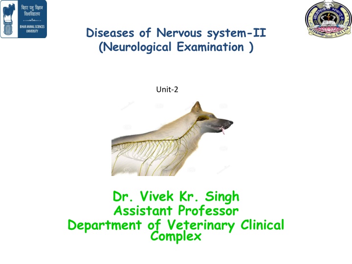
Diseases of Nervous system-II (Neurological Examination ) Unit-2 Dr. Vivek Kr. Singh Assistant Professor Department of Veterinary Clinical Complex
Introduction Neurological rewarding in dogs and cats because all the techniques can be easily employed examination is most In examination is less fruitful because all the techniques can t be employed and poor response to many techniques large animals neurological
Neurological examination includes- Recording History General and detailed clinical examination of nervous system Examination of CSF Radiographic examination EEG Brain Biopsy
History Duration of signs Mode of onset (acute or Chronic) Progression of disease Description of sign Access to poison and traumatic injury
General Clinical Examination Manifestation of mental state Involuntary movement Abnormality in posture and gait Disturbance in sensation Nystagmus Balance Muscle tone
Detailed Clinical Examination Examination of Cranial nerves Postural reactions Examination of spinal Nerves
Olfactory nerve (1st Cranial Nerve ) Optic nerve (2nd ) Usually fail to give an result Menace response, Ability to avoid objects in its path Oculomotor, Trochlear and abducent nerves (3rd ,4th and 6th) Movement of eye ball Trigeminal nerve (5th) Facial Nerve (7th) Unable to masticate, Drop jaw Dropping of ear, jaw, eye remains open Head tilt , falling to the side, rolling, circling, Auditory nerve (8th) Glossopharyngeal nerve (9th) Vagus nerve (10th) Dysphagia, inspiratory dyspnea (due to laryngeal paralysis), voice change and regurgitation Spinal accessory nerve (11th) Deviation of the neck Hypoglossal nerve (12th) Asymmetry of the tongue
Postural Reactions Postural proprioceptive fibers of- Peripheral nerves Spinal cord Brain stem Cerebrum Cerebellum Inner ear Place the patient s limb in an abnormal position to see if the patient returns the limb to a normal position reactions test the
Proprioceptive positioning It is performed by making the animal stand on a limb knuckled over at paw and observe its ability to correct this abnormal position
Hopping reaction Hopping reaction is performed by holding the patient in a way so that the majority of its weight is placed on one limb
Hemihopping/ Hemistanding/ Hemiwalking Patient s limbs on one side are held off the ground, and the patient is forced to walk sideways on its remaining two limbs
Wheel Barrowing Initially patient s thoracic and then pelvic limbs are held off the ground, and animal is make to walk forward and backward on its two legs
Spinal segmental reflex These reflexes directly test reflex arc of spinal cord Proprioceptive reflexes Nociceptive reflexes Special reflexes
Proprioceptive reflexes 1. Triceps reflex- Tests the radial nerve which arises from C7-T1
2. Biceps reflex- Evaluates the musculo- cutaneous nerve which arise from spinal cord segment C6-C7
3. Patellar reflex Evaluates the femoral nerve in the segment of the spinal cord L4-L6
4. Gastrocnemius reflex- Evaluates the tibial branch sciatic originates from L6-S1 nerve which
Nociceptive reflexes Initiated by nociceptive (Painful) stimuli Loss of nociceptive indicates LMN lesions Flexor reflex Perineal reflex Panniculus reflex (Cutaneous trunic reflex)
Analysis of CSF Electroencephaography Computerized tomography Magnet resonance imaging
