Understanding Lower Genital Tract Infections: Causes, Symptoms, and Treatment
Lower genital tract infections encompass conditions like vulvitis, vaginitis, and cervicitis, with causes ranging from irritants to infections. Symptoms include itching, discharge, and swelling. Treatment involves identifying and addressing the underlying cause, whether infectious or non-infectious, while managing symptoms effectively and monitoring for potential complications.

Download Presentation

Please find below an Image/Link to download the presentation.
The content on the website is provided AS IS for your information and personal use only. It may not be sold, licensed, or shared on other websites without obtaining consent from the author. Download presentation by click this link. If you encounter any issues during the download, it is possible that the publisher has removed the file from their server.
E N D
Presentation Transcript
THE LOWER GENITAL TRACT INFECTION DR.Alaa Ibrahim
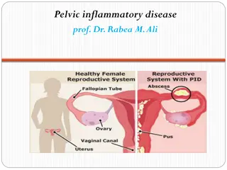
CLASSIFICATION OF GENITAL TRACT INFECTION: -Depending on the site and affection of the infective organism 1. Lower genital tract infections: - vulvitis Vaginitis cervicitis. 2. Upper genital tract infection:- (Pelvic Inflammatory Disease)
Anatomy and physiology: -The vaginal epithelium is lined by stratified squamous epithelium during the reproductive age group under the influence of the estrogen. -The normal vaginal flora is mostly aerobic, with the most common of which is hydrogen peroxide producing lactobacilli.
- -factors that affect the ability of bacteria to survive includes vaginal pH and the availability of glucose for bacterial metabolism. -The pH level of the normal vagina is lower than 4.5, which is maintained by the production of lactic acid by estrogen-stimulated vaginal epithelial cells.
Normal vaginal secretions: Vaginal secretions are floccular in consistency, white in color , and usually located in the dependent portion of the vagina (posterior fornix), mainly consists of mucous , desquamated epithelial cells, bacteria(lactobacillius).
Vulvitis 1- Non-infectious 30%: due to irritation or hypersensitivity. It is more common in premenarchal and post-menopausal women. 2- Infectious: common in reproductive years, most often due to infection ,Bacterial vaginitis , Candidal vaginitis , Trichomonas vaginitis, Chlamydial, gonorrhea infection , Mixed infection. Symptoms: - Erythema, edema of vulva and perineum, itching, dyspareunia, malodour discharge, redness and swelling of the vulvar skin , thickening of vulvar skin, may associated with vaginal discharge.
Treatment: Discontinue the use of any potential irritants - -If discharge from a vaginal infection is the cause of vulvitis, the source of the vaginal infection should be treated and concurrent topical cortisone for vulval itching. -If treatment of vulvitis is not very effective- biopsy of the skin to rule out the potential of vulval dystrophy (a chronic vulvar skin condition) or vulval dysplasia,(a precancerous condition).
Vaginitis- vaginal infection: 1. Bacterial Vaginosis: -It is common condition characterized by the presence of foul smelling discharge without obvious inflammation. - previously known as nonspecific vaginitis or Gardnella vaginitis. It is an alteration of normal vaginal bacterial flora that results in the loss of lactobacilli and an overgrowth of predominantly anaerobic bacteria, causing increasing in vaginal pH making it more alkaline. -Common species: Gardnella vaginalis, Mycoplasma hominis, Bacteroids spp., mobilinius spp.
- -Aetiology and risk factors: repeated alkalinization of the vagina is the major cause, which occur with: 1. frequent sexual intercourse 2. early age of sexual intercourse. 3. sex during menses 4 . Frequent vaginal douching 5. Intrauterine device.
Adveres effects: women with BV are at increased risk for: 1.pelvic inflammatory disease (PID). postabortal PID. 2.Pregnant women with BV are at risk for premature rupture of the membranes preterm labor and delivery chorioamnionitis, and postcesarean endometritis . -perioperative treatment with metronidazole eliminates this increased risk .
-Sign and symptoms: -A fishy vaginal odour, which is particularly noticeable following coitus, and menstruation. - Vaginal secretions are creamy or gray and thinly coat the vaginal walls. -Asymptomatic carriers.
Diagnostic features: Amsel criteria: -A fishy vaginal odour on addition of alkali. - Creamy grayish white discharge and thinly coat the vaginal walls. - Vaginal pH is higher than 4.5. -Presence of clue cells on microscopic examination. The finding of both clue cells and a positive whiff test is pathognomonic, even in asymptomatic patients.
CLUE CELLS SUGGESTIVE OF BACTERIAL VAGINOSIS
Treatment: Ideally, treatment of BV should inhibit anaerobes but not vaginal lactobacilli. 1.Metronidazole: excellent activity against anaerobes, is the drug of choice, dose of 500 mg administered orally twice a day for 7 days should be used or gel, 0.75%, one applicator (5 g) intravaginally once or twice daily for 5 days, may also be prescribed. 2.Clindamycin : 300 mg, orally twice daily for 7 days, or cream, 2%, one applicator full (5 g) at . intravaginally at bedtime for7days.
2.Trichomonas Vaginitis: -Is most common curable STD caused by flagellated parasite, Trichomonas vaginalis and can cause sever vulvovaginites. -Recurrence is common if male is not treated simultaneously.
Signs and symptoms: -Vulval sorness and itching. -Foul smell vaginal discharge, some times frothy yellowish green in nature. -Dysuria, and abdominal discomfort. -Appearance of strawberry cervix due to the presence of punctuate hemorrhages. -Asymptomatic carriers. - Men with trichomoniasis may feel itching or irritation inside the penis, burning after urination or ejaculation, or some discharge from the penis.
TRICHOMONAS VAGINALIS flagellated parasite, flagellated parasite, Trichomonas Trichomonas vaginalis vaginalis
Diagnosis: -The pH of the vaginal is usually higher than 5.0. -Wet mount test reveals motile trichomonads with typical flagellae, and increased numbers of leukocytes. Clue cells may be present because of the common association with BV. -The whiff test may be positive.
Treatment: - partners should be treated and screened for other STI. -Metronidazole is the drug of choice for treatment in a single-dose (2 g orally) and a multidose (500 mg twice daily for 7 days) regimen are highly effective and have cure rates of about 95%. -Tinidazole, 2 g, in a single dose of 2 g for 5 days is equally effective. -Treatment failure- routine administration changed (rectal rather than oral) or higher doses is given.
3.Vulvovaginal candidiasis: -Is one of the most common genital infections , is caused by Candida albicans in the vagina in around 80-90%. -Other species of Candida, such as C. glabrata and C. tropicalis, can cause vulvovaginal symptoms and tend to be resistant to therapy. -Overgrowth of Candida albicans occurs most commonly with: Pregnancy Higher dose Occp and oestrogen-based hormone replacement therapy. A course of broad spectrum antibiotics such as tetracyclin or amoxiclav. Diabetes millitus,Iron deficency anemia. Immunodeficiency e.g., HIV infection
Signs and Symptoms : Vulval Itching, soreness and/or burning discomfort in the vagina and vulva, Discrete pustulopapular peripheral lesions may be present. -Heavy white curd-like vaginal discharge -Vulval oedema , excoriation, redness,erythema. - The cervix appears normal.
Diagnosis: -The pH of the vagina is usually normal (<4.5). -The results of saline preparation of the vaginal secretions usually are normal, Fungal elements, either budding yeast forms or mycelia, appear in as many as 80%of cases. -The whiff test is negative. -A fungal culture is recommended to confirm the diagnosis.
Treatment: 1.Topically applied azole drugs are the most commonly available treatment for VVC, Symptoms usually resolve in 2 to 3 days like Clotrimazole 1% cream, 5g intravaginally for 7 14 days or 100-mg vaginal tablet for 7 days. 2. An oral fluconazole, used in a single 150- mg dose have equal efficacy. 3. Complicated VVC( in pregnancy , diabetes, immunocompromizied) topical treatment can be extended to up to 2 weeks ,plus weak topical steroid, 1% hydrocortisone cream.
4. Recurrent Vulvovaginal Candidiasis- dfined as four or more episodes in a year, presents persistent irritative symptoms , Burning replaces itching as the prominent symptom in patients with RVVC. Treatment with fluconazole (150 mg every 3 days for 3 doses), then maintained on a suppressive dose of fluconazole, 150 mg weekly for 6 months. 90% will remain in remission.
Cervicitis : -is inflammation of the cervix, which can be due to irritation, infection, injury of cells that line the cervix,these cells may become red, swollen, and ooze mucus and pus, they may also bleed easily when touched
Aetiology: 1- usually caused by infections that are passed during sexual activity, Gonorrhea, Chlamydia,Genital herpes, trichomoniasis 2-Allergies to chemicals in spermicides, douches, or to the latex rubber in condoms. 3-Irritation or injury from tampons, pessaries, or from birth control devices like diaphragms 4-Hormonal imbalance; having relatively low estrogen or high progesterone may interfere with the body's ability to maintain healthy cervical tissue. Endocervical infection is commonly by:
1. Neisseria gonorrhoea endocervicitis: is sexually transmitted disease ,caused by neisseria gonorrhoea, gram ve diplococcus, has high affinity to infect cuboidal and columner epitheliumin cervix and urethra. Signs and symptoms: -Asymptomatic -Increased vaginal discharge with lower abdominal pain -Dysuria with urethral discharge. -Proctitis with rectal bleeding , discharge , and pain. -Endocervical mucopurulant discharge and contact bleeding. - mucopurulant urethral discharge. -Pelvic tenderness with cervical excitation.
Diagnostic tests: -Gram staining-visualization of g-ve intracellular diplococcus( small cotton swab is placed into the endocervical canal ). -Culture medium using agar medium containing antimicrobial to reduce growth of other organism. -Nucleic acid amplification tests. - Nucleic acid hybridization tests. Treatment: -Cefixime, 400 mg orally (single dose), or Ceftriaxone, 250 mg intramuscularly (single dose), or Ciprofloxacin, 500 mg orally (single dose).
2.Chlamydia trachomatis endocervicitis: -Is one of the most common STI, caused by an obligate intracellular bacteria Chlamydia, affecting columner epithelium of the genital tract. -Majority is asymptomatic with slow insidious onset. There are several serovars of Chlamydia :D-k infect the GUT.
Signs and symptoms: -Asymptomatic. -Vaginal discharge and lower abdominal pain. -Poscoital bleeding. -Intermenstrual bleeding/ -Mucoprulant cervical discharge with contact bleeding. -Dysuria with urethral discharge. Complications: -Pelvic inflammatory disease. -Perihepatitis: Fitz-Hugh _Curtis syndrome. Neonatal conjunctivitis, and pneumonia. -Reiter's syndrome, reactive arthritis.
Diagnostic tests: 1.Nuclic acid amplification test->90% sensitive, repeated twice to improve specifity of the test. 2.Culture is expensive with limited availability, not routinely recommended. Treatment: Azithromycin, 1 g orally (single dose), or Doxycycline, 100 mg orally twice daily for 7 days, or Ofloxacin, 300 mg orally twice daily for 7 days Cervicitis is commonly associated with BV, which, if not treated concurrently, leads to significant persistence of the symptoms and signs of cervicitis.
Approach to diagnosis in patients with vaginal discharge: -History : quantity and quality of discharge, order, itching, dyspareunia, dysuria, postcoital vulvovaginal prurites, abdominal discomfort. -Physical examination: gross appearance of the discharge should be noticed, vulval erythema. - -Investigations Investigations High Vaginal swab High Vaginal swab : microscopical examination.
1-Wet-mount preparation: Sample of vaginal secretions is suspended in 0.5 mL of normal saline in a glass tube, and assessed by microscopy. normal vaginal secretions reveals many superficial epithelial cells, few white blood cells . if any, clue cells which are superficial vaginal epithelial cells with adherent bacteria, usually occur in infection e.gGardnerella vaginalis. 2-Addition of 10 % KOH to the vaginal secretions (the whiff test) releases a volatile amines that have fishy like odor produced by anaerobic metabolism, in bacterial vaginosis
3 -Gram stain : reveals normal superficial epithelial cells and a predominance of gram-positive rods (lactobacilli). supplementary tests : vaginal culture, DNA amplification test for Chlamydia.