Comprehensive Eye Assessment Lecture by Prof. Salma Khadim Jehad
This detailed eye assessment lecture by Prof. Salma Khadim Jehad covers the objectives of demonstrating safe and accurate eye examinations, documenting data, and the equipment needed. Guidelines for using the ophthalmoscope are provided, along with steps for examining the eye and subjective data to consider. External eye structure assessment is also discussed, emphasizing normal findings. The lecture aims to equip students with the skills necessary for thorough eye assessments.

Download Presentation

Please find below an Image/Link to download the presentation.
The content on the website is provided AS IS for your information and personal use only. It may not be sold, licensed, or shared on other websites without obtaining consent from the author. Download presentation by click this link. If you encounter any issues during the download, it is possible that the publisher has removed the file from their server.
E N D
Presentation Transcript
Eye Assessment Prof Dr. Salma Khadim Jehad Dr. Ali Faris M.Sc Hassanain Mohammed Kadhim Lecture -4-
Objectives: students will be able to: 1. Demonstrate the ability to safely & accurately complete a comprehensive examination of the eye. 2. Demonstrate the ability to accurately document eye assessment data in organized manner.
Equipment Needed 1 . Snellen Chart 2. Near-vision chart 3. Cover card 4. Penlight 5. Ophthalmoscope 6. Ruler
Guidelines for using the ophthalmoscope: 1 . Red numbers indicate a negative diopter & are used for nearsighted clients. 2. Black numbers indicate a positive diopter & are used for farsighted clients. 3. The zero lens is used if neither the examiner nor the client has a refractive error. 4. Turn ophthalmoscope on & select the aperture with the large round beam of white light. 5. Ask the client to remove glasses, remove your glasses. Contact lenses can be left in the eyes of the client or the examiner. .
6. Ask the client to fix gaze on an object that is straight ahead & slightly upward 7. Darken the room to allow pupils to dilate. 8. Hold the ophthalmoscope in your right hand with your index finger on the lens wheel & place the instrument to your right eye. Examine the client's right eye. use your left hand & left eye to examine the client's left eye. 9. Begin about 10-1 5 inches from the client at a 15 degree angel to the client's side. 10. Keep focuses on the red reflex as you move in closer, then rotate the diopter setting to see the optic disc
Subjective data: 1. Vision difficulty (decrease acuity) 2. Redness, swelling 3. Glaucoma (blurring, blind spots) 4. Pain 5, Watering, discharge 6. Use of glasses or contact lenses 7. Strabismus, diplpia 8. Past history of ocular problems 9. Self-care behaviors
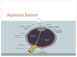
External Eye Structures: Eye brow for shape, movement , hair distribution : normal finding revealed symmetrical in shape intact skin and evenly hair distribution , symmetrical movement Eye lashes : evenly hair distribution ,curl directed outward Eye lids : inspect for color ,lesion and movement : color same as the face no discoloration , free of lesion and discharge when lids closed it should be symmetrical complete , sclera not visible
Assess bulbar conjunctiva ( cover the eye ball and sclera : transparent ,sclera white or yellow in skinny person . capillary appears free of lesion( retract) Assess palpebral conjunctiva line the eye lid normal finding pink to red ,shiny smooth free of discharge and lesion
By using gauze or cotton touch the client cornea blinking indicate that the trigeminal (5th) nerve is intact
Inspect pupil : black in color , equal in size normally 3-7mm in diameter and round ,iris flat and rounded Assess pupil for: Direct reaction to light: pupil constrict ( penlight) Reaction to Accommodation : Penlight placed 10cm or 4 in near nose bridge , normal finding pupil constrict when looking on near object and dilated on far object. Documentation: PERRLA (pupils equal round reacting to light and accommodation)
Assess Lacrimal Gland Sac and Nasolacrimal duct Inspect and use index to palpate : Free of edema ,no tenderness ,no excessive tearing Lacrimal gland in outer eye canthus Lacrimal sac and duct inner canthus of eye
Exteraoccular Muscle Test Stand direct in front of the client use penlight within 1ft distance (30 cm) ask the client to follow the object move toward 6 cardinal direction . Normal finding both eye coordinated movement Abnormal: eye fail to follow the object squint and strabismus ( cross eye) Nystagmus : caused by nerve impairment
How is visual field testing done? Visual field testing is performed on one eye at a time, with the opposite eye completely covered to avoid errors. In all testing, the patient must look straight ahead at all times in order to avoid testing the central vision rather than the periphery.
The reasons for this test are the following : * visual field testing is useful screening for glaucoma, *testing patient with glaucoma for treatment response, *screening and testing for lid droop or ptosis, particularly for insurance approval of lid lift surgical procedures
Normally the results are: * Temporal can detected within 90 degree central * Upward detected with 50 because of orbital ridge * Downward detected with 70 cheek bone * Nasal filed within 50 because of nose Normal : client can see the object Abnormal: small field glaucoma ,
Visual acuity: Near vision: within 30-36cm distance ask the client to read newspaper or magazine with keeping glass or lenses
Distance vision test within 20 feet or 6 meter by using snellen chart the client read the chart line from top to bottom . First number 20 indicate the distance Between the chart &client Second number 20 indicate The distance that normal Eye can read Glass and lenses kept
Distance from the chart D (distant) for the evaluation done at 20 feet (or 6 meters). N (near) for the evaluation done at 14 inches (or 36 cm). Eye evaluated OD (Latin oculus dexter) for the right eye. OS (Latin oculus sinister) for the left eye. OU (Latin oculi uterque) for both eyes. CC (Latin cum correctors) with correctors. SC (Latin sine correctors) without correctors
Performing functional vision test 1- light perception: shin penlight from lateral than off ask client about light if he recognize documentation : LP+ 2- Hand movement within 30 cm 1 feet move hand back ,front than stop ask client when it stopped doc: H\M 1ft + 3- Counting finger within 30 cm or 1 ft ask the client number of finger doc: F\C+
Ophthalmoscope:A lighted instrument, one of the most important tools for examining the optic disc . Normal orange to pink with vessel appearance Abnormal pale red spot