Understanding the Structures and Functions of the Eye
Explore the main structures of the eye including the iris, lens, cornea, retina, and more. Discover how these components work together to facilitate vision and light focusing. Engage in activities to enhance your knowledge of eye anatomy and function.

Download Presentation

Please find below an Image/Link to download the presentation.
The content on the website is provided AS IS for your information and personal use only. It may not be sold, licensed, or shared on other websites without obtaining consent from the author. Download presentation by click this link. If you encounter any issues during the download, it is possible that the publisher has removed the file from their server.
E N D
Presentation Transcript
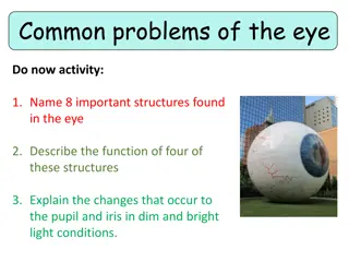
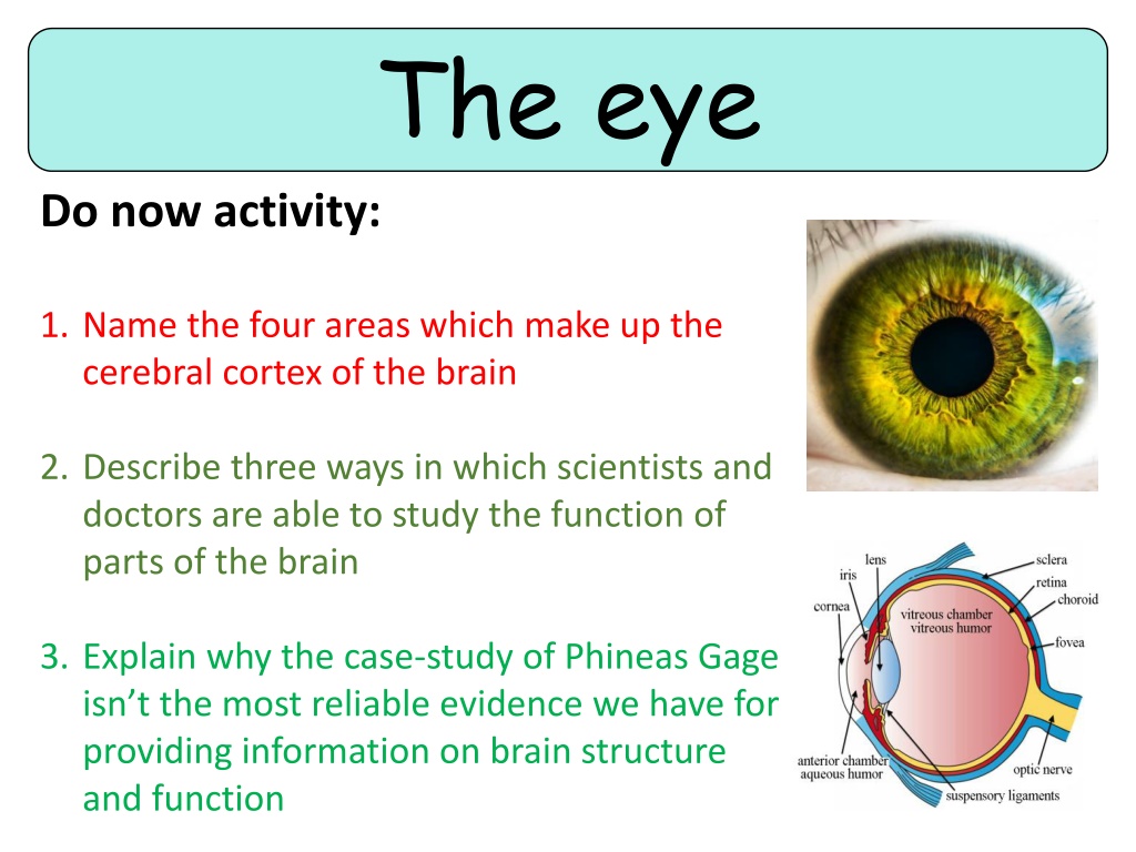
The eye Do now activity: 1. Name the four areas which make up the cerebral cortex of the brain 2. Describe three ways in which scientists and doctors are able to study the function of parts of the brain 3. Explain why the case-study of Phineas Gage isn t the most reliable evidence we have for providing information on brain structure and function
Progress indicators GOOD PROGRESS: Name and label the main structures of the eye Describe the functions of the main structures of the eye OUTSTANDING PROGRESS: Explain how the pupil reflex works and how the eye is able to focus light on the retina
Stimuli ~ Changes that occur in an animal s environment Receptors ~ Cells clustered together in sense organs which detect changes in the environment (stimuli). Think > Pair > Share: What do photoreceptor cells do? Where are they situated? What other structures in the eye help to focus the light on the retina?
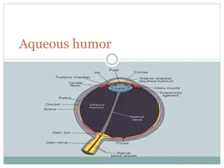
Task: In pairs, try and identify the names of the structures of the eye highlighted in the diagram: Optic nerve Lens Cornea Retina Iris Blind Spot Pupil Suspensory ligament Ciliary muscle Sclera 1. 2. 8. 3. 7. 4. 5. 9. 6. 10. You might not know all of them, but give it a go!
Self-assess your work! How many did you know? Ciliary muscle Suspensory ligament Iris Sclera Retina Pupil Lens Blind spot Cornea Optic nerve
The main structures of the eye Task: You have each been given a slip of information outlining the function of one of the structures listed below. Talk to each other to complete the following table in your books: Structure Function Iris Pupil Lens Cornea Suspensory ligament Ciliary muscle Retina Sclera Optic nerve Blind Spot
Self-assess: Structure Function Iris Made up of muscles which contract and relax to control the size of the pupil and therefore the amount of light that enters the eye. Pupil A hole located in the centre of the iris which allows light to strike the retina Lens The lens is a clear disc which is controlled by the ciliary muscles and suspensory ligaments. The lens focuses the light rays in order to produce a clear image on the retina. Cornea This is a transparent area of the sclera at the front of the eyeball. This lets light into the eye and has an important role of focusing the light rays onto the retina. Suspensory ligament The suspensory ligaments sit either side of the lens and are important for holding the lens in position Ciliary muscle The muscles are attached to the suspensory ligaments and by contracting/relaxing control the position of the lens so that it can focus the light. Retina This is the area at the back of the eye which contains clusters of light-sensitive cells. Sclera This is the white, outer layer of the eye. The sclera is quite tough and also strong so that little damage can come to the eyeball. Optic nerve When light-sensitive cells in the retina are stimulated the impulses are sent along sensory neurones in the optic nerve to the brain to be interpreted. Blind Spot The point where the optic nerve leaves the eye has no retina, therefore no light- sensitive cells.
The pupil reflex Equipment: Posters/blinds to black out the windows, torch METHOD: 1. Black out the room and allow a few minutes for the eyes to adjust. 2. Working in pairs, one person is the experimenter and the other the subject. The subject covers one eye. 3. Slowly bring the light source in from the side to within 5 10 cm of the subject s face. Remove the light if the subject feels any discomfort. 4. Describe any changes in pupil diameter observed.
What did you observe? Dim light Bright light & radial muscles relax & circular muscles relax
Explaining the pupil reflex Key Words: pupil, circular, iris, damage, enlarged, light, retina, radial The ____ is made out of muscle which contracts and relaxes to change the size of the _______ and so control the amount of ______ reaching the retina. In dim light, the pupil is ________ so as much light as possible can enter the eye. In order to enlarge the pupil the ______ muscles relax as the _______ muscles contract, causing the _____ to dilate. In bright light, the pupil is very small. This reduces the amount of light entering the eye so the light does not ______ the light- sensitive cells within the ______. In order to constrict the pupil the ______ muscles relax as the _______ muscles contract.
Self-assessment: The iris is made out of muscle which contracts and relaxes to change the size of the pupil and so control the amount of light reaching the retina. In dim light, the pupil is enlarged so as much light as possible can enter the eye. In order to enlarge the pupil the circular muscles relax as the radial muscles contract, causing the pupil to dilate. In bright light, the pupil is very small. This reduces the amount of light entering the eye so the light does not damage the light-sensitive cells within the retina. In order to constrict the pupil the radial muscles relax as the circular muscles contract.
Focusing the light The light that enters the eye must be focused clearly on the retina in order for a clear image to be produced. The light is focused by the process of refraction, where light changes direction as it passes through the eye. The cornea and the jelly in the eyeball change the direction of the light rays, but they always refract it the same amount. The ciliary muscles can also contract or relax to control the shape of the lens, this effects how the much the lens refracts the light.
Task: 1. Annotate the diagram to show how an image is formed on the retina 2. Explain how the structures of the eye work together to focus the light on the retina, use the list of key words in your answer. Key Words: Ciliary muscle Lens Retina Light- sensitive cells Refraction Light rays Cornea
Plenary: Exam-style question The diagram below shows a reflex in the iris of a human eye in response to changes in light levels. A B Describe the changes in the pupils and the iris going from A to B in Figure 1. Explain how these changes occur and refer to the changes in light levels in your answer. (4 marks)
Mark Scheme: At A the pupils is constricted/at B the pupil has dilated 1 The pupils dilates in low light levels/dim light 1 The pupils dilates because the circular muscles of the iris relax 1 As the radial muscles of the iris contract 1
Photocopy and cut up for a card sort: Iris Made up of muscles which contract and relax to control the size of the pupil and therefore the amount of light that enters the eye. Pupil A hole located in the centre of the iris which allows light to strike the retina Lens The lens is a clear disc which is controlled by the ciliary muscles and suspensory ligaments. The lens focuses the light rays in order to produce a clear image on the retina. Cornea This is a transparent area of the sclera at the front of the eyeball. This lets light into the eye and has an important role of focusing the light rays onto the retina. Suspensory ligament The suspensory ligaments sit either side of the lens and are important for holding the lens in position Ciliary muscle The muscles are attached to the suspensory ligaments and by contracting/relaxing control the position of the lens so that it can focus the light. Retina This is the area at the back of the eye which contains clusters of light-sensitive cells. Sclera This is the white, outer layer of the eye. The sclera is quite tough and also strong so that little damage can come to the eyeball. Optic nerve When light-sensitive cells in the retina are stimulated the impulses are sent along sensory neurones in the optic nerve to the brain to be interpreted. Blind Spot The point where the optic nerve leaves the eye has no retina, therefore no light- sensitive cells.