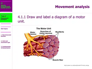
Upper and Lower Motor Neurons
 undefined
Dr Abdulrahman Alhowikan
Collage of medicine
Physiology Dep.
Upper and lower motor lesions
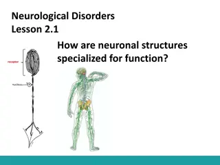
What is upper motor neuron and lower motor
neuron.
What are its function.
Know about damage of UMNL cause loss of
motor function, hyper reflexia, increased deep
reflexes, and presence of babinski’s sign.
Know about damage of LMNL cause loss of
power, wasting loss of muscle tone, decreased or
absent deep reflexes.
What effects are produced if damaged.
They should also know the progress of
hemiplegia, paraplegia.
Objectives:
Upper motor neuron:
A
neuron whose axons
descend from the
cerebral cortex or
brainstem to the spinal
cord (or to a cranial
nerve motor nucleus) to
influence in the lower
motor neurons
a lesion of the neural
pathway above the
anterior horn cell or
motor nuclei of the
cranial nerves.
Pyramidal system
=
lateral and anterior
corticospinal tracts +
corticobulbar tract
Extra-pyramidal
system
= rubrospinal
+ olivospinal +
lateral and medial
reticulospinal +
tectospinal +
vestibulospinal tracts
Corticospinal
–Principal pathway of
production of
skilled voluntary
movements.
Vestibulospinal
–Postural changes to
compensate for tilts and movements
of the body and head
Reticulospinal
–Major alternative
route (to the pyramidal tract) by which
spinal motor neurons are controlled,
both influencing motor neurons
directly, and
regulating the sensitivity
of spinal reflex arcs.
Tectospinal
–assumed to be
important in reflex
turning of the head
in response to visual
and perhaps
other stimuli, but little is actually
known about the function in humans
Rubrospinal
– main pathway for the
mediation (link) of motor movement
Major Descending Neuronal Tracts Involved
Lower motor neuron
are classified based on the
type of muscle fiber they innervate:
Alpha motor neurons (α-MNs)
innervate extrafusal muscle fibers, the most
numerous type of muscle
Gamma motor neurons (γ-MNs)
innervate intrafusal muscle fibers, which together
with sensory afferents compose muscle spindles.
lower motor neuron lesion
,
which affects nerve
fibers travelling from the anterior horn of the
spinal cord to the relevant muscle(s).
Upper Motor Neuron
Lesion ( UMNL)
Can result from
(1) Haemorrhage
,
thrombosis or embolism
in the internal capsule
(2) Spinal cord transection
or hemisection
Lower Motor Neuron
Lesion,( LMNL)
Can result from
(1) Spinal root lesions or
peripheral nerve lesion
( e.g. nerve injury by
trauma or compressive
lesion
(2) Anterior horn cell
lesions
( e.g. , poliomyelitis,
motor neuron disease
UMNL:
(1) No muscle wasting, except from disuse ( disuse atrophy)
(3) Spasticity ( hypertonia ) , called
clasp-knife spasticity ”
(4) Clonus present
(5) Brisk ( exaggerated ) tendon jerks
(6) Extensor plantar reflex , Babinski sign ( dorsiflexion of
the big toe and fanning out of the other toes )
(7) Absent abdominal reflexes
(8) No fasciculations
(
involuntary muscle contraction and
relaxation visible under the skin)
(9) No fibrillation potential in EMG
(rapid, irregular, and
unsynchronized contraction of muscle fibers
{
clasp-knife spasticity
https://www.youtube.com/watch?v=ovQkcw86pMo
------------ ---------
Clonus
https://www.youtube.com/watch?v=9XWmpBz4BVo
------------ ------
Babinski sign
https://www.youtube.com/watch?v=QHV9uig0Kuw
1.
Marked muscle wasting (atrophy )
2.
Flacidity (Hypotonia ) , hence given the
name “ flaccid paralysis ”
3.
No clonus
4.
Diminished or absent tendon reflexes
5.
Absent abdominal reflexes
6.
Fasciculations may occur
7.
Fibrillation potentials present .
Features :
(
1)UMNL involving the half of the body contralateral to the
site of the lesion .
(2) Hypertonia causes the limbs to acquire a specific
posture
Causes :
cerebral heamorrhage ,
thrombosis or embolism
(Difference???)
results in
paralysis of the opposite half of the body
.The commonest cause of cerebral
haemorrhage is hypertension ,
usually associated with rupture of
the lenticulo-striate branch of the
middle cerebral artery in the
internal capsule .
upper limb is
(a) adducted to the side of the trunk , (b)
flexed at the elbow ,(c) the forearm is
semipronated,(d) with flexion of the wrist and
fingers.
lower limb
is
(a) adducted and (b) extended at the knee
and ankle.
(3) Loss of sensation on the opposite side of the body
(Hemianesthesia),
due to damage of the
thalamocortical fibers.
(4)
Homonymous hemianopia
( loss of vision in two
corresponding halves of the visual fields in both
eyes),
may occur if the optic radiation is lesioned
Due to complete spinal cord transection ( E.G. Following
tumor , trauma ( E.G . bullet injury , fractures spine , etc )
The higher the level of the section, the more serious are the
consequences.
If the transection is in the upper cervical region
immediate death follows, due to paralysis of all respiratory
muscles
In the lower cervical region below the 5th cervical segment
diaphragmatic respiration is still possible, but the
patient suffers complete paralysis of all four limbs
(quadriplegia)
.
Transection lower down in the thoracic region allows
normal respiration but the patient ends up with paralysis of
both lower limbs (paraplegia).
Paraplegia
14
Spinal shock
Recovery of reflex activity
Paraplegia in extension
In the immediate period following transection there is :
(1) complete loss of spinal reflex activity below the level of
the lesion .
(2) Loss of all sensations (
anesthesia)
and voluntary
movement ( paralysis) below the level of the lesion , due
to interruption of all sensory and motor tracts
(3) Loss of tendon reflexes and superficial reflexes
(abdominal , plantar & withdrawal reflexes ) .
(5) The loss of muscle tone (flaccidity) and absence of any
muscle activity (muscle pump ) lead to decreased
venous return
causing the lower limbs to become
cold and blue in cold weather
Spinal shock
16
(6) The wall of the urinary bladder becomes
paralysed and urine is retained. This is known as
retention with overflow.
(bladder is full but is not emptied
completely)
(7)Loss of vasomotor tone occurs, due to
interruption of fibers that connect the vasomotor
centres in the medulla oblongata with the lateral
horn cells of the spinal cord, which project
sympathetic vasoconstrictor impulses to blood
vessels.
vasodilatation causes a fall in blood
pressure; the higher the level of the section, the
lower the blood pressure.
This stage varies in duration but usually lasts a
maximum of 2-6 weeks, after which some reflex
activity recovers.
17
As the spinal shock ends , spinal reflex activity
appears again this partial recovery may be due to:
1.
increase in the natural degree of excitability of
the spinal cord neurons
below the level of the
section, presumably to make up for the loss of
supraspinal facilitatory influences.
2.
It may also be due to sprouting (growth) of
fibres from remaining inputs.
18
1.
Gradual rise of
arterial blood pressure due to return of
spinal vasomotor activity
in the lateral horn cells. But,
since vasomotor control from the medulla is absent, the
blood pressure is not stable.
2.
Return of spinal reflexes: Flexor reflexes
return earlier
than extensor ones.
Babiniski
sign
is one of the earliest
signs of this stage . The return of the stretch reflex ( &
cosequently muscle tone) , and vasoconstrictor tone in
arterioles and venules
improve the circulation
through the limbs.
3.
Recovery of visceral reflexes: return of
micturition,
(urination ) defecation (pass stool)
& erection
reflexes.
4.
However , voluntary control over micturition and
defecation , and the sensation of bladder and rectal
fullness are permanently lost.
19
5.
Mass reflex appears in this stage:
A minor painful
stimulus to the skin of the lower limbs will not only
cause withdrawal of that limb but will evoke many
other reflexes through spread of excitation
(by
irradiation) to many autonomic centers. So the bladder
and rectum will also empty, the skin will sweat, the
blood pressure will rise
6.
Since effective regeneration never occurs in the
human central nervous system, patients with complete
transection never recover fully
. Voluntary movements
and sensations are permanently lost
; however,
patients who are rehabilitated and properly managed
may enter into a more advanced stage of recovery.
20
1.
During this
stage the tone in extensor muscles
returns gradually to exceed that in the flexors
.
The lower limbs become extended. Extensor
reflexes become exaggerated, as shown by
brisk tendon jerks and by the appearance of
clonus. The positive supportive reaction
becomes well developed and the patient can
stand on his feet with appropriate support.
2.
The
flexor withdrawal reflex which appeared in
the earlier stage is associated during this stage
with the crossed extensor reflex.
Reference book
Guyton & Hall: Textbook of Medical Physiology 12E
T
e
x
t
b
o
o
k
:
R
e
v
i
e
w
o
f
M
e
d
i
c
a
l
P
h
y
s
i
o
l
o
g
y
(
G
a
n
o
n
g
)
Thank you
Upper motor neurons descend from the cerebral cortex or brainstem to influence lower motor neurons, while lower motor neurons innervate muscle fibers. Damage to upper motor neurons results in hyperreflexia and Babinski's sign, whereas lower motor neuron lesions lead to muscle weakness and decreased reflexes. Explore the functions and effects of these neurons in hemiplegia and paraplegia.

Download Presentation

Please find below an Image/Link to download the presentation.
The content on the website is provided AS IS for your information and personal use only. It may not be sold, licensed, or shared on other websites without obtaining consent from the author.If you encounter any issues during the download, it is possible that the publisher has removed the file from their server.
You are allowed to download the files provided on this website for personal or commercial use, subject to the condition that they are used lawfully. All files are the property of their respective owners.
The content on the website is provided AS IS for your information and personal use only. It may not be sold, licensed, or shared on other websites without obtaining consent from the author.
E N D
Presentation Transcript
Upper and lower motor lesions Dr Abdulrahman Alhowikan Collage of medicine Physiology Dep.
Objectives: Objectives: What is upper motor neuron and lower motor neuron. What are its function. Know about damage of UMNL cause loss of motor function, hyper reflexia, increased deep reflexes, and presence of babinski s sign. Know about damage of LMNL cause loss of power, wasting loss of muscle tone, decreased or absent deep reflexes. What effects are produced if damaged. They should also know the progress of hemiplegia, paraplegia.
Upper motor neuron: A neuron whose axons descend from the cerebral cortex or brainstem to the spinal cord (or to a cranial nerve motor nucleus) to influence in the lower motor neurons a lesion of the neural pathway above the anterior horn cell or motor nuclei of the cranial nerves.
Pyramidal system lateral and anterior corticospinal tracts + corticobulbar tract Extra system + olivospinal + lateral and medial reticulospinal + tectospinal + vestibulospinal tracts Pyramidal system = Extra- -pyramidal system = rubrospinal pyramidal
Major Descending Neuronal Tracts Involved Corticospinal Principal pathway of production of skilled voluntary movements. Vestibulospinal Postural changes to compensate for tilts and movements of the body and head Reticulospinal Major alternative route (to the pyramidal tract) by which spinal motor neurons are controlled, both influencing motor neurons directly, and regulating the sensitivity of spinal reflex arcs. Tectospinal assumed to be important in reflex turning of the head in response to visual and perhaps other stimuli, but little is actually known about the function in humans Rubrospinal main pathway for the mediation (link) of motor movement
Lower motor neuron type of muscle fiber they innervate: Alpha motor neurons ( -MNs) innervate extrafusal muscle fibers, the most numerous type of muscle Gamma motor neurons ( -MNs) innervate intrafusal muscle fibers, which together with sensory afferents compose muscle spindles. lower motor neuron lesion fibers travelling from the anterior horn of the spinal cord to the relevant muscle(s). Lower motor neuron are classified based on the lower motor neuron lesion, which affects nerve
Lower Motor Neuron Lesion,( LMNL) Can result from (1) Spinal root lesions or peripheral nerve lesion ( e.g. nerve injury by trauma or compressive lesion (2) Anterior horn cell lesions ( e.g. , poliomyelitis, motor neuron disease Upper Motor Neuron Lesion ( UMNL) Can result from (1) Haemorrhage , thrombosis or embolism in the internal capsule (2) Spinal cord transection or hemisection
UMNL: (1) No muscle wasting, except from disuse ( disuse atrophy) (3) Spasticity ( hypertonia ) , called clasp-knife spasticity (4) Clonus present (5) Brisk ( exaggerated ) tendon jerks (6) Extensor plantar reflex , Babinski sign ( dorsiflexion of the big toe and fanning out of the other toes ) (7) Absent abdominal reflexes (8) No fasciculations (involuntary muscle contraction and relaxation visible under the skin) (9) No fibrillation potential in EMG (rapid, irregular, and unsynchronized contraction of muscle fibers {
clasp-knife spasticity https://www.youtube.com/watch?v=ovQkcw86pMo ------------ --------- Clonus https://www.youtube.com/watch?v=9XWmpBz4BVo ------------ ------ Babinski sign https://www.youtube.com/watch?v=QHV9uig0Kuw
1. Marked muscle wasting (atrophy ) 2. Flacidity (Hypotonia ) , hence given the name flaccid paralysis 3. No clonus 4. Diminished or absent tendon reflexes 5. Absent abdominal reflexes 6. Fasciculations may occur 7. Fibrillation potentials present .
Causes : cerebral heamorrhage , thrombosis or embolism (Difference???) results in paralysis of the opposite half of the body .The commonest cause of cerebral haemorrhage is hypertension , usually associated with rupture of the lenticulo-striate branch of the middle cerebral artery in the internal capsule . Features : (1)UMNL involving the half of the body contralateral to the site of the lesion . (2) Hypertonia causes the limbs to acquire a specific posture
upper limb is (a) adducted to the side of the trunk , (b) flexed at the elbow ,(c) the forearm is semipronated,(d) with flexion of the wrist and fingers. lower limb is (a) adducted and (b) extended at the knee and ankle. (3) Loss of sensation on the opposite side of the body (Hemianesthesia), due to damage of the thalamocortical fibers. (4) Homonymous hemianopia ( loss of vision in two corresponding halves of the visual fields in both eyes), may occur if the optic radiation is lesioned
Paraplegia Due to complete spinal cord transection ( E.G. Following tumor , trauma ( E.G . bullet injury , fractures spine , etc ) The higher the level of the section, the more serious are the consequences. If the transection is in the upper cervical region immediate death follows, due to paralysis of all respiratory muscles In the lower cervical region below the 5th cervical segment diaphragmatic respiration is still possible, but the patient suffers complete paralysis of all four limbs (quadriplegia). Transection lower down in the thoracic region allows normal respiration but the patient ends up with paralysis of both lower limbs (paraplegia).
Spinal shock Spinal shock Recovery of reflex activity Recovery of reflex activity Paraplegia in extension Paraplegia in extension 14
Spinal shock In the immediate period following transection there is : (1) complete loss of spinal reflex activity below the level of the lesion . (2) Loss of all sensations (anesthesia) and voluntary movement ( paralysis) below the level of the lesion , due to interruption of all sensory and motor tracts (3) Loss of tendon reflexes and superficial reflexes (abdominal , plantar & withdrawal reflexes ) . (5) The loss of muscle tone (flaccidity) and absence of any muscle activity (muscle pump ) lead to decreased venous return causing the lower limbs to become cold and blue in cold weather
(6) The wall of the urinary bladder becomes paralysed and urine is retained. This is known as retention with overflow. (bladder is full but is not emptied completely) (7)Loss of vasomotor tone occurs, due to interruption of fibers that connect the vasomotor centres in the medulla oblongata with the lateral horn cells of the spinal cord, which project sympathetic vasoconstrictor impulses to blood vessels. vasodilatation causes a fall in blood pressure; the higher the level of the section, the lower the blood pressure. This stage varies in duration but usually lasts a maximum of 2-6 weeks, after which some reflex activity recovers. 16
As the spinal shock ends , spinal reflex activity appears again this partial recovery may be due to: 1. increase in the natural degree of excitability of the spinal cord neurons below the level of the section, presumably to make up for the loss of supraspinal facilitatory influences. 2. It may also be due to sprouting (growth) of fibres from remaining inputs. 17
1.Gradual rise of arterial blood pressure due to return of spinal vasomotor activity in the lateral horn cells. But, since vasomotor control from the medulla is absent, the blood pressure is not stable. 2. Return of spinal reflexes: Flexor reflexes return earlier than extensor ones. Babiniski sign is one of the earliest signs of this stage . The return of the stretch reflex ( & cosequently muscle tone) , and vasoconstrictor tone in arterioles and venules improve the circulation through the limbs. 3.Recovery of visceral reflexes: return of micturition,(urination ) defecation (pass stool) & erection reflexes. 4.However , voluntary control over micturition and defecation , and the sensation of bladder and rectal fullness are permanently lost. 18
Mass reflex appears in this stage: A minor painful stimulus to the skin of the lower limbs will not only cause withdrawal of that limb but will evoke many other reflexes through spread of excitation (by irradiation) to many autonomic centers. So the bladder and rectum will also empty, the skin will sweat, the blood pressure will rise 5. Since effective regeneration never occurs in the human central nervous system, patients with complete transection never recover fully. Voluntary movements and sensations are permanently lost; however, patients who are rehabilitated and properly managed may enter into a more advanced stage of recovery. 6. 19
During this stage the tone in extensor muscles returns gradually to exceed that in the flexors. The lower limbs become extended. Extensor reflexes become exaggerated, as shown by brisk tendon jerks and by the appearance of clonus. The positive supportive reaction becomes well developed and the patient can stand on his feet with appropriate support. 1. The flexor withdrawal reflex which appeared in the earlier stage is associated during this stage with the crossed extensor reflex. 2. 20
Reference book Guyton & Hall: Textbook of Medical Physiology 12E Textbook : Review of Medical Physiology (Ganong) Thank you Thank you


")

 adducted to the side of the trunk , (b)")


 The wall of the urinary bladder becomes")