Neurological Assessment Essentials for Upper and Lower Limb Function
Learn about the motor and sensory nerve roots essential for evaluating upper and lower limb function, limb reflexes, and pathways of sensation. Understand key differences between UMN and LMN signs, common viva questions, and tips for preparing for finals.

Uploaded on Sep 17, 2024 | 0 Views
Download Presentation

Please find below an Image/Link to download the presentation.
The content on the website is provided AS IS for your information and personal use only. It may not be sold, licensed, or shared on other websites without obtaining consent from the author.If you encounter any issues during the download, it is possible that the publisher has removed the file from their server.
You are allowed to download the files provided on this website for personal or commercial use, subject to the condition that they are used lawfully. All files are the property of their respective owners.
The content on the website is provided AS IS for your information and personal use only. It may not be sold, licensed, or shared on other websites without obtaining consent from the author.
E N D
Presentation Transcript
Limbs: Tutor Information PULSE: Preparation for Finals Tutor name
Resource summary Common viva questions/topics Case-based additional information (Cases 1 9)
Common questions Things you might pick up and questions you will get asked
UMN vs LMN LMN UMN Inspection Wasting, fasciculation Decreased Pronator drift Tone Increased Power Decreased Decreased Reflexes Reduced Brisk, clonus Plantars Down Up
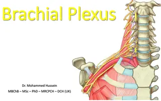
What are the motor nerve roots for the upper limb? There is a difference between assessing nerve roots and nerves; but in general they can both be done in the same series of movements C5 = shoulder abduction (axillary) C6 = elbow flexion (musculocutaneous) C7 = wrist extension (radial) C8 = finger flexion (median/ulnar) T1 = finger abduction (ulnar) T1 = thumb abduction (median)
What are the motor nerve roots for the lower limb? L1/2 = hip flexion L3/4 = knee extension L4 = ankle dorsiflexion L5 = big toe extension S1 = ankle plantarflexion L5/S1 = knee flexion L5/S1 = hip extension
What are the nerve roots for the limb reflexes? S1/2 = ankle L3/4 = knee C5/6 = biceps, supinator C7/8 = triceps Reinforcement if can t find reflex
What are the main pathways of sensation? Again remember to use sternum as reference point, and get patient to close their eyes Different modalities; Light touch and proprioception = dorsal column Pain and temperature = spinothalamic tract
What are the nerve roots for sensation in the upper limb? C4 = shoulder tip C5 = regimental badge C6 = tip of thumb (6 shooter) C7 = tip of middle finger (7-up) C8 = tip of little finger T1 = medial mid-forearm
What are the nerve roots for sensation in the lower limb? L2 = upper thigh L3 = above knee (3 the knee) L4 = medial mid-leg L5 = dorsum of foot S1 = lateral sole of foot (stand on S1)
What are the causes of peripheral neuropathy? ABCDE-O Alcohol (mixed) B12 deficiency (mixed) Chronic renal failure (sensory) Diabetes (sensory) Every vasculitis (mixed) Others - Guillain-Barre (motor), lead poisoning (motor), paraneoplastic (mixed), amyloid (mixed), Lyme disease
What are the causes of proximal myopathy? Muscular dystrophy Poly and dermatomyositis Cushings, Acromegaly, thyrotoxicosis Diabetic amyotrophy Etoh, statins, steroids Paraneoplastic
What is Froments sign? Froments = ulnar nerve palsy; loss of thumb adduction FrOments - makes an O shape
What are the causes of a positive prayer sign? Fixed flexion deformity of the fingers RA Scleroderma Diabetes Ulnar nerve palsy (partial claw hand) T1 palsy (complete claw hand) Dupytren s contracture
What are the manifestations of a T1 nerve root lesion? Seen in e.g. Pancoast tumours Horners syndrome - ptosis, miosis, anhydrosis Pain/sensory loss in axilla Complete claw hand Wasting of interossei
To complete my examination Full cranial nerve examination (homonymous hemianopia, visual defects) Assessment of higher cognitive function speech (dominant hemisphere) and sensory inattention (non-dominant hemisphere) Cardiovascular examination including BP, Carotid bruits and murmurs, AF, Cardiovascular history - modifiable risk factors
Differential Diagnoses Vascular ischaemia, infarction, embolism, heamorrhage SOL malignancy, abcess, hydrocephalus Inflammatory MS, Consider the age of the patient when ranking these.
Where is the lesion? Classify by site and/or by artery involved . Site of lesion Cerebral Hemisphere Pattern Contra-lateral UMN motor weakness +/- higher order cognitive deficit. Internal Capsule Contra-lateral UMN weakness only Brain-stem Contra-lateral UMN lesion with Ipsilateral LMN cranial nerve palsies. Spinal Cord Ipsilateral UMN motor weakness. Motor function and reflexes preserved above the lesion. LMN features at level of lesion, UMN features below.
Artery Carotid Middle Cerebral Artery Pattern of stroke Similar to MCA. Contrateral hemiplegia, hemisensory loss mainly of face and arm. Supplies front and middle of cerebrum causing symptoms in the contralateral leg. Spares the face Contralateral homonymous hemianopia Cerebellar signs, LMS Anterior cerebral artery Posterior Cerebral Vertebrobasilar circulation
Causes of Stroke Ischaemic (85%) Haemorrhagic (15%) Atherosclerosis HTN Dissection Aneurysms Vasculitis Elderly Injury (inc iatrogenic) Malformations Spasm (migraine) Autoimmune (vasculitis) Embolus (cardiac AF, IE) Toxin (warfarin) Other haemorrhagic transformation Occlusion Mets/primary brain tumour Hypercoaguable states (e.g. hereditary, OCP, polycythaemia, neoplasia) Accident head injury
What are the causes of a peripheral neuropathy? Physical trauma Compression Pinching Cutting Projectile injuries Strokes including prolonged occlusion of blood flow Others Shingles Malignant disease HIV Radiation Chemotherapy Genetic Toxic Alcohol Drugs - Fluoroquinolones - Vincristin - Phenytoin - Nitrofurantoin - Isoniazid Organic metals Heavy metals Excess Vit B6 (pyridoxine) Vit deficiency B12 A E B1 Charcot-Marie-Tooth syndrome Friedrich s ataxia Metabolic/endocrine Inflammatory disease Guillain-Barr syndrome SLE Leprosy Sjogren s DM Chronic renal failure Porphyria Amyloidosis Liver failure Hypothyroidism Most common causes: 1. DM 2. Alcohol 3. Vitamin deficiency
Causes Degenerative disease of the spine Brown-Sequard Syndrome Syringomyelia Motor changes Ipsilateral UMN signs below the hemisection (corticospinal tract) LMN signs at the level of hemisection on the same side Cord tumour Haematomyelia MS Sensory changes Contralateral pain and temperature loss (upper level of sensory loss usually a few segments below the level of the lesion) spinothalamic tract Ipsilateral vibration and proprioception loss (dorsal column) In segment of lesion ipsilateral anaesthesia and zone of hyperaesthesia Angioma Trauma, e.g. bullet or stab wounds Myelitis Post-radiation myelopathy
O/E Lower limbs Increased tone and brisk reflexes Upgoing plantars bilaterally Upper limbs Decreased pin-prick in hands Atrophy and weakness of hand muscles Cervical Myelopathy Diagnosis
Localise the Lesion Peripheral Neuropathy Glove and Stocking Likely cause: EtOH XS (+/- B1 deficiency) Particular causative diseases have predilection for specific nerves or fibre types and for certain components of the nerve course Mononeuropathy (tend to be unilateral) CTS, CPNP Mononeuritis multiplex vasculitis, diabetes Polyneuropathy (diffuse, symmetrical fashion) Autonomic neuropathy diabetes, amyloidosis, GBS
Peripheral Neuropathy Aetiology Tests Inflammatory Guillain Barr Syndrome, CIDP, RA, SLE, vasculitis Bloods to Ix cause (GBS/CIDP) ?Lumbar puncture Infective - Leprosy Nerve conduction studies Toxic EtOH (+ thiamine deficiency), Drugs (chemo) Metabolic Diabetes, Hypothyroid, CKD Treatment Idiopathic (one third of cases cause unknown) Neuropathic analgesia, Mx underlying cause Neoplastic paraneoplastic, MGUS, myeloma Deficiency vitamins B12, B1, B6 Genetic HSMN (Charcot-Marie Tooth), Friedrich s Ataxia
O/E Large sore tongue Lower limbs Spastic paresis Romberg positive, wide based gait Bilateral Babinski sign Loss of sensation in a stocking distribution Absent ankle reflexes Diagnosis Subacute Combined Degeneration of Spinal Cord
O/E Lower limb Paraplegia, with bilateral Babinski sign Bilateral loss of pain and temp Normal JPS and vibration Diagnosis Anterior Spinal Artery Syndrome
Infarction of anterior spinal artery Aetiology Aortic insufficiency Vasculitis Trauma/Neoplasia Ischaemia Complete motor paralysis ASA Syndrome Loss of pain and temp (STT) Bladder & Bowel dysfunction Dorsal columns intact
O/E Left ptosis Diplopia on left and right gazes Left eye doesn t fully elevate Mild facial weakness Weakness of neck extension Proximal muscle weakness (MRC 4-) Diagnosis Myasthenia Gravis
O/E Flaccid paralysis in arms and legs Widespread areflexia Absent sensation in arms and legs Evidence of sacral sparing voluntary anal sphincter contraction Hypotension Spinal Shock Diagnosis