Understanding Teleost Gill Histology and Function
This presentation delves into the intricate histology of teleost gills, showcasing the structure of primary and secondary lamellae, epithelial cells, mucous cells, and more. It illustrates the branchial anatomy in bony fish, detailing the arrangement of holobranchs and hemibranchs. Additionally, it explores the essential functions of gills in gaseous exchange, acid-base balance, and osmoregulation.
Download Presentation

Please find below an Image/Link to download the presentation.
The content on the website is provided AS IS for your information and personal use only. It may not be sold, licensed, or shared on other websites without obtaining consent from the author. Download presentation by click this link. If you encounter any issues during the download, it is possible that the publisher has removed the file from their server.
E N D
Presentation Transcript
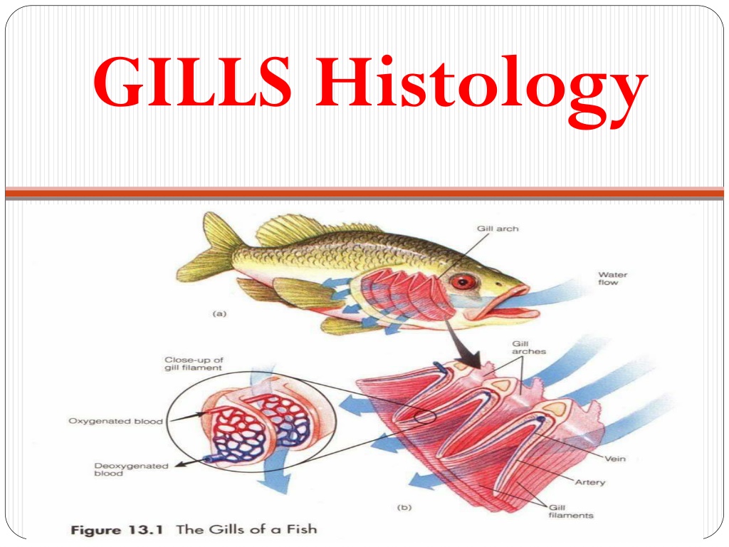
Normal Teleost gill form (anatomy) Branchia in greek = gills In bony fish (Teleosts): Gills lie in a branchial cavity covered by the operculum: Usually two sets of four holobranchs Each holobranch consists of two hemibranchs ( half gill ): Anterior and posterior Hemibranchs consist of a row of long filaments (primary lamellae) with semilunar folds (secondary lamellae). Lamellae or filaments: Connective tissue scaffold (epithelial cells) framing a vascular network providing blood flow primarily used for gas and ion exchange. Primary and secondary.
1. gill 1. gill raker 2. mucosal 2. mucosal epithelium; 3. epithelium; 3. basement basement membrane; 4. membrane; 4. submucosa submucosa; ; 5. bone; 5. bone; 6. adipose tissue; 6. adipose tissue; 7. efferent 7. efferent branchial arterioles; arterioles; 8. afferent 8. afferent branchial artery; artery; 9. primary lamellae; 9. primary lamellae; 10. secondary 10. secondary lamellae. lamellae. raker; ; branchial branchial
Teleost gill structure Holobranch Gill filaments Hemibranch (anterior)
Gill lamellae Secondary lamellae Fish Gill Filaments Primary lamellae
GILLS structure 1. primary lamella; 2. secondary lamella; 3. epithelial cell; 4. mucous cell; 5. pillar cell; 6. lacuna (capillary lumen); 7. erythrocyte within capillary lumen; 8. undifferentiated basal cell; 9. central venous sinus
1. primary lamella 1. primary lamella ; 2. extracellular ; 2. extracellular cartilaginous matrix; cartilaginous matrix; 3. chondrocytes; 3. chondrocytes; 4. secondary lamella; 4. secondary lamella; 5. epithelial 5. epithelial cell; cell; 6. mucous cell; 6. mucous cell; 7. chloride cell; 7. chloride cell; 8. pillar cell; 8. pillar cell; 9. lacuna (capillary 9. lacuna (capillary lumen); lumen); 10. red blood cells 10. red blood cells within lacun within lacuna.
Normal Teleost gill function Gill functions: Gaseous exchange = O2 via 2o lamellae. Acid-base balance = equilibrium between acidity/alkalinity. Osmoregulation = adjustment of internal osmotic pressure in relation to surrounding medium. Excretion of nitrogenous waste = ammonia. Gills sensitive to a range of environmental pollutants. Gaseous exchange = secondary lamellae: Consist of an envelope of epithelial cells Usually one layer thick Supported/separated by specialised cells: Pillar cells = regulate blood flow. Chloride cells = maintain internal ionic homeostasis.
References Deeds, J.R., Reimschuessel, R. and Place, A.R. (2006). Histopathological Effects in Fish Exposed to the Toxins from Karlodinium micrum. Journal of Aquatic Animal Health. 18(2), 136-148. Evans, D.H., Piermarini, P.M. and Choe, K.P. (2005). The Multifunctional Fish Gill: Dominant Site of Gas Exchange, Osmoregulation, Acid-Base Regulation, and Excretion of Nitrogenous Waste. Physiological Review: 85, 97 177. Hallegraeff, G., Mooney, B. and Evans, K. (2010). What triggers Fish-Killing Karlodinium veneficum Dinoflagellate Blooms in the Swan Canning River System?. Swan Canning Research and Innovation Program. H.W. Ferguson. (2006). Systemic pathology of fish. A Text and Atlas of Normal Tissues in Teleosts and their Responses in Disease. Scotian Press, London. McGavin, M.D. and Zachary, J.F. (2007). Pathological basis of Veterinary Disease. 4th Ed. Mosby Elsevier, Miissouri. Roberts. R.J. (1989). Fish Pathology. 2nd Ed. W.B. Saunders, London. University of Maryland College Park Campus, Aquatic Pathobiology Centre, Virginia-Maryland Regional College of Veterinary Medicine, Atlas of Flathead Minnow Normal Histology. Retreived from the World Wide Web August 2012: http://aquaticpath.umd.edu/fhm/resp.html
