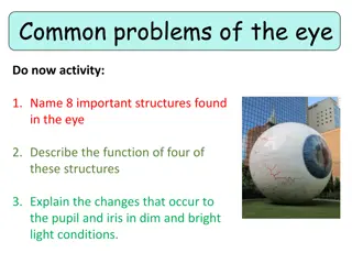
Anatomy and Histology of the Eye: A Comprehensive Study

1
C
o
r
n
e
a
I
r
i
s
A
n
t
e
r
i
o
r
c
h
a
m
b
e
r
P
o
s
t
e
r
i
o
r
c
h
a
m
b
e
r
C
i
l
i
a
r
y
b
o
d
y
a
n
d
p
r
o
c
e
s
s
e
s
V
i
t
r
e
o
u
s
b
o
d
y
H
y
a
l
o
i
d
c
a
n
a
l
O
p
t
i
c
n
e
r
v
e
C
e
n
t
r
a
l
r
e
t
i
n
a
l
a
r
t
e
r
y
&
v
e
i
n
R
e
t
i
n
a
C
h
o
r
o
i
d
S
c
l
e
r
a
#
E
y
e
-
2
U
C
S
F
#
1
6
4
C
C
o
o
r
r
n
n
e
e
a
a
:
:
I
t
i
s
t
r
a
n
s
p
a
r
e
n
t
,
a
v
a
s
c
u
l
a
r
a
n
d
h
i
g
h
l
y
i
n
n
e
r
v
a
t
e
d
a
n
t
e
r
i
o
r
p
o
r
t
i
o
n
o
f
t
h
e
f
i
b
r
o
u
s
.
I
I
t
t
i
i
s
s
c
c
o
o
m
m
p
p
o
o
s
s
e
e
d
d
o
o
f
f
5
5
d
d
i
i
s
s
t
t
i
i
n
n
c
c
t
t
l
l
a
a
y
y
e
e
r
r
s
s
:
:
Bowman's membrane
Corneal stroma
Descemet's Membrane
Corneal
endothelium
Stratified squamous
Epithelium
Corneal fibroblasts
S
S
C
C
L
L
E
E
R
R
A
A
•
IT covers the
Posterior 5/6 of the
fibrous tunic.
•
Consists of bundles
of Type 1 collagen
(dense irregular
C.T.).
•
Melanocytes are
located in the
deeper regions.
I
I
R
R
I
I
S
S
I
t
i
s
f
o
r
m
e
d
o
f
5
l
a
y
e
r
s
R
R
e
e
t
t
i
i
n
n
a
a
I
t
i
s
f
o
r
m
e
d
o
f
1
0
d
i
s
t
i
n
c
t
l
a
y
e
r
s
F
r
o
m
I
n
s
i
d
e
t
o
o
u
t
s
i
d
e
•
1) Inner limiting layer
•
2) Optic nerve fiber layer
•
3) Ganglion cell layer
•
4) Inner plexiform layer
•
5) Inner nuclear layer
•
6) Outer plexiform layer
•
7) Outer nuclear layer
•
8) Outer limiting
membrane
•
9) Rods and cones layer
•
10) Pigmented epithelium
Types of cells in the retina:
Types of cells in the retina:
1.Pigmented epithelium
2.Nerve cells
:
A. Photoreceptor cells .
B. Bipolar neurons
C. Ganglion cells
3.Neuroglial cells
:
A. Muller’s cells
B. Astrocytes
Inside to outside
Inside to outside
C
C
O
O
N
N
J
J
U
U
N
N
C
C
T
T
I
I
V
V
A
A
It’s the
transparent mucous
membrane
lining the inner
surface of the eyelids (palpebral
conjunctiva) and reflecting onto
the sclera of the anterior surface
of the eye (bulbar conjunctiva).
1- Epithelium:
A. Stratified columnar epithelium
B. numerous goblet cells.
2- Lamina propria:
. Loose C.T.
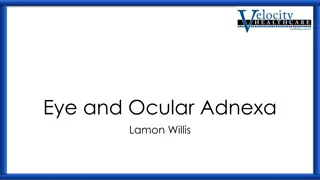
An in-depth exploration of the anatomy and histology of the eye, covering structures such as the cornea, sclera, choroid, iris, and more. Detailed descriptions of each component, including layers, composition, and functions, are provided alongside accompanying images for visual reference.

Download Presentation

Please find below an Image/Link to download the presentation.
The content on the website is provided AS IS for your information and personal use only. It may not be sold, licensed, or shared on other websites without obtaining consent from the author. Download presentation by click this link. If you encounter any issues during the download, it is possible that the publisher has removed the file from their server.
E N D
Presentation Transcript
Mustansiriyah University College of science Biology Dept. Zoology 4thclass Organs Histology LAB. (7) NAME : 1
Cornea Cornea Anterior chamber Anterior chamber UCSF UCSF #164 #164 Iris Iris Posterior chamber Posterior chamber Ciliary body and processes Ciliary body and processes Vitreous body Vitreous body Hyaloid canal Hyaloid canal Sclera Sclera Choroid Choroid # Eye # Eye- -2 2 Retina Retina Optic nerve Optic nerve Central retinal Central retinal artery artery & vein & vein
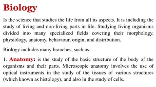
Cornea: Cornea: It is transparent ,avascular and highly innervated anterior portion of the fibrous. It is composed of 5 distinct layers: It is composed of 5 distinct layers: 1. Corneal Epithelium A.NON- keratinized stratified squamous tissue. B. Contains numerous free nerve endings. 2. Bowman s membrane A. It is homogenous non-cellular layer. B. Contains type 1 collagen fiber. 3. Stroma * Thickest layer (about 90%) Composed of Parallel Lamella of DENSE C.T. 4. Descemet s membrane A. Thick Basement membrane. B. Formed by : Corneal endothelium (the layer under it). 5. Corneal endothelium Simple squamous epithelium.
Stratified squamous Epithelium Bowman's membrane Corneal endothelium Corneal stroma Corneal fibroblasts Descemet's Membrane
SCLERA SCLERA IT covers the Posterior 5/6 of the fibrous tunic. Consists of bundles of Type 1 collagen (dense irregular C.T.). Melanocytes are located in the deeper regions.
CHOROID CILIARY BODY 1. IT is the vascular pigmented Posterior portion of the middle vascular tunic. 1. It is the anterior continuation of the choroid, it surrounds the lens. 2.Structure: IT composed mainly of Loose C.T. with Melanocytes. 2.Structure: IT formed of Loose vascular and pigment C.T.
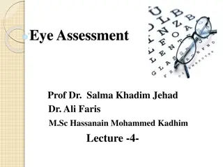
1) Anterior border layer Incomplete layer of: fibroblasts melanocytes. IRIS IRIS It is formed of 5 5 layers layers Poorly vascularized C.T with: fibroblasts melanocytes. 2) Stroma 3) Vessel layer Well-vascularized loose C.T. Centrally, it contains circularly arranged smooth muscle fibers (sphincter pupillae muscle). 4) Dilator pupillae muscle layer Contains radially arranged myoepithelial cells. 5) Posterior surface layer It is composed of two rows of pigmented epithelial cells.
Retina Retina It is formed of 10 distinct layers It is formed of 10 distinct layers From Inside to outside From Inside to outside 1) Inner limiting layer 2) Optic nerve fiber layer 3) Ganglion cell layer 4) Inner plexiform layer 5) Inner nuclear layer 6) Outer plexiform layer 7) Outer nuclear layer 8) Outer limiting membrane 9) Rods and cones layer 10) Pigmented epithelium
Inside to outside Types of cells in the retina: 1.Pigmented epithelium 2.Nerve cells: A. Photoreceptor cells . B. Bipolar neurons C. Ganglion cells 3.Neuroglial cells: A. Muller s cells B. Astrocytes
CONJUNCTIVA CONJUNCTIVA It s the transparent mucous membrane lining the inner surface of the eyelids (palpebral conjunctiva) and reflecting onto the sclera of the anterior surface of the eye (bulbar conjunctiva). 1- Epithelium: A. Stratified columnar epithelium B. numerous goblet cells. 2- Lamina propria: . Loose C.T.


 Anterior border layer")