Understanding H&E Staining in Histology
H&E staining, a widely used histological technique, involves staining cell nuclei blue and cytoplasmic components pink to aid in tissue structure analysis. This method provides essential insights into normal and pathological tissue changes, assisting in diagnoses and further testing decisions. The procedure involves steps like fixative application, deparaffinization, staining with hematoxylin and eosin, and mounting for examination. The advantages of H&E staining include its simplicity, quick results, and accurate differentiation of tissue structures. Overall, H&E staining is a crucial tool in histology for examining tissue architecture and identifying various cellular components.
Download Presentation

Please find below an Image/Link to download the presentation.
The content on the website is provided AS IS for your information and personal use only. It may not be sold, licensed, or shared on other websites without obtaining consent from the author. Download presentation by click this link. If you encounter any issues during the download, it is possible that the publisher has removed the file from their server.
E N D
Presentation Transcript
Staining HAEMATOXYLIN & EOSIN Supervised by : Dr.Sherifa Prepared by: Reem Aldossari Lama Alabdi
H&E staining: The best known and most widely used staining procedure, hematoxylin-eosin staining (H&E), uses hematoxylin solutions for nuclear staining and eosin solutions for cytoplasmic staining. In the first step, the nuclei are stained with a hematoxylin solution. The nuclei stain blue, dark violet to black. The second step is counterstaining with eosin Y or eosin B. In this process cytoplasm, collagen, keratin stain red.
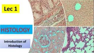
H&E staining is the standard staining method used in histology. It gives an overview of the structure of the tissue, enabling differentiation of the structures being examined as normal, inflammed ,degenerative and pathological changes . It is often possible to make a diagnosis based on the results of H&E staining, or it can be determined which special staining techniques are still necessary for subtyping.
APPLICATION Gives a general overview of the tissue structure. Can be used as a diagnostic tool.
Advantages of H&E staining Combination of the two stains gives outstanding results. Ready to use reagents . Easy to use. Results in just two steps.
Procedure for H & E Staining of Paraffin Sections: 1-(Fixative: Any general fixative is adequate. Ex.formalin) 2--Deparaffinization in : 3 - labeled Xylene I ( 1-3 minutes) 4- EtOH (ethanol alcohol) 100% (2-3 minutes 5-EtOH(ethanol alcohol) 96% (2-3 minutes) 6-EtOH(ethanol alcohol) 80% (2-3 minutes) 7-EtOH(ethanol alcohol) 70% (2-3 minutes) 8-Dist. water, 2 minutes. 9. Hematoxylin, approximately 10 minutes. 10-EtOH(ethanol alcohol) 70% (2-3 minutes) 11-EtOH(ethanol alcohol) 80% (2-3 minutes) 12-EtOH(ethanol alcohol) 96% (2-3 minutes) 13-Eosin Y solution, 2 to 5 minutes. 16- Absolute 100% ethanol, 1-2 minutes. 18-Xylene I, 1-2 minutes. 21-Mounting with DPX
