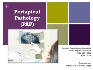
Understanding Periapical Pathology in Dentistry
Periapical pathology encompasses various conditions such as periapical abscess, granuloma, periodontal cyst, radicular cyst, and more. These pathologies result from different factors like infections, inflammation, or developmental issues. Identifying and treating these conditions is crucial in maintaining oral health.
Download Presentation

Please find below an Image/Link to download the presentation.
The content on the website is provided AS IS for your information and personal use only. It may not be sold, licensed, or shared on other websites without obtaining consent from the author. Download presentation by click this link. If you encounter any issues during the download, it is possible that the publisher has removed the file from their server.
E N D
Presentation Transcript
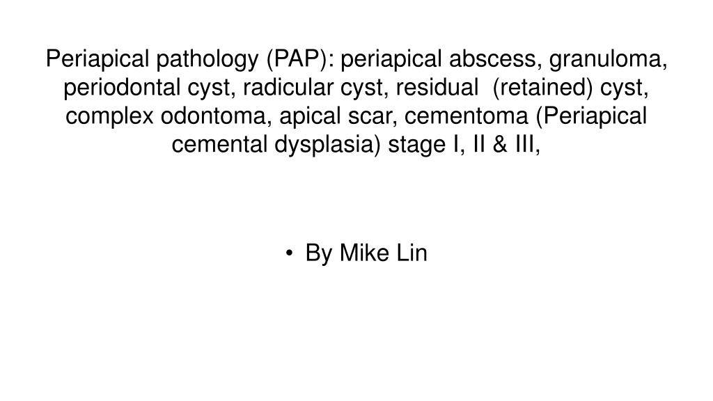
Periapical pathology (PAP): periapical abscess, granuloma, periodontal cyst, radicular cyst, residual (retained) cyst, complex odontoma, apical scar, cementoma (Periapical cemental dysplasia) stage I, II & III, By Mike Lin
Periapical Abscess A periapical abscess is a collection of pus at the root of a tooth, usually caused by an infection that has spread from a tooth to the surrounding tissues.
Granuloma It is a localized mass of chronic inflamed granulaiton tissue at the apex of a non vital tooth, due to pulp death & necrosis.
Periodontal cyst non-inflammatory developmental cyst that arises from the epithelial post-functional dental lamina, which is a remnant from odontogenesis. It is more common in middle-aged males.
Radicular cyst A radicular cyst is generally defined as a cyst arising from epithelial residues (cell rests of Malassez) in the periodontal ligament as a consequence of inflammation, usually following the death of the dental pulp.
Residual (retained) cyst residual cyst is used most often for retained radicular cyst from teeth that has been removed. Residual cysts are among most common cysts of the jaws.
Complex odontoma Tooth components are less well organized: toothlike structures are not formed.
Apical scar An apical scar is a scar that is found at the apex of a tooth from dense connective tissue. It is generally found after there has been a surgical procedure or endodontic treatments.
Cementoma (Periapical cemental dysplasia) stage I, II & III, Cementoma (stage 1) The involved tooth is vital and often painful. First appears as Fibrous tissue stage.
Cementoma (Periapical cemental dysplasia) stage I, II & III, Cementoma stage 2 Teeth are vital. Single or multiple circumscribed lesions at the apices of the teeth. Most Frequently involved the anterior mandibular region. Female > Male. No need for treatment
Cementoma (Periapical cemental dysplasia) stage I, II & III, Cementoma Stage III Multiple circumscribed radiopaque lesion at the apices of the teeth
Reference Garg, E. (2015, April 08). Periapical pathology. Retrieved from https://www.slideshare.net/ektagarg11/periapical-pathology Iannucci, J. M., & Howerton, L. J. (2017). Dental radiography: Principles and techniques. St. Louis, MO: Elsevier/Saunders. Neville, B. W. (2016). Oral and maxillofacial pathology. St. Louis: Elsevier.
