Understanding Bacterial Taxonomy and Staining Techniques
Bacterial taxonomy involves classifying and identifying bacteria, while staining techniques such as Gram staining help differentiate between Gram-positive and Gram-negative bacteria. This article discusses the importance of distinguishing bacterial strains, preparing smears, and performing differential staining methods. Detailed instructions for the Gram staining procedure are provided along with visuals to aid in the understanding of these fundamental microbiology practices.
Download Presentation

Please find below an Image/Link to download the presentation.
The content on the website is provided AS IS for your information and personal use only. It may not be sold, licensed, or shared on other websites without obtaining consent from the author. Download presentation by click this link. If you encounter any issues during the download, it is possible that the publisher has removed the file from their server.
E N D
Presentation Transcript
Lab -1-2- distinguish among strains and to group them by criteria of interest to microbiologists and other scientists. Bacterial Taxonomy Bacteria are classified and identified to identification of unknown bacteria
The Staining **Preparation of a smear and heat fixing Using a sterilized inoculating loop, transfer loopful of liquid suspension containing bacteria to a clean slide isolated colony from a culture plate to a slide with a water drop. Disperse the bacteria on the loop in the drop of water on the slide and spread the drop. It should be a thin, even smear. Allow the smear to dry thoroughly. Heat-fix the smear cautiously by passing the underside of the slide through the burner flame two or three times. It fixes the cell in the slide. Do not overheat the slide as it will distort the bacterial cells. Simple stains Diagnostic microbiology laboratory generally do not perform simple staining method ( using one stain ) . Differential staining such as Gram Staining and AFB Staining are commonly used to identify and differentiate the bacterial isolates. Simple staining can be useful in the some circumstances such as ( To differentiate bacteria from yeast cells: When endocervical swab or high vaginal swab culture is done in Blood agar both Staphylococcus spp and yeast cells may give similar looking colonies in Blood agar). or transfer an
Differential stains Gram Staining Gram procedure in Microbiology, was developed by Danish physician Hans Christian Gram in 1884. Gram staining is still the identification and taxonomic division. This differential staining procedure separates most bacteria into two groups on the basis of cell wall composition: Gram positive bacteria (thick layer of peptidoglycan- 90% of cell wall)- stains purple Gram negative bacteria (thin layer of peptidoglycan- 10% of cell wall and high lipid content) stains red/pink staining method, the most important cornerstone of bacterial
**Gram Staining Procedure: Flood air-dried, heat-fixed smear of cells for 1 minute with crystal violet staining reagent. Please note that the quality of the smear (too heavy or too light cell concentration) will affect the Gram Stain results. Wash slide in a gentle and indirect stream of tap water for 2 seconds. Flood slide with the mordant: Gram s iodine. Wait 1 minute. Wash slide in a gentle and indirect stream of tap water for 2 seconds. Flood slide with decolorizing agent (Acetone-alcohol decolorizer). Wait 10-15 seconds or add drop by drop to slide until decolorizing agent running from the slide runs clear . Flood slide with counterstain, safranin. Wait 30 seconds to 1 minute. Wash slide in a gentile and indirect stream of tap water until no color appears in the effluent and then blot dry with absorbent paper. Observe the results of the staining procedure under microscope. Gram-negative bacteria will stain pink/red and Gram-positive bacteria will stain blue/purple.
gram-stain-mine Procedure of Gram Staining; note the color change after each steps
Acid fast stain or acid fast bacilli test ( AFB ) The main aim of this staining is to differentiate bacteria into acid fast group and non-acid fast groups.This method is used for those microorganisms which are not staining by simple or Gram staining method, particularly the most Mycobacterium tuberculosis which are resistant and can only be medically important AFB visualized by acid-fast staining. When the smear is stained with carbol fuchsin, it solubilizes the lipoidal material present in the Mycobacterial cell wall but by the application of heat, carbol fuchsin further penetrates through lipoidal wall and enters into cytoplasm. Then after all cell appears red. Then the smear is decolorized with decolorizing agent (3% HCL in 95% alcohol) but the acid fast cells are resistant due to the presence of large amount of lipoidal material in their cell wall which prevents the penetration of decolorizing solution. The non-acid fast organism lack the lipoidal material in their cell wall due to which they are easily decolorized, leaving the cells colorless. Then the smear is stained with counterstain, methylene blue. Only decolorized cells absorb the counter stain and take its color and appears blue while acid-fast cells retain the red color.
**Procedure of Acid-Fast Stain Prepare bacterial smear on clean slide, using sterile technique. Cover the smear with carbol fuchsin stain. Heat the stain until vapour just begins to rise (i.e. about 60 C). Do not overheat. Allow the heated stain to remain on the slide for 5 minutes. Wash off the stain with clean water. Cover the smear with 3% v/v acid alcohol for 5 minutes or until the smear is sufficiently decolorized, i.e. pale pink. Wash well with clean water. Cover the smear with methylene blue stain for 1 2 minutes, using the longer time when the smear is thin. Wash off the stain with clean water. Wipe the back of the slide clean, and place it in a draining rack for the smear to air-dry. Examine the smear microscopically, using the 100 X oil immersion objective.
Interpretation of Acid-Fast Stain Acid fast: Bright red to intensive purple (B), Red, straight or slightly curved rods, occurring singly or in small groups, may appear beaded Non-acid fast: Blue color (A)
Special stains ( structural stains) Capsule Staining ( negativestaining) The main purpose of capsule stain is to distinguish capsular material from the bacterial cell. A capsule is a gelatinous outer layer secreted by bacterial cell and that surrounds and adheres to the cell wall. The capsule stain employs an acidic stain and a basic stain to detect capsule production. Negative staining methods contrast a darker colored, background with stained cells but an unstained capsule. The background is formed with india ink or nigrosin or congo red. A positive capsule stain requires a mordant that precipitates the capsule. By counterstaining with dyes like crystal violet or methylene blue, bacterial cell wall takes up the dye. Capsules appear colourless with stained cells against dark background.
**Procedure of Capsule Staining Place a small drop of a negative stain (India Ink, Congo Red, Nigrosin) on the slide. Using sterile technique, add a loopful of bacterial culture to slide, smearing it in the dye. Use the other slide to drag the ink-cell mixture into a thin film along the first slide and let stand for 5-7 minutes. Allow to air dry (do not heat fix). Flood the smear with crystal violet stain (this will stain the cells but not the capsules) for about 1 minutes. Drain the crystal violet by tilting the slide at a 45 degree angle and let stain run off until it air dries . Examine the smear microscopically (100X) for the presence of encapsulated cells as indicated by clear zones surrounding the cells. Note: negative staining is a mild technique that may not destroy the microorganisms, and is therefore unsuitable for studying pathogens
Result of Capsule Staining Capsule: Clear halos zone against dark background No Capsule: No Clear halos zone
Endospore Staining The Schaeffer-Fulton method is used to distinguish between the vegetative cells and the endospores. A primary stain (malachite green) is used to stain the endospores. Because endospores resist staining, the malachite green will be forced into (i.e, malachite green penetrate the spore wall) the endospores by heating. In this technique heating acts as a mordant. There is no need of using any decolorizer in this spore staining as the primary dye malachite green bind relatively weakly to the cell wall and spore wall .In fact If washed well with water the dye come right out of cell wall however not from spore wall once the dye is locked in. Water is used to decolorize the vegetative cells. ( Note: In Gram Staining and AFB Staining we use Alcohol or Acid Acohol or Acid as a decolorizer but in spore staining water is sufficient ( to be used as decolorizer) because: malachite green dye is water-soluble and does not adhere well to the cell wall vegetative cells have been disrupted by heat, because of these reasons, the malachite green rinses easily from the vegetative cells. ) As the endospores are resistant to staining, the endospores are equally resistant to de-staining and will retain the primary dye while the vegetative cells will lose the stain. The addition of a secondary stain (safranin) is used to stain the decolorized vegetative cell
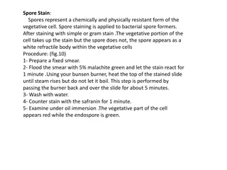
**Procedure of endospore stain: Prepare smears of organisms to be tested for presence of endospores on a clean microscope slide and air dry it.. Cover the smears with a piece of absorbent paper cut to fit the slide and place the slide on a wire gauze on a ring stand . Saturate the paper with malachite green and holding the Bunsen burner in the hand heat the slide until steam can be seen rising from the surface. Remove the heat and reheat the slide as needed to keep the slide steaming for about three minutes. As the paper being to dry add a drop or two malachite green to keep it moist, but do not add so much at one time that the temperature is reduced. Remove the paper with tweezers and rinse the slide thoroughly with tap water. After 5 minutes carefully remove the slide from the rack using a clothespin Remove the blotting paper and allow the slide to cool to room temperature for 2 minutes. Rinse the slide thoroughly with tap water (to wash the malachite green from both sides of the microscope slide). Stain the smear with safranin for 2 minutes. Rinse both side of the slide to remove the secondary stain and blot the slide/ air dry and exam. The vegetative cells will appear red and the spores will appear green
bacillus cereus_endospore stain Result of endospores Staining
Spore staining procedure Spore staining procedure


