Tissues: Types, Characteristics, and Functions

Histology
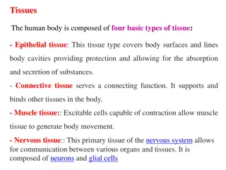
Tissues
•
Groups of cells similar in
structure
and
function
•
- the structure plays a role into the function
•
The structure and function complement each
other
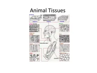
Types of Tissues
•
Epithelial tissue
•
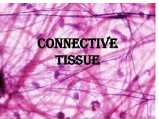
Connective tissue
•
Muscle tissue
•
Nerve tissue
•
Atoms to Molecules to Cells
Tissue Characteristics
•
Differences between the tissue classes
•
Types and functions of cells
•
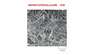
Characteristics of matrix (extracellular matrix)
•
Fibrous proteins
•
Ground substance – thick or thin
•
Clear gels (ECF, tissue fluid, interstitial fluid,
tissue gel)
•
Rubbery or stony in cartilage or bone
Tisssue Characteristics
•
Space occupied by cells versus matrix
•
Connective tissue cells are widely separated
•
Little matrix between epithelial and muscle
cells
Cardiac Muscle
- one nucleus in cardiac muscle
Bone tissue
Skeletal muscle
- alternating dark and white patterns
in skeletal muscles
Smooth Muscle
- they do not have alternating dark and
light muscles
Smooth Muscle
Epithelial Tissue
•
Two main types
•
- Covering and lining epithelia
•
- on external and internal surfaces
•
Glandular epithelia
•
Secretory tissue in glands
Glandular Epithelia
ge – glandular epithelia
lp – lamina propria
Epithelial Tissue
•
Layers of closely adhering cells
•
Flat sheet with upper surfaces exposed to the
environment or an internal body cavity
•
No blood vessels
•
Underlying connective tissue supplies oxygen
•
Rests on basement membrane
•
Thin layer of collagen and adhesive proteins
•
Anchors epithelium to connective tissue
Basement Membrane
Basement Membrane
•
Not only an anchoring gel but also provides
nutrients to the epithelium
•
Squamous – covering
•
Cuboidal and Columnar – absorption
•
The nucleus of columnar cells are found on
the lower third of a cell
Squamous
Simple Squamous
•
1 layer of flat cells
Serous Membranes – watery
membrane
•
Small intestine – has epithelial tissue lining
the inside and outside
•
Outside of small intestine – serous membrane
•
Outer layer of the heart – serous membrane
•
What is the common feature of the small
intestine and the heart?
•
Heart is constantly pumping – the serous
membrane also reduces wear and tear
•
Small intestine – produces watery secretions
•
- the water in the serous membrane reduces
friction and therefore reduces wear and tear
Simple Squamous – Alveoli
Simple squamous – alveoli
•
Surrounding the alveoli air sacs are capillaries
•
Alveoli – capillaries for oxygenation and
removal of carbon dioxide
•
Allows for rapid exchange if you have simple
squamous epithelium
Simple Cuboidal Picture
Simple Cuboidal Epithelium
•
Found in areas of secretion and excretion
•
Kidneys
Renal Tubules
Renal Tubules
Simple Cuboidal Epithelium
•
One layer of cube-shaped cells
Simple Columnar Epitheliumm
Simple Columnar Epithelium
•
Tallest of the epithelial cells
•
Found in small intestines – almost all of
absorption of nutrients is found here
Small Intestine: Villi
Villi – finger-like extensions
Lumen – hollow opening within an
organ
Crypts
Villi – in the apical surface are
microvilli
•
Microvilli – they form the brush border
•
Small extensions of the cell
•
The taller the cells the higher the chance of
absorption
•
The microvilli increases the absorptive area by
increasing the height of cells
•
Whenever you see cells with microvilli you
should think that these cells are used to
absorb everything
Villi and Goblet Cells
Goblet cells – produce mucus
Pseudostratified Columnar Epithelium
•
Stratified – multi-layered
•
- every single cell originates on one exact
membrane but they are of different heights
that is why they look stratified
•
Respiratory mucosa
Ciliated Epithelium
– respiratory mucosa
- lines the conducting airway of the
lungs
Respiratory Mucosa
Goblet Cells
•
Secrete mucus and line the conducting
airways
•
Very sticky secretion
•
Release a protein called Mucin
•
Mucin combines with water and it becomes
mucus
•
We want to clear the air before it reaches the
our air sacs so that it would not damage our
lungs
Fallopian tubes in females
•
Which one moves? Cilia or microvilli?
Stratified Squamous Epithelium
•
2 major types
•
a. keratinized
•
b. non-keratinized
Stratified Squamous Non Keratinized –
wet areas of the body
Stratified Squamous Epithelium Non
Keratinized – all cells are alive
•
Oral mucosa
•
Inner lining of the vagina
•
Lining of the esophagus
•
Wet, slippery areas
•
Epithelial Tissue – high turnover meaning
rapid change of cells like every 2-3 days
•
- mitosis is fast
Stratified Squamous Keratinized
•
Melanin – shields us from ultraviolet rays
Keratinocytes
•
The most abundant type of cells
•
Dead tough cells
•
Dead cells will exfoliate off
Stratified Cuboidal
Stratifed Cuboidal
•
Quite rare in the body
•
Found in some sweat and mammary glands
•
Testicles
•
Typically two cell layers thick
Stratified Cuboidal: Sweat Glands
Stratified Cuboidal: Seminiferous
Tubules of the Testes
Transitional Epithelium
•
Coolest of them all
•
Either round or flat
Transitional Epithelium
•
Change depending on how much stress you
apply
•
Found in the urinary system
•
- bladder, ureters, urethra
Glandular Epithelia
•
A gland is one or more cells that makes and
secretes an aqeuous fluid
•
Classified by:
•
- Site of product released
•
- Structure of the gland (endocrine, exocrine)
•
- Distance travelled by secretion
•
- Relative number of cells forming the gland –
unicellular (e.g., goblet cells) or multicellular
•
Endocrine glands – secrete into blood vessels
•
Exocrine glands – have ducts – ducts open into
body surfaces
Pancreas
•
Is a mixed gland
•
It has endocrine and exocrine functions
•
Most of it is exocrine gland
•
98% is exocrine in function, secretes digestive
enzymes – pancreatic secretions go into the
stomach
Pancreas
•
Pancreatic secretion should not be going
outside of the body
•
2% of the pancreas is endocrine – secretion of
insulin and glucagon
•
Main job of a cell is to produce proteins
•
For every amino acid you make you have to
burn ATP
•
Golgi Apparatus – packages proteins
•
for goblet cells it is the packaged mucin
•
Goblet Cell – endocrine or exocrine?
•
- short distance of travel of mucin
Exocrine Glands
•
Sebaceous glands – oil releasing glands
Explore the intricate world of histology and tissues, where groups of cells with similar structures and functions come together to support various bodily processes. From epithelial to nerve tissues, delve into the differences in tissue classes, cell types, and extracellular matrices. Discover the unique features of cardiac muscle, bone tissue, and skeletal muscle patterns, unraveling the essential role each plays in maintaining the body's structural integrity and function.

Download Presentation

Please find below an Image/Link to download the presentation.
The content on the website is provided AS IS for your information and personal use only. It may not be sold, licensed, or shared on other websites without obtaining consent from the author.If you encounter any issues during the download, it is possible that the publisher has removed the file from their server.
You are allowed to download the files provided on this website for personal or commercial use, subject to the condition that they are used lawfully. All files are the property of their respective owners.
The content on the website is provided AS IS for your information and personal use only. It may not be sold, licensed, or shared on other websites without obtaining consent from the author.
E N D
Presentation Transcript
Tissues Groups of cells similar in structure and function - the structure plays a role into the function The structure and function complement each other
Types of Tissues Epithelial tissue Connective tissue Muscle tissue Nerve tissue Atoms to Molecules to Cells
Tissue Characteristics Differences between the tissue classes Types and functions of cells Characteristics of matrix (extracellular matrix) Fibrous proteins Ground substance thick or thin Clear gels (ECF, tissue fluid, interstitial fluid, tissue gel) Rubbery or stony in cartilage or bone
Tisssue Characteristics Space occupied by cells versus matrix Connective tissue cells are widely separated Little matrix between epithelial and muscle cells
Cardiac Muscle - one nucleus in cardiac muscle
Skeletal muscle - alternating dark and white patterns in skeletal muscles
Smooth Muscle - they do not have alternating dark and light muscles
Epithelial Tissue Two main types - Covering and lining epithelia - on external and internal surfaces Glandular epithelia Secretory tissue in glands
Glandular Epithelia ge glandular epithelia lp lamina propria
Epithelial Tissue Layers of closely adhering cells Flat sheet with upper surfaces exposed to the environment or an internal body cavity No blood vessels Underlying connective tissue supplies oxygen Rests on basement membrane Thin layer of collagen and adhesive proteins Anchors epithelium to connective tissue
Basement Membrane Not only an anchoring gel but also provides nutrients to the epithelium
Squamous covering Cuboidal and Columnar absorption The nucleus of columnar cells are found on the lower third of a cell
Simple Squamous 1 layer of flat cells
Serous Membranes watery membrane
Small intestine has epithelial tissue lining the inside and outside Outside of small intestine serous membrane Outer layer of the heart serous membrane What is the common feature of the small intestine and the heart?
Heart is constantly pumping the serous membrane also reduces wear and tear Small intestine produces watery secretions - the water in the serous membrane reduces friction and therefore reduces wear and tear
Surrounding the alveoli air sacs are capillaries Alveoli capillaries for oxygenation and removal of carbon dioxide Allows for rapid exchange if you have simple squamous epithelium
Simple Cuboidal Epithelium Found in areas of secretion and excretion Kidneys
Simple Cuboidal Epithelium One layer of cube-shaped cells
Simple Columnar Epithelium Tallest of the epithelial cells Found in small intestines almost all of absorption of nutrients is found here
Villi in the apical surface are microvilli
Microvilli they form the brush border Small extensions of the cell
The taller the cells the higher the chance of absorption The microvilli increases the absorptive area by increasing the height of cells Whenever you see cells with microvilli you should think that these cells are used to absorb everything
Pseudostratified Columnar Epithelium Stratified multi-layered - every single cell originates on one exact membrane but they are of different heights that is why they look stratified Respiratory mucosa
Ciliated Epithelium respiratory mucosa - lines the conducting airway of the lungs
Respiratory Mucosa Goblet Cells Secrete mucus and line the conducting airways Very sticky secretion Release a protein called Mucin Mucin combines with water and it becomes mucus We want to clear the air before it reaches the our air sacs so that it would not damage our lungs
Stratified Squamous Epithelium 2 major types a. keratinized b. non-keratinized
Stratified Squamous Non Keratinized wet areas of the body