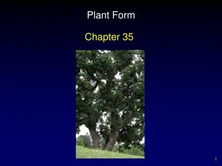
Plant Anatomy: Understanding Tissues and Growth in Plants
Plants have various types of tissues, including meristematic and permanent tissues, which play crucial roles in their growth and structure. Meristematic tissues are responsible for growth in specific regions, while permanent tissues undergo differentiation to perform specific functions. Understanding plant tissues is essential in comprehending the primary structure of dicot stems, roots, and leaves, as well as secondary thickening in dicot stems.

Download Presentation

Please find below an Image/Link to download the presentation.
The content on the website is provided AS IS for your information and personal use only. It may not be sold, licensed, or shared on other websites without obtaining consent from the author.If you encounter any issues during the download, it is possible that the publisher has removed the file from their server.
You are allowed to download the files provided on this website for personal or commercial use, subject to the condition that they are used lawfully. All files are the property of their respective owners.
The content on the website is provided AS IS for your information and personal use only. It may not be sold, licensed, or shared on other websites without obtaining consent from the author.
E N D
Presentation Transcript
Unit - III (Plant Anatomy) Tissues simple and complex. Primary structure of dicot stem, root and leaf. Secondary thickening in dicot stem.
Tissues Plants are stationary or fixed they don t move. Most of the tissues they have are supportive, which provides them with structural strength. Most of the plant tissues are dead, since dead cells can provide mechanical strength as easily as live ones, and need less maintenance. Animals on the other hand move around in search of food, mates and shelter. They consume more energy as compared to plants. Most of the tissues they contain are living. Another difference between animals and plants is in the pattern of growth. The growth in plants is limited to certain regions, while this is not so in animals. There are some tissues in plants that divide throughout their life. These tissues are localised in certain regions.
Based on the dividing capacity of the tissues, various plant tissues can be classified as growing or meristematic tissue and permanent tissue. Cell growth in animals is more uniform. So, there is no such demarcation of dividing and non-dividing regions in animals. The structural organisation of organs and organ systems is far more specialised and localised in complex animals than even in very complex plants. This fundamental difference reflects the different modes of life pursued by these two major groups of organisms, particularly in their different feeding methods. Also, they are differently adapted for a sedentary existence on one hand (plants) and active locomotion on the other (animals), contributing to this difference in organ system design.
Meristematic Tissue The growth of plants occurs only in certain specific regions. This is because the dividing tissue, also known as meristematic tissue, is located only at these points. Depending on the region where they are present, meristematic classified as lateral and intercalary. New cells produced by meristem are initially like those of meristem itself, but as they grow and characteristics slowly change and they become differentiated as components of other tissues. tissues are apical, mature, their
1.Apical meristem is present at the growing tips of stems and roots and increases the length of the stem and the root. 2.The girth of the stem or root increases due to lateral meristem (cambium). 3.Intercalary meristem is the meristem at the base of the leaves or internodes (on either side of the node) on twigs. As the cells of this tissue are very active, they have dense cytoplasm, thin cellulose walls and prominent nuclei. They lack vacuoles.
Permanent Tissue What happens to the cells formed by meristematic tissue? They take up a specific role and lose the ability to divide. As a result, they form a permanent tissue. This process of taking up a permanent shape, size, and a function is called differentiation. Cells of meristematic tissue differentiate to form different types of permanent tissue. Simple Permanent Tissue Parenchyma A few layers of cells form the basic packing tissue. This tissue is parenchyma, a type of permanent tissue. It consists of relatively unspecialised cells with thin cell walls. They are live cells. They are usually loosely packed, so that large spaces between cells (intercellular spaces) are found in this tissue
Chlorenchyma This tissue provides support to plants and also stores food. In some situations, it contains chlorophyll and performs photosynthesis, and then it is called chlorenchyma. Aerenchyma In aquatic plants, large air cavities are present in parenchyma to give buoyancy to the plants to help them float. Such a parenchyma type is called aerenchyma. The parenchyma of stems and roots also stores nutrients and water. Collenchyma The flexibility in plants is due to another permanent tissue, collenchyma. It allows easy bending in various parts of a plant (leaf, stem) without breaking. It also provides mechanical support to plants. We can find this tissue in leaf stalks below the epidermis. The cells of this tissue are living, elongated and irregularly thickened at the corners. There is very little intercellular space.
Sclerenchyma Yet another type of permanent tissue is sclerenchyma. It is the tissue which makes the plant hard and stiff. We have seen the husk of a coconut. It is made of sclerenchymatous tissue. The cells of this tissue are dead. They are long and narrow as the walls are thickened due to lignin (a chemical substance which acts as cement and hardens them). Often these walls are so thick that there is no internal space inside the cell. This tissue is present in stems, around vascular bundles, in the veins of leaves and in the hard covering of seeds and nuts. It provides strength to the plant parts.
Epidermis What you observe is the outermost layer of cells, called epidermis. The epidermis is usually made of a single layer of cells. In some plants living in very dry habitats, the Epidermis may be thicker since protection against water loss is critical. The entire surface of a plant has this outer covering of epidermis. It protects all the parts of the plant. Epidermal cells on the aerial parts of the plant often secrete a waxy, water-resistant layer on their outer surface. This aids in protection against loss of water, mechanical injury and invasion by parasitic fungi. Since it has a protective role to play, cells of epidermal tissue form a continuous layer without intercellular spaces.
Complex Permanent Tissue The different types of tissues we have discussed until now are all made of one type of cells, which look like each other. Such tissues are called simple permanent tissue. Yet another type of permanent tissue is complex tissue. Complex tissues are made of more than one type of cells. All these cells coordinate to perform a common function. Xylem and phloem are examples of such complex tissues. They are both conducting tissues and constitute a vascular bundle. Vascular or conductive tissue is a distinctive feature of the complex plants, one that has made possible their survival in the terrestrial environment.
Xylem Xylem consists of tracheids, vessels, xylem parenchyma and xylem fibres. The cells have thick walls, and many of them are dead cells. Tracheids and vessels are tubular structures. This allows them to transport water and minerals vertically. The parenchyma stores food and helps in the sideways conduction of water. Fibres are mainly supportive in function. Phloem Phloem is made up of four types of elements: sieve tubes, companion cells, phloem fibres and the phloem parenchyma. Sieve tubes are tubular cells with perforated walls. Phloem is unlike xylem in that materials can move in both directions in it. Phloem transports food from leaves to other Parts of the plant. Except for phloem fibres, phloem cells are living cells.
Primary structure of dicot stem Epidermis: It is protective in function and forms the outermost layer of the stem. It is a single layer of parenchymatous rectangular cells. The cells are compactly arranged without intercellular spaces. The outer walls of the epidermal cells have a layer called cuticle. The cuticle checks the transpiration. The cuticle is made up of a waxy substance known as cutin. Stomata may be present here and there. Epidermal cells are living. Chloroplasts are usually absent. A large number of multicellular hairs occur on the epidermis. Cortex: Cortex lies below the epidermis. The cortex is differentiated into three zones. Below the epidermis, there are a few layers of collenchyma cells. This zone is called hypodermis. It gives mechanical strength to the stem. These cells are living and thickened at the corners. Inner to the hypodermis, a few layers of chlorenchyma cells are present with conspicuous intercellular spaces. This region performs photosynthesis. Some resin ducts also occur here. The third zone is made up of parenchyma cells. These cells store food materials.
Stele: The central part of the stem inner to the endodermis is known as stele. It consists of pericycle, vascular bundles and pith. In dicot stem, vascular bundles are arranged in a ring around the pith. This type of stele is called eustele. Pericycle: Pericycle is the layers of cells that occur between the endodermis and vascular bundles. In the stem of sunflower (Helianthus), a few layers of sclerenchyma cells occur in patches outside the phloem in each vascular bundle. This patch of sclerenchyma cells is called bundle cap or hard bast. The bundle caps and the parenchyma cells between them constitute the pericycle in the stem of sunflower. Vascular bundles: The vascular bundles consist of xylem, phloem and cambium. Xylem and phloem in the stem occur together and form the vascular bundles. These vascular bundles are wedge shaped. They are arranged in the form of a ring. Each vascular bundle is conjoint, collateral, open and endarch.
Phloem: Primary phloem lies towards the periphery. It consists of protophloem and metaphloem. Phloem consists of sieve tubes, companion cells and phloem parenchyma. Phloem fibres are absent in the primary phloem. Phloem conducts organic food materials from the leaves to other parts of the plant body. Cambium: Cambium consists of brick shaped and thin walled meristematic cells. It is two to three layers in thickness. These cells are capable of forming new cells during secondary growth. Xylem: Xylem consists of xylem fibres, xylem parenchyma, vessels and tracheids. Vessels are thick walled and arranged in a few rows. Xylem conducts water and minerals from the root to the other parts of the plant body. Pith: The large central portion of the stem is called pith. It is composed of parenchyma cells with intercellular spaces. The pith is also known as medulla. The pith extends between the vascular bundles. These extensions of the pith between the vascular bundles are called primary pith rays or primary medullary rays. Function of the pith is storage of food.
Primary structure of dicot root I. Epidermis: It is single-layered and composed of thin- walled cells. The outer walls of epidermal cells are not cutinised. Many epidermal cells prolong to form long hairy bodies, the typical unicellular hairs of roots. Epidermis of root is also called epiblema or piliferous layer (pilus = hair; ferous bearing). II. Cortex: It is quite large and extensive in roots. Cortex is made of thin-walled living parenchymatous cells with leucoplasts, which convert sugar into starch grains. The last layer of cortex is endodermis. It is of universal occurrence in roots. Endodermis is composed of one layer of barrel-shaped cells which are closely arranged without having intercellular spaces. The endodermal cells have thickened radial walls, which are called Casparian strips, after the name of Caspary, the gentleman who first noted them.
III. Stele or Central Cylinder: Next to endodermis there is a single-layered pericycle made up of thin-walled parenchyma cells. Pericycle is the seat of the origin of lateral roots. Vascular bundles are typically radial in roots. Xylem and phloem form separate patches and are intervened by non-conducting cells. In dicotyledonous roots the number of bundles is limited. Xylem has protoxylem towards circumference abutting on pericycle and metaxylem towards centre. This is called exarch arrangement (of endarch arrangement of stems). Phloem with sieve tubes, etc., form patches arranged alternately with xylem. A small patch of sclerenchyma cells is present outside every group of phloem. Conjunctive Tissue: Thin-walled parenchymatous cells lying in between xylem and phloem groups constitute the conjunctive tissue. Pith: At the centre there is a small parenchymatous pith. It may be even absent in dicotyledonous roots.
Primary structure of dicot leaf Epidermis A dicotyledonous leaf is generally dorsiventral. It has upper and lower epidermis. The epidermis is usually made up of a single layer of cells that are closely packed. The cuticle on the upper epidermis is thicker than that of lower epidermis. The minute openings found on the epidermis are called stomata. Stomata are more in number on the lower epidermis than on the upper epidermis. A stoma is surrounded by a pair of bean shaped cells called guard cells. Each stoma opens into an air chamber. These guard cells contain chloroplasts, whereas other epidermal cells do not contain chloroplasts. The main function of the epidermis is to give protection to the inner tissue called mesophyll. The cuticle helps to check transpiration. Stomata are used for transpiration and gas exchange.
Mesophyll The entire tissue between the upper and lower epidermis is called the mesophyll (Gk meso=in the middle; phyllome=leaf). There are two regions in the mesophyll. They are palisade parenchyma and spongy parenchyma. Palisade parenchyma cells are seen beneath the upper epidermis. It consists of vertically elongated cylindrical cells in one or more layers. These cells are compactly arranged without intercellular spaces. Palisade parenchyma cells contain more chloroplasts than the spongy parenchyma cells. The function of palisade parenchyma is photosynthesis. Spongy parenchyma lies below the palisade parenchyma. Spongy cells are irregularly shaped. These cells are very loosely arranged with numerous airspaces. As compared to palisade cells, the spongy cells contain lesser number of chloroplasts. Spongy cells facilitate the exchange of gases with the help of air spaces. The air space that is found next to the stoma is called respiratory cavity or sub-stomatal cavity.
Vascular tissues: Vascular tissues are present in the veins of leaf. Vascular bundles are conjoint, collateral and closed. Xylem is present towards the upper epidermis, while the phloem towards the lower epidermis. Vascular bundles are surrounded by a compact layer of parenchymatous cells called bundle sheath or border parenchyma. Xylem consists of metaxylem vessels and protoxylem vessels. Protoxylem vessels are present towards the upper epidermis. Phloem consists of sieve tubes, companion cells and phloem parenchyma. Phloem fibres are absent. Xylem consists of vessels and xylem parenchyma. Tracheids and xylem fibres are absent.
Secondary thickening in dicot stem. Primary growth produces growth in length and development of lateral appendages. Secondary growth is the formation of secondary tissues from lateral meristems. It increases the diameter of the stem. In woody plants, secondary tissues constitute the bulk of the plant. They take part in providing protection, support and conduction of water and nutrients. Secondary tissues are formed by two types of lateral meristems, vascular cambium and cork cambium or phellogen. Vascular cambium produces secondary vascular tissues while phellogen forms periderm. Secondary growth occurs in perennial gymnosperms and dicots such as trees and shrubs. It is also found in the woody stems of some herbs. In such cases, the secondary growth is equivalent to one annual ring, e.g., Sunflower.
A. Formation of Secondary Vascular Tissues: They are formed by the vascular cambium. Vascular cambium is produced by two types of meristems, fascicular or intra-fascicular and inter-fascicular cambium. Intra-fascicular cambium is a primary meristem which occurs as strips in vascular bundles. Inter-fascicular cambium arises secondarily from the cells of medullary rays which occur at the level of intra-fascicular strips. Vascular Rays:The vascular rays or secondary medullary rays are rows of radially arranged cells which are formed in the secondary vascular tissues. They are a few cells in height. Depending upon their breadth, the vascular rays are uniseriate (one cell in breadth) or multiseriate (two or more cells in breadth). Vascular rays may be homo-cellular (having one type of cells) or hetero-cellular (with more than one type of cells). The cells of the vascular rays enclose intercellular spaces Secondary Phloem (Bast): It forms a narrow circle on the outer side of vascular cambium. Secondary phloem does not grow in thickness because the primary and the older secondary phloem present on the outer side gets crushed with the development of new functional phloem The crushed or non-functioning phloem may, however, have fibres and sclereids.
3. Secondary Xylem: It forms the bulk of the stem and is commonly called wood. The secondary xylem consists of vessels, tracheids (both tracheary elements), wood fibres and wood parenchyma. Annual rings are formed due to sequence of rapid growth (favourable season, e.g., spring), slow growth (before the onset of un-favourable period, e.g., autumn) and no growth (un-favourable season, e.g., winter). Annual rings are not distinct in tropical areas which do not have long dry periods. Annual Rings (Growth Rings). It is the wood formed in a single year. It consists of two types of wood, spring wood and autumn wood (Fig. 6.31). The spring or early wood is much wider than the autumn or late wood. It is lighter in colour and of lower density. Spring wood consists of larger and wider xylem elements.
Softwood and Hardwood: Softwood is the technical name of gymnosperm wood because it is devoid of vessels. Several of the softwoods are very easy to work with (e.g., Cedrus, Pinus species). However, all of them are not soft . The softness depends upon the content of fibres and vascular rays. 90-95% of wood is made of tracheids and fibres. Vascular rays constitute 5-10% of the wood. Sapwood and Heartwood: The wood of the older stems (dalbergia, Acacia) gets differentiated into two zones, the outer light coloured and functional sapwood or alburnum and the inner darker and nonfunctional heartwood or duramen (Fig. 6.33). The tracheids and vessels of the heart wood get plugged by the in growth of the adjacent parenchyma cells into their cavities through the pits. These ingrowths are called tyloses
B. Formation of Periderm: In order to provide for increase in girth and prevent harm on the rupturing of the outer ground tissues due to the formation of secondary vascular tissues, dicot stems produce a cork cambium or phellogen in the outer cortical cells. Rarely it may arise from the epidermis (e.g., Teak, Oleander), hypodermis (e.g., Pear) or phloem parenchyma. Phellogen cells divide on both the outer side as well as the inner side (bipolar) to form secondary tissues. The secondary tissue produced on the inner side of the phellogen is parenchymatous or collenchymatous. It is called secondary cortex or phelloderm. Its cells show radial arrangement.
Lenticels: Lenticels are aerating pores in the bark of plants. They appear on the surface of the bark as raised scars containing oval, rounded or oblong depressions (Fig. 6.34 A). They occur in woody trees but not in climbers. Normally they are formed in areas with underlying rays for facilitating gas exchange. Lenticels may occur scattered or form longitudinal rows.
Bark: In common language and economic botany, all the dead cells lying outside phellogen are collectively called bark. The outer layers of the bark are being constantly peeled off on account of the formation of new secondary vascular tissues in the interior. The peeling of the bark may occur in sheets (sheets or ring bark, e.g., Eucalyptus) or in irregular strips (scaly bark). Significance of Secondary Growth: 1. Secondary growth adds to the girth of the plant. It provides support to increasing weight of the aerial growth. 2. Secondary growth produces a corky bark around the tree trunk that protects the interior from abrasion, heat, cold and infection. 3. It adds new conducting tissues for replacing old non-functioning ones as well as for meeting increased demand for long distance transport of sap and organic nutrients.
")