The Bones of Upper and Lower Limbs
Explore the classification and main features of bones in the upper and lower limbs, including the arm, forearm, hand, thigh, leg, foot, and pectoral girdle. Learn about the clavicle, scapula, and humerus in detail, identifying key structures and functions to enhance your knowledge of human anatomy.

Download Presentation

Please find below an Image/Link to download the presentation.
The content on the website is provided AS IS for your information and personal use only. It may not be sold, licensed, or shared on other websites without obtaining consent from the author.If you encounter any issues during the download, it is possible that the publisher has removed the file from their server.
You are allowed to download the files provided on this website for personal or commercial use, subject to the condition that they are used lawfully. All files are the property of their respective owners.
The content on the website is provided AS IS for your information and personal use only. It may not be sold, licensed, or shared on other websites without obtaining consent from the author.
E N D
Presentation Transcript
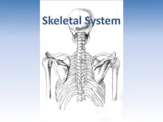
BONES OF THE UPPER and LOWER LIMBS
OBJECTIVES At the end of the lecture the students should be able to: Classify the bones of the three regions of the upper and lower limb. Memorize the main features of the Bones of the arm (humerus), of the thigh (femur & patella) Bones of the forearm (radius & ulna ), of the leg (tibia & Fibula). Bones of the hand ( carpal, metacarpal, phalanges), of the foot (tarsals, metatarsals and phalanges) Recognize the side and position of each bone
The Bones of UL are: Pectoral Girdle. Arm : Humerus. Forearm : Radius & Ulna. Wrist : Carpal bones Hand: Metacarpals & Phalanges
Pectoral Girdle Formed of Two Bones: Clavicle (anteriorly) and Scapula (posteriorly). It is very light and allows the upper limb to have exceptionally free movement.
Clavicle It is a doubly curved long bone lying horizontally across the root of the neck It is subcutaneous throughout its length. It has Two Ends: Medial (Sternal) : enlarged & triangular. Lateral (Acromial) : flattened. Body (shaft): Its medial 2/3 is convex forward. Its lateral 1/3 is concave forward. Surfaces: Superior : smooth as it lies just deep to the skin. Inferior : rough because strong ligaments bind it to the 1strib.
Scapula (Shoulder Blade) It is a triangular Flat bone. Extends between the 2nd_ 7thribs. It has : Three Processes: (1)Spine, (2) Acromion, (3) Coracoid Three Borders: Superior, Medial (Vertebral) & Lateral (Axillary) Three Angles: Superior, Lateral (forms the Glenoid cavity), Inferior. Two Surfaces: Convex Posterior, Smaller Supraspinous Fossa (above the spine) and the larger Infraspinous Fossa (below the spine). Concave Anterior (Costal)
Humerus Typical Long bone. Proximal End: Head, Neck, Greater & Lesser Tubercles. Intertubercular Groove. Anatomical neck: formed by a groove separating the head from the tubercles. Surgical Neck: a narrow part distal to the tubercles. Shaft (Body): Has two prominent features: 1. Deltoid tuberosity: 2. Spiral (Radial) groove: Distal End: Medial (can be felt) and Lateral Epicondyles. surgical
Structures at Distal end: Anteriorly: Trochlea: (medial) for articulation with the ulna Capitulum: (lateral) for articulation with the radius. Coronoid fossa: above the trochlea. Radial fossa: above the capitulum. Posteriorly: Olecranon fossa: above the trochlea.
Ulna It is the stabilizing bone of the forearm. It is the medial & longer of the two bones of the forearm. Proximal End 1. Olecranon Process : 2. Coronoid Process : 3.Tuberosity of Ulna: 4.Trochlear Notch: 5.Radial Notch : Shaft : Thick & cylindrical superiorly but diminishes in diameter inferiorly It has Three Surfaces (Anterior, Medial & Posterior). Sharp Lateral Interosseous border. Distal End: Small rounded 1. Head: lies distally at the wrist. . 2. Styloid process: Medial.
Radius It is the shorter and lateral of the two forearm bones. Proximal End: 1. Head: small & circular Its upper surface is concave for articulation with the Capitulum. 2. Neck. 3. Radial (Biciptal) Tuberosity : medially directed and separates the proximal end from the body. Shaft: Has a lateral convexity. It gradually enlarges as it passes distally. Distal (Lower) End: It is rectangular 1. Ulnar Notch : a medial concavity to accommodate the head of the ulna. 2.Radial Styloid process: extends from the lateral aspect. 3.Dorsal tubercle: projects dorsally.
Carpal Bones Composed of Eight short bones Proximal row (from lateral to medial): Scaphoid, Lunate, Triquetral & Pisiform bones. Distal row (from lateral to medial): Trapezium, Trapezoid, Capitate & Hamate. Five Metacarpal bones, each has a Base, Shaft, and a Head. Each digit has Three Phalanges Except the Thumb which has only Two
The Bones of LL are: Pelvic Girdle: Hip bone &Sacrum Thigh: Femur& Patella. Leg: Tibia & Radius. Ankle: Tarsal bones Foot : Metatarsal & Phalanges.
BONES OF THIGH (Femur and Patella) Femur: Articulates above with acetabulum of hip bone to form the hip joint. Articulates below with tibia and patella to form the knee joint. Femur : Consists of : Upper end Shaft Lower end
UPPER END OF FEMUR Head : It articulates with acetabulum of hip bone to form hip joint. Neck : It connects head to the shaft. Greater & lesser trochanters : Anteriorly, connecting the 2 trochanters, the inter- trochanteric line, where the iliofemoral ligament is attached. Posteriorly, the inter- trochanteric crest, on which is the quadrate tubercle (Qudratus femoris muscle).
SHAFT OF FEMUR It has 3 surfaces Anterior Medial Lateral http://anatomy2.wikispaces.com/file/view/femur_3.JPG/96409766/femur_3.JPG It has 3 borders Two roundedmedial and lateral One thick posterior border or ridge called linea aspera Anterior view Posterior view
LOWER END OF FEMUR Has lateral and medial condyles, separated anteriorly by articular patellar surface, and posteriorly by intercondylar notch or fossa. The 2 condyles take part in the knee joint. Above the condyles are the medial & lateral epicondyles. Anterior Posterior
PATELLA https://encrypted-tbn3.gstatic.com/images?q=tbn:ANd9GcT0DQNbeJOUKMB13by-xrVL7RQbSJv96FpMMLPPpycKUHR7-O95 http://www.greatrivermedical.org/files/Image/Ortho_images/Healthy_Knee.jpg It is a largest sesamoid bone (lying inside the Quadriceps tendon in front of knee joint). Its anterior surface is rough and subcutaneous. Its posterior surface articulates with the condyles of the femur to form knee joint. Its apex lies inferiorly and is connected to tuberosity of tibia by ligamentum patellae. Its upper, lateral, and medial margins give attachment to Quadriceps femoris muscles.
BONES OF LEG (TIBIA AND FIBULA) Tibia : It is the medial bone of leg. Fibula : It is the lateral bone of leg. Each of them has upper end, shaft, and lower end.
TIBIA Upper end has: 2 tibial condyles: Medial condyle : is larger and articulate with medial condyle of femur. It has a groove on its posterior surface for semimembranosus muscles. Lateral condyle : is smaller and articulates with lateral condyle of femur. It has facet on its lateral side for articulation with head of fibula to form proximal tibio-fibular joint. Intercondylar area : is rough and has intercondylar eminence. http://images.slideplayer.com/16/5198169/slides/slide_83.jpg
TIBIA Shaft has: Tibial tuberosity : Its upper smooth part gives attachment to ligamentum patellae. Its lower rough part is subcutaneous. 3 borders : Anterior boder : sharp and subcutaneous. Medial border. Lateral border interosseous border. 3 surfaces : Medial : subcutaneous. Lateral Posterior has oblique line, soleal line for attachment of soleus muscle
TIBIA Lowe end: Articulates with talus for formation of ankle joint. Medial malleolus: Its medial surface is subcutaneous. Its lateral surface articulate with talus. Fibular notch: lies on its lateral surface of lower end to form distal tibiofibular joint.
FIBULA upper end of fibula It is the selender lateral bone of the leg. It takes no part in articulation of knee joint. Its upper end has: Head : articulates with lateral condyle of tibia. Styloid process. Neck. Shaft has: 4 borders : its medial interosseous border gives attachment to interosseous membrane. 4 surfaces. Lower end forms: Lateral malleolus: is subcutaneous, Its medial surface is smooth for articulation with talus to form ankle joint.
BONES OF FOOT 7 Tarsal bones: 1. Calcaneum. 2. Talus . 3. Navicular. 4. Cuboid. 5. 3 cuneiform bones. Only Talus articulates with tibia & fibula at ankle joint. Calcaneum: the largest bone of foot, forming the heel. 5 Metatarsal bones: They are numbered from medial (big toe) to lateral. 1st metatarsal bone is large and lies medially. Each metatarsal bone has a base (proximal). a shaft and a head (distal). 14 phalanges: 2 phalanges for big toe (proximal & distal) 3 phalanges for each of the lateral 4 toes (proximal, middle & distal) 1 2 3 4 5


")