Development of Limbs: From Initiation to Bone Formation
Limb development begins in the fourth week of embryonic development, with limbs forming from paraxial and lateral plate mesoderm. The process involves mesenchyme condensation, chondrocyte differentiation, and endochondral bone formation. Limb defects can occur, affecting approximately 1 in 200 live human births. Understanding the stages of limb development is crucial for studying limb abnormalities and disorders.

Uploaded on Sep 17, 2024 | 2 Views
Download Presentation

Please find below an Image/Link to download the presentation.
The content on the website is provided AS IS for your information and personal use only. It may not be sold, licensed, or shared on other websites without obtaining consent from the author.If you encounter any issues during the download, it is possible that the publisher has removed the file from their server.
You are allowed to download the files provided on this website for personal or commercial use, subject to the condition that they are used lawfully. All files are the property of their respective owners.
The content on the website is provided AS IS for your information and personal use only. It may not be sold, licensed, or shared on other websites without obtaining consent from the author.
E N D
Presentation Transcript
Limb Development Matthew Velkey matt.velkey@duke.edu 380 Seeley Mudd Suggested Reading: Langman s Medical Embryology (13th ed): Chapter 10 (143-144, 152-155) Chapter 11 (156-159) Chapter 12 (163-174) OR Langman s Medical Embryology (12th ed): Chapter 11 (145-50) Chapter 12 (151-61) OR Langman s Medical Embryology (11th ed): Chapter 10 (133-34; 142-44) Chapter 11 (145-147) Chapter 12 (151-161)
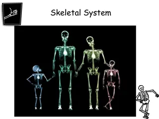
Human Limb Development 5 weeks 6 weeks 8 weeks
Limbs develop (mostly) from paraxial (somitic) and lateral plate mesoderm From somites: From lateral plate mesoderm: Bones, blood vessels, and associated connective tissue From neural crest: melanocytes in epidermis, Schwann cells in nerves Dermal connective tissue derived from dermatome Limb muscles derived from ventrolateral (abaxial) myotome
EARLY LIMB PATTERNING: Limb formation initiates during the fourth week of development as the primary antero-posterior axis (AP) is still elongating. First the forelimb and then the hindlimb begin as protrusions from the lateral plate mesoderm at the sides of the embryo. Limb buds consist of a core of mesenchyme and an outer covering of ectoderm. Forelimb bud Hindlimb bud 1 in 200 live human births display limb defects.
While external shape is being established, the mesenchyme begins condensing and chondrocyte differentiation ensues. The first cartilage models are formed in the sixth week of development. Joints form at regions of arrested chondrogenesis by cell death. early 6 weeks late 6 weeks early 8 weeks
Endochondral bone formation in the limbs: A. Mesenchyme from lateral plate mesoderm condenses into chondrocytes B. Chondrocytes form a model of the prospective bone C. Blood vessels invade the center of the model, where osteoblasts localize, and proliferation is restricted to the ends (epiphyses) D. Chondrocytes toward the shaft (diaphysis) undergo hypertrophy and apoptosis as they mineralize the surrounding matrix. 12 weeks Achondroplasia: defects in cartilage proliferation at the growth plate result in premature closure of the plate and shortened limb bones. Growth of the long bones continues into early adulthood and is maintained by proliferation of chondrocytes in the growth plates (long bones have two growth plates, in smaller bones (phalanges), there is only one at the tip)
At 6 weeks, the terminal portion of the limb buds flatten to form hand- and footplates. Fingers and toes (digits) are formed when cell death in the AER separates the plate into five parts. Digits continue to grow and cell death in intervening mesenchymal tissue delineates the digits. Bmp signaling 41 days 51 days 56 days
Limb patterning and growth is spatially and genetically regulated Proximodistal: shoulder/hip to fingers/toes Anteroposterior (or craniocaudal): thumb/big toe to pinky/little toe Dorsoventral: dorsum to palm/sole Bone identity regulated by HOX genes Predictable limb defects arise when one or more of these mechanisms is disrupted.
Limb malformations Amelia Absence of an entire limb (early loss of Fgf signaling) Meromelia .. Absence of part of a limb (later or partial loss of Fgf signaling) Phocomelia .Short, poorly formed limb (partial loss of Fgf, Hox disruption) Adactyly .Absence of digits (even later loss of Fgf) Ectrodactyly Lobster-Claw deformity (variant of adactyly middle digit is lost) Polydactyly .Extra digits (disruption of Shh pathway) Syndactyly Fusion of digits (BMP disruption)
At the tip of the limb bud a signaling structure, the apical ectodermal ridge (AER) forms. It signals mesenchyme to proliferate and the limb grows. Removal of the AER inhibits outgrowth of the limb Transplantation of a second AER duplicates the limb; diplopodia.
Proximodistal (PD) growth - regulated by AER (Fgfs): Embryological manipulation in the developing chick limb established that the apical ectodermal ridge (AER) is necessary for maintenance of PD outgrowth in the limb bud. Removal of the AER at early stages causes severe truncations of the limb skeletal elements. Progressively later removal of the AER causes progressively more distal truncations of limb elements. Removal of AER in chick at progressively later HH stages of development
Removal of AER: arrests limb development at stage at which it was removed
Failure to develop a limb: amelia Loss of AER (e.g. clipped off by amniotic banding) or FGF signaling (e.g. mutation)
Forelimb-Hindlimb Identity: the ectoderm (AER) promotes outgrowth, but mesoderm controls limb identity. ectoderm, which is FORELIMB-derived, differentiates to produce SCALES, not feathers
Post-specification: What molecular signals distinguish forelimb from hindlimb are poorly defined Expression of Tbx4 defines lower limb. Expression of Tbx5 defines upper limb.
The Zone of Polarizing activity establishes POSTERIOR (caudal) identity (duplication results in a mirror image) P2 A2 A1 P1 low ZPA signaling = make a thumb high ZPA signaling = make a pinky
ZPA also controls proliferation at AER Factors secreted by ZPA indirectly induce proliferation of the AER Loss of the ZPA therefore not only affects A-P pattern (e.g. loss of ulnar structures in the forelimb), but will also result in shortened limbs
Formation of digits: a balance between cell proliferation at AER maintained at digit tips versus cell death in interdigital regions where AER regresses.
Syndactyly: fusion of fingers and toes due to inadequate cell death between digital rays (probably also affected by Shh, note most fusions involve digits 3,4, and/or 5)
Polydactyly: duplication of digits (very often posterior) Disrupted (usually gain of function) Shh/Gli3 signaling
Maturation of limb elements Sequence is proximo-distal (e.g. humerus before radius) and postero-anterior (e.g. ulna before radius) humerus, ulna, d4, d5 are posterior (post-axial) elements radius, d3, d2, d1 are anterior (pre-axial) elements (radial hemiamelia)
The Hox code is used for proximo- distal patterning of the limbs knock out Hox11d: no radius or ulna There are well known HOX mutations in humans, so some limb defects are genetic. However, HOX gene expression is also influenced by exogenous substances (e.g. retinoic acid, ethanol) so environmental factors can also contribute.
Phocomelia: partial loss of limb Multifactorial: FGF disruption, HOX disruption, angiogenesis disruption (e.g. thalidomide)
The limbs rotate inward: forelimb ~90 degrees hindlimb ~180 degrees
Development of body musculature Primaxial myotome (dorsomedial portion closest to neural tube) cells are generally less migratory and therefore contribute to epaxial musculature and proximal hypaxial musculature (e.g. scalenes, geniohyoid, prevertebral, intercostals, rhomboids, levator scapulae, and latissimus dorsi). Abaxial myotome (ventrolateral portion closer to lateral plate mesoderm) cells are more migratory and therefore go into limbs and ventral body wall. The innervation of the adult muscle reflects the segment from which is was derived. primaxial myoblasts abaxial myoblasts
Abnormal muscle development Poland Syndrome absence/reduction of pectoralis minor/major muscles. Unknown etiology, but often associated with subclavian artery anomalies (suggests that likely cause is ischemia during time of pectoralis muscle development).
Abnormal muscle development Prune belly syndrome --atrophy or lack of abdominal wall musculature Congenital joint contracture observed with in utero denervation or atrophy of muscle (suggests that proper joint/skeleton development requires force feedback from muscles)


 from paraxial (somitic)")


 growth - regulated by AER (Fgfs):")


 promotes")