Pioneering Innovation in Brain Imaging: The Center for Morphometric Analysis (CMA)
Dr. Verne Caviness, along with colleagues David Kennedy and Pauline Filipek, founded the Center for Morphometric Analysis (CMA) in 1988 at MGH, playing a significant role in the development of computer-assisted programs for precise volumetric imaging of the human brain using MRI. The CMA's early achievements include the co-registration program for the first demonstration of fMRI in humans and the pioneering use of DTI for studying patterns of axonal distribution in the brain.

Download Presentation

Please find below an Image/Link to download the presentation.
The content on the website is provided AS IS for your information and personal use only. It may not be sold, licensed, or shared on other websites without obtaining consent from the author.If you encounter any issues during the download, it is possible that the publisher has removed the file from their server.
You are allowed to download the files provided on this website for personal or commercial use, subject to the condition that they are used lawfully. All files are the property of their respective owners.
The content on the website is provided AS IS for your information and personal use only. It may not be sold, licensed, or shared on other websites without obtaining consent from the author.
E N D
Presentation Transcript
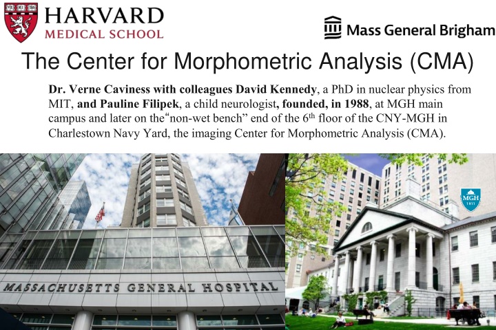
The Center for Morphometric Analysis (CMA) Dr. Verne Caviness with colleagues David Kennedy, a PhD in nuclear physics from MIT, and Pauline Filipek, a child neurologist, founded, in 1988, at MGH main campus and later on the non-wet bench end of the 6thfloor of the CNY-MGH in Charlestown Navy Yard, the imaging Center for Morphometric Analysis (CMA).
The CMA Snapshot (the founders) Dr. Verne Caviness with colleagues David Kennedy, a PhD in nuclear physics from MIT, and Pauline Filipek, a child neurologist, founded, in 1988, at MGH main campus and later on the non-wet bench end of the 6thfloor of the CNY-MGH in Charlestown Navy Yard, the imaging Center for Morphometric Analysis (CMA), among the first centers internationally to develop computer assisted programs for precise volumetric imaging of human brain MRI.
The CMA Snapshot (brief history) Among the early achievements of the CMA, Dr. David Kennedy developed the co-registration program for the first demonstration of fMRI in humans by the MGH-NMR team headed by Drs. Jack Belliveau and Bruce Rosen. Belliveau, Kennedy, et al., Science, 1992
The CMA Snapshot (brief history) Dr. Nikos Makris, trained primarily in psychiatry, early became a member of the CMA and later became its director. Working with Drs. Deepak Pandya, Edith Kaplan, David Kennedy, Andy Worth, Van Wedeen, Tim Davis, Greg Sorensen, and George Papadimitriou, in 1997 Dr. Makris published the first use and validation of DTI for the study of patterns of associative axonal distribution in the human brain. This was the first peer-reviewed publication of the RGB (Red-Green-Blue) color-coding scheme for diffusion tensor visualization of XYZ spatial dimensions. This quickly became the standard convention in the field of research and clinical diffusion tensor imaging (DTI). Makris, Kennedy, Pandya, et al., Ann Neurol, 1997
The CMA Snapshot (brief history) A specific brain data visualization and analysis program, Cardviews (Cardinal Views), developed in the CMA by James Meyer, has been the basis for landmark studies, inter alia, of patterns of normal brain development and sexual dimorphism in the brain, the earliest studies establishing morphometric features of autistic brains and developmental language disorders, and the morphometric profiles of early onset bipolar disorder and schizophrenia.The principal investigators in these programs were Dr. Martha Herbert, a graduate of the Pediatric Neurology training program, Dr. Jean Frazier, a child psychiatrist, and Drs. Larry Seidman and Jill Goldstein from the Harvard Department of Psychiatry.
The CMA Snapshot (brief history) Pioneering studies of morphometric analysis in obsessive-compulsive disorder (OCD) were conducted by Dr. Scott Rauch, then Director of Psychiatric Neuroimaging at MGH and currently the President and Psychiatrist-in-Chief of McLean Hospital in Boston.
The CMA Snapshot (brief history) The close collaboration of neurobiologically and clinically-oriented scientists with technologists (CMA and NMR Center at MGH) became the basis for landmark studies in brain imaging such as in cocaine addiction with colleagues Hans Breiter, Greg Gasic and Steven Hyman. Breiter, Hyman, et al., Neuron, 1997 Breiter, Gasic, et al., Neuron, 2004 Breiter, Gasic, et al., Neuron, 2008
The CMA Snapshot (brief history) By 1999, a description of the MRI brain volumetrics field of science (Caviness et al., 1999) was developed, consisting of a theoretical framework for quantitative brain imaging-based anatomy. Kennedy, Caviness, Makris et al., Cer Cor, 1998 Makris, Meyer, Caviness, et al., NI, 1999
The CMA Snapshot (brief history) In the early 2000s, the CMA framework generated the human Harvard Oxford Atlas (HOA). Probabilistic atlases covering 48 cortical and 21 subcortical structural areas, derived from structural data and segmentations kindly provided by Dr. David Kennedy of the Harvard Center for Morphometric Analysis (CMA) in the early 2000s.
The CMA Snapshot (brief history) In the early 2000s, the CMA framework generated the human Harvard Oxford Atlas (HOA). Probabilistic atlases covering 48 cortical and 21 subcortical structural areas, derived from structural data and segmentations kindly provided by Dr. David Kennedy of the Harvard Center for Morphometric Analysis (CMA) in the early 2000s.
The CMA Snapshot (brief history) In the early 2000s, the CMA framework generated the human Harvard Oxford Atlas (HOA). Probabilistic atlases covering 48 cortical and 21 subcortical structural areas, derived from structural data and segmentations kindly provided by Dr. David Kennedy of the Harvard Center for Morphometric Analysis (CMA) in the early 2000s.
The CMA Snapshot (brief history) The CMA-NMR/A.A. Martinos Center for Biomedical Imaging partnership became the basis for validation of the FreeSurfer automated structural MRI segmentation methodologies (Fischl, et al. 2002, 2004).
The CMA Snapshot (brief history) The CMA-NMR/A.A. Martinos Center for Biomedical Imaging partnership became the basis for validation of the FreeSurfer automated structural MRI segmentation methodologies (Fischl, et al. 2002, 2004).
")
")
")
")
")
")
")
")
")
")
")
")
")
")
")