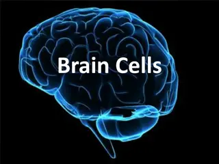
Neurological Communication: Neurons and Signals
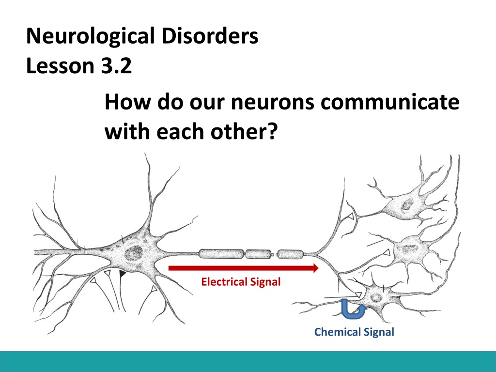
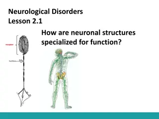
Neurological Disorders
Lesson 3.2
How do our neurons communicate
with each other?
Chemical Signal
Electrical Signal
Do Now:
•
Sleeping Beauty just pricked her
finger and is feeling a lot of pain.
Model the neurons involved in
Sleeping Beauty sensing this pain.
•
In order for one neuron in this
pathway to send information to the
next, how would you change the
electrical signal of the axon into a
chemical signal at the synapse ?
Pain Pathway
Projection neuron
Interneuron
Motor neuron
Sensory neuron
Converting an Electrical Signal to Chemical
Signal
Chemical Signal
Electrical Signal
Synaptic Transmission
Electrical Signal
Neurotransmitter
Electrical Signal
Synaptic Transmission
Neurotransmitter
The Stage:
Presynaptic cell
Synapse
Postsynaptic Cell
The Characters:
Voltage-gated
Ca
2+
channels
Synaptic vesicles
Neurotransmitters (NT)
Receptors
Action
Potential
Reuptake Transporters
Ca
2+
sensitive
proteins
Your most common neurotransmitters
How do the characters work together to
complete synaptic transmission?
Card Sort Activity
The Play:
1. Action
Potential
The Play:
1. Action
Potential
2. Voltage-gated Ca
2+
channels open.
The Play:
1. Action
Potential
Ca
2+
2. Voltage-gated Ca
2+
channels open.
3. Ca
2+
flows into cell
The Play:
1. Action
Potential
Ca
2+
2. Voltage-gated Ca
2+
channels open.
4. Ca
2+
sensitive proteins
fuse synaptic vesicles to
membrane.
3. Ca
2+
flows into cell
The Play:
1. Action
Potential
Ca
2+
2. Voltage-gated Ca
2+
channels open.
4. Ca
2+
sensitive proteins
fuse synaptic vesicles to
membrane.
3. Ca
2+
flows into cell
5. NTs are released into
synaptic cleft
The Play:
1. Action
Potential
Ca
2+
2. Voltage-gated Ca
2+
channels open.
6. NTs bind to postsynaptic
receptors.
4. Ca
2+
sensitive proteins
fuse synaptic vesicles to
membrane.
3. Ca
2+
flows into cell
5. NTs are released into
synaptic cleft
The Play:
1. Action
Potential
Ca
2+
7. Ion channels open on
postsynaptic membrane,
allowing ions to flow into cell.
8. Excess NTs are degraded
by enzymes or pumped
back into presynaptic cell.
2. Voltage-gated Ca
2+
channels open.
6. NTs bind to postsynaptic
receptors.
4. Ca
2+
sensitive proteins
fuse synaptic vesicles to
membrane.
3. Ca
2+
flows into cell
5. NTs are released into
synaptic cleft
What would happen if…
You took a drug that destroyed
the Ca
2+
sensitive proteins that
fuse synaptic vesicles to the
membrane???
You wouldn’t be able to release
synaptic vesicles.
That’s How Botox Works!
Botox destroys the proteins that fuse
synaptic vesicles with the membrane.
By stopping vesicle release, Botox
prevents muscle contraction which
prevents wrinkles!
Synaptic Transmission
1. Action
Potential
2. Voltage-gated Ca
2+
channels open.
Ca
2+
flows into cell
Ca
2+
4. NTs bind to postsynaptic
receptors.
5. Ion channels open on
postsynaptic membrane,
allowing ions to flow into cell.
6. Excess NTs are degraded
by enzymes or pumped
back into presynaptic cell.
3. Ca
2+
sensitive proteins
fuse synaptic vesicles to
membrane, releasing NTs
into synaptic cleft
Does it matter which ions flow into the
postsynaptic cell?
Sodium (Na
+
)
Calcium (Ca
2+
)
Chloride (Cl
-
)
Positive
Positive
Negative
Neurons communicate through electrical signals that are converted into chemical signals at synapses. Explore the pathway of sensory neurons in responding to pain, understand the conversion of signals, and learn about common neurotransmitters like acetylcholine, glutamate, GABA, epinephrine, dopamine, and serotonin.

Download Presentation

Please find below an Image/Link to download the presentation.
The content on the website is provided AS IS for your information and personal use only. It may not be sold, licensed, or shared on other websites without obtaining consent from the author.If you encounter any issues during the download, it is possible that the publisher has removed the file from their server.
You are allowed to download the files provided on this website for personal or commercial use, subject to the condition that they are used lawfully. All files are the property of their respective owners.
The content on the website is provided AS IS for your information and personal use only. It may not be sold, licensed, or shared on other websites without obtaining consent from the author.
E N D
Presentation Transcript
Neurological Disorders Lesson 3.2 How do our neurons communicate with each other? Electrical Signal Chemical Signal
Do Now: Sleeping Beauty just pricked her finger and is feeling a lot of pain. Model the neurons involved in Sleeping Beauty sensing this pain. In order for one neuron in this pathway to send information to the next, how would you change the electrical signal of the axon into a chemical signal at the synapse ?
Pain Pathway Sensory neuron Projection neuron Motor neuron Interneuron
Converting an Electrical Signal to Chemical Signal Electrical Signal Chemical Signal
Synaptic Transmission Electrical Signal Neurotransmitter
Synaptic Transmission Electrical Signal Neurotransmitter
The Stage: Presynaptic cell Synapse Postsynaptic Cell
The Characters: Voltage-gated Ca2+ channels Synaptic vesicles Neurotransmitters (NT) Action Potential Receptors Ca2+ sensitive proteins Reuptake Transporters
Your most common neurotransmitters Neurotransmitter Function Acetylcholine Gets us going. It excites cells, activates muscles, and is involved in wakefulness, attentiveness, anger, aggression, and sexuality. Alzheimer s disease is associated with a shortage of acetylcholine. Glutamate Is a major excitatory neurotransmitter. It is dispersed widely throughout the brain. It s involved in learning and memory. GABA Is your brain s main inhibitory neurotransmitter. It slows everything down and helps keep your system in balance. It helps regulate anxiety. Epinephrine Also known as adrenaline, keeps you alert and your blood pressure balanced, and it jumps in when you need energy. It s produced and released by the adrenal glands in time of stress. Too much can increase anxiety or tension. Dopamine Is vital for voluntary movement, attentiveness, motivation and pleasure. It s a key player in addiction. Serotonin Helps regulate body temperature, memory, emotion, sleep, appetite, and mood. Many antidepressants work by regulating serotonin.
How do the characters work together to complete synaptic transmission? Card Sort Activity
The Play: 1. Action Potential
The Play: 2. Voltage-gated Ca2+ channels open. 1. Action Potential
The Play: 2. Voltage-gated Ca2+ channels open. 3. Ca2+ flows into cell Ca2+ 1. Action Potential
The Play: 2. Voltage-gated Ca2+ channels open. 4. Ca2+ sensitive proteins fuse synaptic vesicles to membrane. 3. Ca2+ flows into cell Ca2+ 1. Action Potential
The Play: 2. Voltage-gated Ca2+ channels open. 4. Ca2+ sensitive proteins fuse synaptic vesicles to membrane. 3. Ca2+ flows into cell 5. NTs are released into synaptic cleft Ca2+ 1. Action Potential
The Play: 2. Voltage-gated Ca2+ channels open. 4. Ca2+ sensitive proteins fuse synaptic vesicles to membrane. 6. NTs bind to postsynaptic receptors. 3. Ca2+ flows into cell 5. NTs are released into synaptic cleft Ca2+ 1. Action Potential
The Play: 2. Voltage-gated Ca2+ channels open. 4. Ca2+ sensitive proteins fuse synaptic vesicles to membrane. 6. NTs bind to postsynaptic receptors. 3. Ca2+ flows into cell 5. NTs are released into synaptic cleft Ca2+ 1. Action Potential 7. Ion channels open on postsynaptic membrane, allowing ions to flow into cell.
The Play: 2. Voltage-gated Ca2+ channels open. 4. Ca2+ sensitive proteins fuse synaptic vesicles to membrane. 6. NTs bind to postsynaptic receptors. 3. Ca2+ flows into cell 5. NTs are released into synaptic cleft Ca2+ 1. Action Potential 7. Ion channels open on postsynaptic membrane, allowing ions to flow into cell. 8. Excess NTs are degraded by enzymes or pumped back into presynaptic cell.
What would happen if You took a drug that destroyed the Ca2+ sensitive proteins that fuse synaptic vesicles to the membrane??? You wouldn t be able to release synaptic vesicles.
Thats How Botox Works! Before After Botox destroys the proteins that fuse synaptic vesicles with the membrane. By stopping vesicle release, Botox prevents muscle contraction which prevents wrinkles!
Synaptic Transmission 2. Voltage-gated Ca2+ channels open. Ca2+ flows into cell 3. Ca2+ sensitive proteins fuse synaptic vesicles to membrane, releasing NTs into synaptic cleft 4. NTs bind to postsynaptic receptors. Ca2+ 1. Action Potential 5. Ion channels open on postsynaptic membrane, allowing ions to flow into cell. 6. Excess NTs are degraded by enzymes or pumped back into presynaptic cell.
Does it matter which ions flow into the postsynaptic cell? Sodium (Na+) Calcium (Ca2+) Chloride (Cl-) Positive Positive Negative