Microscope Functionality and Different Types
Learn about the essential parts and functions of microscopes, including magnification, resolution, and different types such as bright-field, dark-field, phase-contrast, dissecting, and inverted microscopes. Discover how parfocal microscopes maintain focus and the roles of ocular lenses, nose pieces, objective lenses, and slide holders in microscopy. Enhance your knowledge of how microorganisms are observed and studied using various microscope techniques.

Download Presentation

Please find below an Image/Link to download the presentation.
The content on the website is provided AS IS for your information and personal use only. It may not be sold, licensed, or shared on other websites without obtaining consent from the author.If you encounter any issues during the download, it is possible that the publisher has removed the file from their server.
You are allowed to download the files provided on this website for personal or commercial use, subject to the condition that they are used lawfully. All files are the property of their respective owners.
The content on the website is provided AS IS for your information and personal use only. It may not be sold, licensed, or shared on other websites without obtaining consent from the author.
E N D
Presentation Transcript
Function Parts and Function Different types
Scale 2
Magnification: Able to see and enlarge microorganisms that could not be seen by naked eye. Resolution: resolve the image.
Resolution: The ability to distinguish between two points at short distances from each other. Resolved Not resolved (same magnification) wavelength of light used is major factor in resolution, shorter wavelength resolution greater
Bright-field microscope Dark-field microscope Phase-contrast microscope Dissecting microscope Inverted microscope All are compound microscopes image formed by action of 2 lenses 5
Produces a dark image against a brighter background Has several objective lenses Uses ordinary bulb light as source of light. Total magnification is 1000x The resolution is 0.2 m It is mainly used to examine stained preparations. 6
Parfocal Microscopes remain in focus when objectives are changed. Total magnification Product of the magnifications of the ocular lens and the objective lens. To determine the magnification ; multiply the ocular lens by the objective lens Ocular 10x Objective 40x :10 x 40 = 400
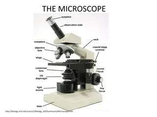
Ocular Lens/Eyepiece Magnifies the specimen image 10x Nose piece The Nose Piece holds the objective lenses and can be turned to increase the magnification Objective lens The Objective Lenses increase magnification (4 or 5 =4x 10x 20x 40x 100x)
Arm Used to support the microscope when carried. Holds the body tube, nose piece and objective lenses Slide holder Holds the specimen in place. It has 2 clips Stage Supports the slide/specimen
Stage control knobs It moves stage forwards, backwards. It moves stage right to left to adjust slide under objective lens. Coarse adjustment Knob It moves the stage rapidly to get approximate focusing. Fine adjustment knob It moves the stage slowly to get definite focusing
Condenser It condenses light rays into a cone shape to enter objective lens for proper illumination. When using X40 or X100 lens raise the condenser up. Iris diaphragm It controls the ring of light that goes into condenser. When using X100 lens open iris diaphragm.
Microscope specimen is on a glass slide. Light passes through glass slide air lens gets refracted At high magnification, this refraction (bending) of the light blurs the image To eliminate refraction between slide and lens: Eliminate the air, replace with immersion oil (Immersion oil same index of refraction as glass)
Produces a bright image of the object against a dark background. A special condenser condenses the light on specimen but of the objective lens A cell or particle will deflect the light into the objective lens thus seen as bright shape against dark background. It is used to observe living, unstained preparations especially to examine motility. 16
Excellent way to observe living cells in wet preparations. It has special condenser and phase- plate which retards light waves that go through cells in specimen. This makes contrast between cells and background. The cells appear darker against a brighter background. 18
It is also called stereo- type microscope It has oculars and stage only. It magnifies x10 only It is useful in Mycology and Parasitology.
Exposes specimen to ultraviolet, violet, or blue light Specimens usually stained with fluorochromes Shows a bright image of the object resulting from the fluorescent light emitted by the specimen. It is mostly in immunology for detection of antigen and antibody. 21
Parts of this microscope are similar to bright field microscope except condenser is located above stage while objectives are below the stage. There is a bigger working distance to allow use of cell culture flasks. It is mostly used in virology.
Electrons are used instead of light waves. 2 types: 1. Transmission electron microscope 2. Scanning electron microscope Total magnification 100,000-300,000 Resolution .0003um It is used to examine viruses and internal cell components.
Clean only with a soft cloth/tissue Make sure it s on a flat surface Be gentle with the microscope Carry it with 2 HANDS one on the arm and the other on the base