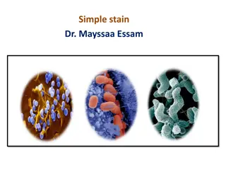
Microbiology Essentials: Gram Stain & Meningitis Overview
Practical Microbiology
Neuropsychiatry Block
•
Doctor’s notess
•
Extra explanation
•
Answers
"
لا حول ولا قوة إلا بالله العلي العظيم
"
وتقال
هذه الجملة إذا داهم الإنسان أمر عظيم لا
يستطيعه ، أو يصعب عليه القيام به .
Done by:
Samar Al-Qahtani & Hamad Al-Khudairy
EXTRA
this slide is just review the basics of microbiology
If you are already familiar with these terms just skip it
.
EXTRA
Key words to help you answer cases’ questions.
Q1
: What is your diagnosis?
Acute Pyogenic Meningitis.
Q2
: What is the most likely infection responsible?
Bacterial infection.
More serious than viral infections.
Q3:
What is your justification for your answer to question two?
↑ WBCs + ↑ Protein + ↓ Glucose + there is Polymorphs.
Q4
: Describe the microorganism’s appearance under microscope?
Gram negative intracellular bean-shaped diplococci + many pus cells.
Oxidase and catalase tests are ( positive )
Q5
: Name the media used for growing such organism?
Thayer-Martin agar or
Chocolate agar.
Q6
: What further investigation would you like to do at this stage?
CSF culture and smear, Blood culture, CBC
(complete blood count)
, Protein and
glucose levels, PCR
(polymerase chain reaction)
(DNA detection),
Serology
(Antigen detection).
antigen = capsule )
Q7
: Mention two recommended empirical antibiotics that can be used in such a
case?
Ceftriaxone with Vancomycin
.
Because they’re bactericidal.
Extra Q:
if the patient received the required vaccination before his travel to Hajj,
how would you explain his infection despite vaccination?
A: he might take the vaccination for 2 serotypes (A&C) and he may be infected by
W135.
B: he should take the vaccination before traveling by tow weeks at least.
Protect against meningitis caused by
Neisseria meningitides
, by
Meningococcal conjugate vaccine for people going to
Hajj
.
Q1
: What is your diagnosis?
Acute Pyogenic Meningitis
Q2
: What is the most likely infection responsible?
Pneumococcal bacterial infection.
Q3
: Describe the microorganism’s appearance under microscope?
gram-positive diplococcic with lanceolate shape and polymorphneoclear leucocyte
Q4
:Name the media used for growing such organism?
Blood agar
.
Q5
: Describe the microorganism’s morphology?
Gray white, Alpha hemolytic streptococci.
Q6
: Describe the microorganism’s reactivity towards the Optochin test?
Optochin sensitive
.
Q7
: What further investigation would you like to do at this stage?
CSF culture and smear, Blood culture, CBC, Protein and glucose levels, PCR
Q8
: Mention two of the recommended antibiotics that can be used as empiric treatment
in such a case?
Vancomycin + ceftriaxone
Q1
: What is your diagnosis?
Acute Pyogenic Meningitis.
Q2
: What is the most likely infection responsible?
Bacterial infection.
Q3
: What is the most probable Pathogen isolated?
Haemophilus Influenzae.
Q4
: Describe the microorganism’s appearance under microscope?
Gram-Negative pleomorphic coccobacilli with many polymorphneuclear leucocyte.
Q5
: Name the media used for growing such organism?
Chocolate agar , Blood agar and Nutrient agar
.
Q6
: Describe the microorganism’s morphology on Chocolate Agar?
Grey mucoid colonies of
Haemophilus Influenzae
due to the presence of X and V factors.
Q7
:Describe the microorganism’s morphology on Nutrient Agar?
H. influenzae
:Growth around XV factors( requires both factors XV)
no growth around X or V alone the optimum growth temperature is (35°C - 37°C in 5% CO2).
In this
culture
haemophilus
has only grown around the paper disc that has been impregnated with X and V
factors. There is no bacterial growth around the discs that only contain either X or V factor.
Q8
:Describe the microorganism’s morphology on Blood Agar?
Growth on blood agar showing
satellitisim
adjacent to a streak of
S.aureus.
S.ureus
producing surplus factor increasing growth of adjacent
H.influenzae.
satellitism is the pattern of arrangement of
heamophillus influenzae
on blood agar strearked with
staphylococcus aureus
in the centre.
Q9
: What further investigation would you like to do at this stage?
CSF culture and smear, Blood culture, CBC, Protein and glucose levels, PCR.
Q10
: Mention two recommended empirical antibiotics that can be used in such a case?
Ceftriaxone with Vancomycin.
Q1
: What is your diagnosis?
Acute Pyogenic Meningitis.
Q2
: What is the most likely infection responsible?
Bacterial infection.
Q3
: What is the most probable Pathogen isolated?
Escherichia Coli.
Q4
: Describe the microorganism’s appearance under microscope?
Gram negative bacilli (rods).
Q5
: Name the media used for growing such organism?
MacConkey’s agar.
Q6
: Describe the microorganism’s morphology on MacConkey Agar?
Lactose fermenter (pink colonies).
Q7
: What further investigation would you like to do at this stage?
CSF culture and smear, Blood culture, CBC, Protein and glucose levels, PCR.
Q8
: Mention two recommended empirical antibiotics that can be used in such a case?
Child: Ceftriaxone with Vancomycin.
Neonate: Ampicillin with Gentamicin.
Q1
: What is your diagnosis?
Aseptic (Lymphocytic) Meningitis.
Q2
: What is the most likely infection responsible?
Viral Infection.
Q3
: What is your justification for your answer to question two?
↑ WBCs + moderate ↑ Protein + normal Glucose + present of
Lymphocytes.
Q4
: What further investigation would you like to do at this stage?
CSF culture and smear, Blood culture, CBC, Protein and glucose levels,
PCR.
CSF Molecular testing is
positive
Q1
: What is your diagnosis?
Chronic Bacterial Meningitis.
Q2
: What is the most likely infection responsible?
Mycobacterial infection.
Q3
: What is your justification for your answer to question two?
↑ WBCs + ↑ Protein + ↓ Glucose + present of Lymphocytes.
Q4
: What is the most probable Pathogen isolated?
Mycobacterium Tuberculosis
.
Q5
: What is the stain used to identify such organism?
Ziehl-Neelsen (ZN) stain for Acid Fast Bacilli (AFB).
Q6
: Describe the microorganism’s appearance under microscope?
Acid Fast Bacilli (AFB) with a blue background
.
Q7
: Name the media used for growing such organism?
Lowenstein-Jensen (LJ) media.
Q8:
describe the culture on Lowenstein-Jensen?
Colonies or growth is rough, tough and buff.
Q9
: what further investigation would like to do at this stage?
CSF culture, blood culture, PCR, CBC, Tuberculin skin test, chest X-ray.
Q10
: Name the drug used to treat such infections?
For the first 2 months: Rifampicin + Isoniazid (INH) + Ethambutol + Pyrazinamide. Then, for 4-6
months: Rifampicin + Isoniazid (INH).
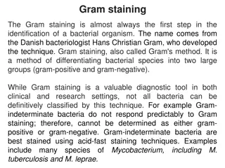
Learn about distinguishing Gram-positive and Gram-negative bacteria, key features of acute and chronic meningitis, and causes of these infections.

Download Presentation

Please find below an Image/Link to download the presentation.
The content on the website is provided AS IS for your information and personal use only. It may not be sold, licensed, or shared on other websites without obtaining consent from the author.If you encounter any issues during the download, it is possible that the publisher has removed the file from their server.
You are allowed to download the files provided on this website for personal or commercial use, subject to the condition that they are used lawfully. All files are the property of their respective owners.
The content on the website is provided AS IS for your information and personal use only. It may not be sold, licensed, or shared on other websites without obtaining consent from the author.
E N D
Presentation Transcript
Practical Microbiology Neuropsychiatry Block Doctor s notess Extra explanation Answers " . " Done by: Samar Al-Qahtani & Hamad Al-Khudairy
EXTRA this slide is just review the basics of microbiology If you are already familiar with these terms just skip it. How can I know if the bacteria is Gram positive or Gram negative? GRAM STAIN IS THE ONLY WAY +ve -ve Purple / Blue Red / Pink Coccus Bacillus Coccus Bacillus Gram +ve Cocci Staphylococcus (in clusters) Streptococcus (in chains or pairs) We differentiate between the two by using the Catalase test Bubbles Catalase Positive No reaction Catalase Negative Streptococcus Alpha Hemolytic ( ) Species Further test Result Streptococcus pneumoniae Streptococcus viridans We differentiate between them using Optochin Test Pneumoniae = Sensitive Viridans = Resistant Beta Hemolytic ( ) Species Further test Result Group A ( Pyogen) Group B (Agalactiae ) Group C We differentiate between them using Bacitracin Test Group A = Sensitive Group B and C = Resistant
EXTRA Key words to help you answer cases questions. Key information Acute Pyogenic Meningitis Acute onset (sudden)+ pus cells+ fever, headache, stiff neck, other meningitis symptoms + Turbid CSF + High protein + Low glucose + Polymorphs. Viral (Aseptic/Lymphocytic) Meningitis Acute onset (sudden)+ Symptoms of meningitis +Clear CSF + Unchanged/high protein + Unchanged/low glucose + Lymphocytes. Chronic Meningitis Chronic onset + chronic headache,facial weakness, double vision, other meningitis symptoms + Turbid CSF + High protein + Low glucose + Lymphocytes. causes of Acute Pyogenic Meningitis it differs based on the age group and all are capsulated Streptococcus pneumoniae Haemophilus influenzae Neisseria meningitidis Escherichia coli Adults&Infants / Children. Gram -ve diplococci.(kidney shaped) Infants / Children. Gram -ve coccobacilli / pleomorphic. Need blood for optimal growth, Hematin (factor X) & NAD (factor V). Type B is invasive and capsulated. The capsule is used as a conjugate vaccine Newborns. Gram -ve bacilli / Lactose fermenter. K1 sialic acid capsule invasion of brain microvascular endothelial cells. Adults&Infants / Children. Gram +ve diplococci. Optochin sensitive. The most invasive pathogen, high mortality rate>30% Glucose & maltose fermenter . Its capsule: Its capsule: produce pnemolysin immunogenic and induce immuneresponse. 1-Produce endotoxin (LPS). Causes skin rash and septic shock. 2-Resists phagocytosis. Management Treatment (10-14 days): Children & Adults: Ceftriaxone + Vancomycin Neonates: Ampicillin + Cefotaxime Causes of Chronic Meningitis Tuberculosis Mycobacterium tuberculosis Brucellosism Br.melitensis Microscopically: Ziehl Neelsen stain acid fast bacilli. Tests: Mantoux test, Tuberculin skin test (TST) Treatment: Start with 4 drugs for 2 months Rifampicin + Isonized (INH) + Ethambutol + Pyrazinamide. Affect people who are in contact with domestic animals Sheep or those who consume raw milk and milk products. Can rarely be transmitted sexually & by inhalation.(lab. Acquired) Treatment: Rifampicin + Cotrimoxazole . Then: Rifam Rifampicin + Isonized (INH) for 4-6 months
CASE 1 Acute Pyogenic Meningitis Clinical Presentation Scenario:A 15-year-old healthy male visited the ER presenting with fever, headache, vomiting and drowsiness for the past week. Physical examination showed decreased level of consciousness, neck stiffness and high temperature of 38 . Cerebrospinal fluid (CSF) examination revealed opening pressure of 20 cmH2O. Microscopy of the cerebrospinal fluid showed gram ve diplococci. The patient showed complete recovery after administration of ceftriaxone for 10 days. (purpura is classic sign for neisseria) The results of the lumber puncture are shown below: CSF Patient s results Normal range Turbid (cloudy) Clear Appearance WBCs (cells/mm3) 8,320 Few (<5) Mainly polymorphonuclear leucocytes (84%) 5.0 0.1-0.4 Protein (g/L) Glucose (mmol/L) 1.3 3.0-4.5 Chloride(mmol/L) 110 115-130
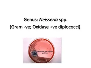
Acute Pyogenic Meningitis 1- Neisseria meningitidis Microscopic Appearance Culture on Thayer-Martin agar Specific for neisseria Protect against meningitis caused by Neisseria meningitides, by Meningococcal conjugate vaccine for people going to Hajj. Q1: What is your diagnosis? Acute Pyogenic Meningitis. Q2: What is the most likely infection responsible? Bacterial infection. More serious than viral infections. Q3: What is your justification for your answer to question two? WBCs + Protein + Glucose + there is Polymorphs. Q4: Describe the microorganism s appearance under microscope? Gram negative intracellular bean-shaped diplococci + many pus cells. Oxidase and catalase tests are ( positive ) Q5: Name the media used for growing such organism? Thayer-Martin agar or Chocolate agar. Q6: What further investigation would you like to do at this stage? CSF culture and smear, Blood culture, CBC (complete blood count) , Protein and glucose levels, PCR (polymerase chain reaction) (DNA detection), Serology (Antigen detection). antigen = capsule ) Q7: Mention two recommended empirical antibiotics that can be used in such a case? Ceftriaxone with Vancomycin. Because they re bactericidal. Extra Q: if the patient received the required vaccination before his travel to Hajj, how would you explain his infection despite vaccination? A: he might take the vaccination for 2 serotypes (A&C) and he may be infected by W135. B: he should take the vaccination before traveling by tow weeks at least.
CASE Scenario: A 59 year-old male farmer with sudden onset of fever, headache, neck stiffness and confusion The results of the lumber puncture are shown below: CSF Patient s results Normal range Appearance Turbid Clear WBCs (cells/mm3) 3520 Few (<5) Neutrophils(100%) Protein (g/L) 3.68 0.1-0.4 Glucose (mmol/L) 0.5 3.0-4.5 2-Streptococcus Pneumoniae Microscopic Appearance blood agar Optochin Test Q1: What is your diagnosis? Acute Pyogenic Meningitis Q2: What is the most likely infection responsible? Pneumococcal bacterial infection. Q3: Describe the microorganism s appearance under microscope? gram-positive diplococcic with lanceolate shape and polymorphneoclear leucocyte Q4:Name the media used for growing such organism? Blood agar. Q5: Describe the microorganism s morphology? Gray white, Alpha hemolytic streptococci. Q6: Describe the microorganism s reactivity towards the Optochin test? Optochin sensitive. Q7: What further investigation would you like to do at this stage? CSF culture and smear, Blood culture, CBC, Protein and glucose levels, PCR Q8: Mention two of the recommended antibiotics that can be used as empiric treatment in such a case? Vancomycin + ceftriaxone
3- Haemophilus Influenzae Microscopic Appearance Nutrient agar chocolate agar Blood agar Q1: What is your diagnosis? Acute Pyogenic Meningitis. Q2: What is the most likely infection responsible? Bacterial infection. Q3: What is the most probable Pathogen isolated? Haemophilus Influenzae. Q4: Describe the microorganism s appearance under microscope? Gram-Negative pleomorphic coccobacilli with many polymorphneuclear leucocyte. Q5: Name the media used for growing such organism? Chocolate agar , Blood agar and Nutrient agar. Q6: Describe the microorganism s morphology on Chocolate Agar? Grey mucoid colonies of Haemophilus Influenzae due to the presence of X and V factors. Q7:Describe the microorganism s morphology on Nutrient Agar? H. influenzae :Growth around XV factors( requires both factors XV) no growth around X or V alone the optimum growth temperature is (35 C - 37 C in 5% CO2). In this culture haemophilus has only grown around the paper disc that has been impregnated with X and V factors. There is no bacterial growth around the discs that only contain either X or V factor. Q8:Describe the microorganism s morphology on Blood Agar? Growth on blood agar showing satellitisim adjacent to a streak of S.aureus. S.ureus producing surplus factor increasing growth of adjacent H.influenzae. satellitism is the pattern of arrangement of heamophillus influenzae on blood agar strearked with staphylococcus aureus in the centre. Q9: What further investigation would you like to do at this stage? CSF culture and smear, Blood culture, CBC, Protein and glucose levels, PCR. Q10: Mention two recommended empirical antibiotics that can be used in such a case? Ceftriaxone with Vancomycin. 4. Escherichia Coli Microscopic Appearance MacConkey agar Q1: What is your diagnosis? Acute Pyogenic Meningitis. Q2: What is the most likely infection responsible? Bacterial infection. Q3: What is the most probable Pathogen isolated? Escherichia Coli. Q4: Describe the microorganism s appearance under microscope? Gram negative bacilli (rods). Q5: Name the media used for growing such organism? MacConkey s agar. Q6: Describe the microorganism s morphology on MacConkey Agar? Lactose fermenter (pink colonies). Q7: What further investigation would you like to do at this stage? CSF culture and smear, Blood culture, CBC, Protein and glucose levels, PCR. Q8: Mention two recommended empirical antibiotics that can be used in such a case? Child: Ceftriaxone with Vancomycin. Neonate: Ampicillin with Gentamicin.
CASE 2 Electron Micrograph of Enterovirus Viral (Aseptic/Lymphocytic) Meningitis Scenario: A 10-year-old boy is brought to the emergency department at KKUH accompanied by his mother. He has fever, headache, and vomiting for the last 2 days. Clinical examination confirmed that he has meningeal irritation. The results of the lumber puncture are shown below: CSF Molecular testing is positive CSF Patient s results Normal range Appearance Clear Clear WBCs (cells/mm3) 1200 Few (<5) Mainly lymphocytes (80%) Protein (g/L) 0.5 0.1-0.4 Glucose (mmol/L) 2.7 3.0-4.5 Chloride (mmol/L) 100 115-130 Q1: What is your diagnosis? Aseptic (Lymphocytic) Meningitis. Q2: What is the most likely infection responsible? Viral Infection. Q3: What is your justification for your answer to question two? WBCs + moderate Protein + normal Glucose + present of Lymphocytes. Q4: What further investigation would you like to do at this stage? CSF culture and smear, Blood culture, CBC, Protein and glucose levels, PCR.
CASE 3 Chronic Meningitis Scenario: A 65-year-old is referred from a general practitioner because of headache, fever, excessive sweating at night, and weight loss over the last 4-5 months. He has lost his appetite for food. On examination, there is neck rigidity. Laboratory tests including blood count, serum and electrolytes, blood urea, creatinine and blood culture are all normal. The results of the lumber puncture are shown below: CSF Patient s results Normal range Appearance Turbid Clear WBCs (cells/mm3) 300 Few (<5) Mainly lymphocytes (80%) Protein (g/L) 0.8 0.1-0.4 Glucose (mmol/L) 2.0 3.0-4.5 Chloride (mmol/L) 115 115-130 Chronic Meningitis Mycobacterium Tuberculosis Microscopic Appearance Lowenstein-Jensen Medium Q1: What is your diagnosis? Chronic Bacterial Meningitis. Q2: What is the most likely infection responsible? Mycobacterial infection. Q3: What is your justification for your answer to question two? WBCs + Protein + Glucose + present of Lymphocytes. Q4: What is the most probable Pathogen isolated? Mycobacterium Tuberculosis. Q5: What is the stain used to identify such organism? Ziehl-Neelsen (ZN) stain for Acid Fast Bacilli (AFB). Q6: Describe the microorganism s appearance under microscope? Acid Fast Bacilli (AFB) with a blue background. Q7: Name the media used for growing such organism? Lowenstein-Jensen (LJ) media. Q8: describe the culture on Lowenstein-Jensen? Colonies or growth is rough, tough and buff. Q9: what further investigation would like to do at this stage? CSF culture, blood culture, PCR, CBC, Tuberculin skin test, chest X-ray. Q10: Name the drug used to treat such infections? For the first 2 months: Rifampicin + Isoniazid (INH) + Ethambutol + Pyrazinamide. Then, for 4-6 months: Rifampicin + Isoniazid (INH).