Leucocytic Counting and Types Under Microscope
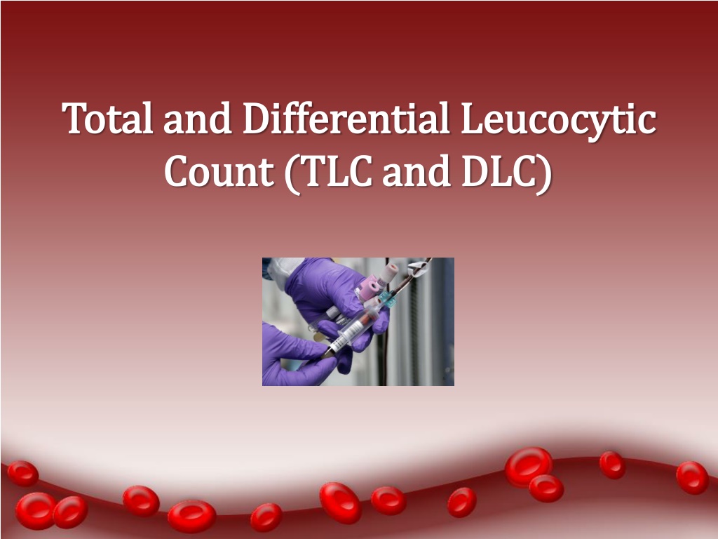

Total and Differential Leucocytic
Total and Differential Leucocytic
Count (TLC and DLC)
Count (TLC and DLC)
Objectives
Objectives
•
To be able to identify the different types of leucocytes
under the microscope
•
To practice the procedure for differential leucocyte
counting.
•
To know the normal values expected for the differential
white cell count.
•
To understand the use of the differential white cell
count in the diagnosis of disease processes.
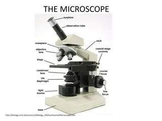
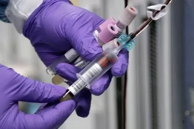
Reagents and apparatus:
Reagents and apparatus:
•
A microscope with an oil immersion objectives.
•
Mineral or cedar oil
•
Various dyes for staining blood films (e.g., Wright’s
stain)
•
Microscope slides.
Procedure
Procedure
•
Prepare blood film and stain
it with Wright’s stain
•
Examine it under the oil
immersion objective lens of
the microscope and identify
the different leucocytes (
count about 100 cells)
WBCs are classified into
Granular :
Granular :
•
Neutrophils
•
Eosinophils
•
Basophils
Agranular:
Agranular:
•
Lymphocytes
•
Monocytes
Neutrophils
Neutrophils
•
Most common type of blood cells (50-70%)
•
They have small cytoplasmic granules and a complex,
multilobed nucleus.
•
Their granules take a neutral (purple or pink) color
with various stains such as Wright’s stain.
Eosinophils
Eosinophils
•
Less common in the blood stream (1-3%)
•
They are characterized by a dumbbell-shaped nucleus
(bi-lobed) and large, prominent, red (eosinophilic)
granules
Basophils
Basophils
•
The rarest of all blood cells (0.4-1%)
•
It is a large cell filled with prominent blue (basophilic)
granules. These large granules contain heparin and
histamine. The nucleus is somewhat hidden behind
these large granules.
Lymphocytes
Lymphocytes
•
About 25-35% of the blood cells
•
Small, spherical cells with large, round nucleus
•
The cytoplasm does not contain any granules.
•
The nucleus occupies most of the volume of the cell,
leaving only a thin rim of the cytoplasm around it .
Monocytes
Monocytes
•
About 4-6% of the blood cells
•
The largest of the blood cells, the cytoplasm has no
granules
•
The nucleus is large and kidney-shaped
Clinical Application
Clinical Application
Differential count provides clues about certain illnesses
1.
Neutrophilia:
pyogenic illness.
2.
Eosinophilia:
Allergy and parasitic infections
3.
Basophilia
: in allergy and malignancy
4.
Lymphocytosis:
viral infections (infectious
mononucleosis)
.
Thank you
Thank you
Explore the total and differential leucocytic count (TLC and DLC) procedure, identifying different types of leucocytes, and their normal values for diagnosis. Learn about granular and agranular white blood cells including Neutrophils, Eosinophils, Basophils, Lymphocytes, and Monocytes. Follow the detailed procedure involving staining blood films, classifying WBCs, and examining under a microscope.

Download Presentation

Please find below an Image/Link to download the presentation.
The content on the website is provided AS IS for your information and personal use only. It may not be sold, licensed, or shared on other websites without obtaining consent from the author.If you encounter any issues during the download, it is possible that the publisher has removed the file from their server.
You are allowed to download the files provided on this website for personal or commercial use, subject to the condition that they are used lawfully. All files are the property of their respective owners.
The content on the website is provided AS IS for your information and personal use only. It may not be sold, licensed, or shared on other websites without obtaining consent from the author.
E N D
Presentation Transcript
Total and Differential Total and Differential Leucocytic Count (TLC and DLC) Count (TLC and DLC) Leucocytic
Objectives Objectives To be able to identify the different types of leucocytes under the microscope To practice the procedure for differential leucocyte counting. To know the normal values expected for the differential white cell count. To understand the use of the differential white cell count in the diagnosis of disease processes.
Reagents and apparatus: Reagents and apparatus: A microscope with an oil immersion objectives. Mineral or cedar oil Various dyes for staining blood films (e.g., Wright s stain) Microscope slides.
P Procedure rocedure Prepare blood film and stain it with Wright s stain Examine it under the oil immersion objective lens of the microscope and identify the different leucocytes ( count about 100 cells)
WBCs are classified into Granular : Granular : Neutrophils Eosinophils Basophils Agranular Agranular: : Lymphocytes Monocytes
Neutrophils Neutrophils Most common type of blood cells (50-70%) They have small cytoplasmic granules and a complex, multilobed nucleus. Their granules take a neutral (purple or pink) color with various stains such as Wright s stain.
Eosinophils Eosinophils Less common in the blood stream (1-3%) They are characterized by a dumbbell-shaped nucleus (bi-lobed) and large, prominent, red (eosinophilic) granules
B Basophils asophils The rarest of all blood cells (0.4-1%) It is a large cell filled with prominent blue (basophilic) granules. These large granules contain heparin and histamine. The nucleus is somewhat hidden behind these large granules.
Lymphocytes Lymphocytes About 25-35% of the blood cells Small, spherical cells with large, round nucleus The cytoplasm does not contain any granules. The nucleus occupies most of the volume of the cell, leaving only a thin rim of the cytoplasm around it .
Monocytes Monocytes About 4-6% of the blood cells The largest of the blood cells, the cytoplasm has no granules The nucleus is large and kidney-shaped
Clinical Application Clinical Application Differential count provides clues about certain illnesses 1. 1. Neutrophilia Neutrophilia: :pyogenic illness. 2. 2. Eosinophilia Eosinophilia: :Allergy and parasitic infections 3. 3. Basophilia Basophilia: in allergy and malignancy 4. 4. Lymphocytosis Lymphocytosis: : viral mononucleosis). infections (infectious
Blood Blood element element % of % of leukocyte leukocyte s s Size Size Cytoplasmic Cytoplasmic staining staining Nucleus Nucleus morphology morphology Erythrocyte Erythrocyte - - 7 7- -8 8 pink, no granules pink, no granules none none Neutrophil Neutrophil 50 50- -70 70 10 10- -12 12 salmon salmon- -colored small colored small granules granules Segmented, Segmented,- -2 2- - 5 lobed 5 lobed Lymphocyte Lymphocyte 25 25- -35 35 7 7- -8 8 Light blue, scant Light blue, scant amount, no granules amount, no granules Single large Single large Oval purple Oval purple Monocyte Monocyte 4 4- -6 6 16 16- -18 18 Basophilic, no granules Basophilic, no granules Large, kidney Large, kidney shaped shaped Eosinophil Eosinophil 1 1- -3 3 13 13- -14 14 Bright red coarse Bright red coarse granules granules bilobed bilobed purplish purplish Basophil Basophil 0 0- -4 4- -1 1 14 14- -15 15 Large, basophilic Large, basophilic granules granules Bilobed Bilobedbluish black black bluish
Thank you Thank you