Gingival Crevicular Fluid in Oral Health
GINGIVAL CREVICULAR
FLUID
P.K.Sasikumar, MDS, PhD
Anatomy of the gingival crevice
The gingival sulcus
is the shallow crevice or
space around the tooth ,
bounded by the surface
of the tooth on one side and
the epithelial lining the
free margin of the
gingiva on the other.
Is GCF a Transduate of interstitial
fluid?
From the work of
Alfano (1974)
and from the
hypothesis postulated by
Pashley (1976)
which
suggested that the initial fluid produced could simply
represent interstitial fluid which appears in the
crevice as a result of an osmotic gradient. This initial,
pre-inflammatory fluid was considered to be a
transduate, and, on stimulation, this changed to
become an inflammatory exudate
The model proposed by
Pashley
predicted that GCF production is governed
by the passage of
fluid
from capillaries into
the tissues (capillary filtrate) and the removal
of this
fluid
by the lymphatic system
(lymphatic uptake). When the rate of
capillary filtrate exceeds that of lymphatic
uptake,
fluid
will accumulate as edema
and/or leave the area as GCF
Factors modulating:
Filtration coefficient of the lymphatic and
capillary endothelium
Osmotic pressure within the different
compartments.
Even in health also, if the osmotic pressure of the
sulcular fluid exceeds that of the tissue fluid, there
will be net increase in the flow of GCF.
GINGIVAL VASCULATURE AND
CREVICULAR FLUID
Brill in 1959
demonstrated that increased vascular
permeability plays an important role in the production
of gingival fluid
Egelberg in 1966
, in a first experiment obtained an
increased permeability of the blood vessels of healthy
gingiva by the use of three different methods:
topical application of histamine,
gentle massage of the gingival by means of a ball-
ended amalgam plugger
scraping of the gingiva crevice by means of a
blunted dental explorer
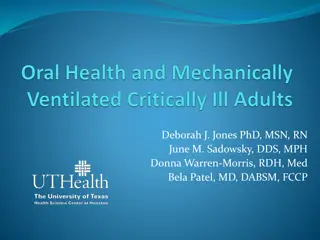
FUNCTIONS :
1) cleanse material from the sulcus
2) contain plasma proteins that may improve
adhesion of the epithelium to the tooth.
3) Possess antimicrobial properties.
4) Exert antibody activity in defense of the gingiva.
PERMEABILITY OF JUNCTIONAL AND
ORAL SULCULAR EPITHELIA
Substances that have been shown to penetrate the
sulcular epithelium include albumin, Endotoxins,
thymidine, histamine, phenytoin, peroxidase.
The main pathway for the transport of substances
across the junctional and sulcular epithelia seems to
be the intercellular spaces which according to
Schroeder and Munzel – Pedrazzoli (1970)
form
18% of the total volume of the junctional epithelium
and 12% that of the oral sulcular epithelium.
According to
Squier (1973)
the degree of
permeability of the oral mucosa does not seem to
depend upon its degree of Keratinization. The
mechanisms of penetration through an intact
epithelium were reviewed by
Squier and Johnson.
Three routes have been described:
Passage form CT into the sulcus
Passage from the sulcus into the CT
Passage of substances through pathological or
experimentally modified gingival sulcus
Passage form CT into the sulcus:
In a series of experiments, Brill verified the
assumption that
Interstitial fluid entered the gingival sulcus
through its epithelial wall
------ by showing that
the tracer material, Sodium fluorescein administered
parenterally or per orally, could be recovered form the
gingival sulcus but not form other oral epithelia.
Gingival washing methods
In this technique the gingival
crevice is perfused with an
isotonic solution, such as
Hanks’ balanced salt solution,
usually of fixed volume.
The fluid collected then
represents a dilution of
crevicular fluid and contains
both cells and soluble
constituents such as plasma
proteins.
2 different techniques have been used.
The simplest technique – instillation and re-aspiration of 10ml of
Hanks’ balanced salt solution at the interdental papilla
Skapski H,
1976.
This process was repeated 12 times to allow thorough mixing of the
transport solution and GCF.
This technique could therefore be applied either to individual
interdental units or to multiple units which were then pooled.
A more complicated method
Oppenheim FG, 1970
involved the
construction of a customized acrylic stent which isolated the
gingival tissues from the rest of the mouth.
The tissues were then irrigated for 15min, with a saline solution,
using a peristaltic pump, and the diluted GCF was removed.
Adavantage and disadvantages
The washing technique is particularly valuable for harvesting cells from
the gingival crevice region.
However, the production of customized acrylic stents is complicated
and technically demanding.
It is technique of limited application and has been restricted to the study
of GCF obtained from only a few individuals.
Only applied to the maxillary arch, because of the difficulties of
producing a technically satisfactory appliance for the mandibular arch.
GCF from individual sites cannot be analyzed.
All fluid may not be recovered during the aspiration and re-aspiration
procedure. Thus accurate quantification of GCF volume or
composition is not possible as the precise dilution factor cannot be
determined.
Absorbent filter paper strip
These strips are placed within the sulcus
(Intrasulcular method) or at its entrance
(Extrasulcular method). The placement of
filter paper strip in relation to the sulcus or
pocket is important.
The Brill technique
places it into the
pocket until resistance is encountered.
DISADVANTAGE ---- This method
introduces a degree of irritation of the
sulcular epithelium than can, by itself trigger
the oozing of fluid.
To minimize this irritation,
Loe and Holm
–
Pedersen 1965
placed the filter paper strip just at the
entrance of the pocket or over the pocket entrance. In
this way, fluid seeping out is picked up by the strip,
but the sulcular epithelium will not be in contact with
the paper.
Method of collection
positioning of papers for the filter paper strip method
of collection: (a) extracrevicular
method; (b) intracrevicular method ‘superficial’ [Löe
& Holm-Pederson], (c) intracrevicular method ‘deep’
[Brill].
Preweighed twisted threads
Threads were placed in the gingival crevice around the
tooth ,and the amount of fluid collected was
estimated by weigh in the sample thread.
Used by
WEINSTEIN et al 1967
Micropipettes/ capillary tubing
KRASSE AND EGELBERG 1958
were first to utilize
capillary tubing.
This permits the collection of fluid by capillary action.
After isolation and drying of collection
site,capillary tubes of known diameter are inserted
into the entrance of gingival crevice, GCF migrates
into the tube by capillary action.
As diameter is known, the amount of GCF
can be calculated by measuring the distance which
the GCF has migrated.
And finally, their content is then centrifuged
and analyzed.
Advantage: provides an undiluted sample of native
GCF
Disadvantages:
Difficult to hold capillary tube at the entrance of
gingival crevice for such lengthy periods
Difficult to remove the complete sample from the
tubing
May cause trauma as excessive holding of pipette
is required
The collection of the fluid is difficult because the
viscosity of the fluid makes the aspiration difficult
Crevicular washings
The Method Of Oppenheim:
This method uses an appliance consisting of a hard
acrylic plate covering the maxilla with soft borders
and a groove following the gingival margins,
connected to four collection tubes.
The washings are obtained by rinsing the crevicular
areas from one side to the other, using a peristaltic
pump.
ADVANTAGES:
Useful for longitudinal studies
Permits collection without disturbing the integrity
of the marginal tissues
Contamination is least
DISADVANTAGES:
Complex procedure
Represents a dilution of crevicular fluid
The Method Of Skapski And Lehner:
This method uses two injection needles fitted one
within the other such that during sampling the
inside, or ejection, needle is at the bottom of the
pocket and the outside, or collecting, one is at the
gingival margin. The collection needle is drained
into a sample tube by continuous suction
ADVANTAGES
Useful for cases of clinically normal gingival
Useful for studying the number and state of cells
and bacteria form the crevicular area
DISADVANTAGES;
Does not permit absolute Quantitative assessment
as the dilution factor cannot be determined
Problems during GCF collection
Contamination
The major sources of contamination of GCF sample
would be blood, saliva, or plaque.
Sampling time
The problem with prolonged collection times is that the
nature of the GCF sample collected is likely to
change with the protein concentration of the initial
GCF collected.
Volume determination
Recovery from strips
Data reporting
Constituents found within GCF samples have either
been reported as absolute amount (mg), concentrations
(mg/ml)
PERIOTRON
An electronic method has been devised for measuring gingival
fluid absorbed on paper strips by Harco electronics called
Periotron (Dental product division Winnipeg, Manitoba, Canada).
An electronic measuring device which measures the
affect on the electric current flow of wetted paper
strip.
It has 2 metal jaws which acts as the plates of an
electrical condenser.
When a dry strip is places
zero reading is obtained
A wet paper strip will increase the capacitance in
proportion to the volume of fluid and this can be
measured as an increased value in the readout.
Three models
600, 6000 and 8000.
Limitations: inability to measure the volume of FCG
greater than 1.0µl.
Enzymes & other compounds in GCF
Acid phosphates
Alkaline phosphatase
Alpha 1 antitrypsin
Arylsulphatse
Aspartate aminotransfarase
Chondroition sulphate
Immunoglobulins (IgG, IgA, IgG4, IgM)
Cystatins
B-glucuronidase
Cathepsin
Matrix metalloproteins
Cytokines – IL-1
α
,
β
IL-6, IL-8
Bacteria derived enzymes
Acid phosphatase
Alkaline phosphatase
Collagenase
Hyaluronidase
Phospholipse-A
Phospholipase-C
Cellular elements
Bacteria
Desquamated epithelial cells
Leucocytes ( PMN’S, monocytes/macrophages)
Electrolytes
Potassium
Calcium
Sodium
Organic compounds
Carbohydrates-glucosehexosamine
-Hexuronic acid
Proteins
Metabolic end products
Lactic acid
Urea
Hydroxyproline
Endotoxins
Cytotoxic substances
Hydrogen sulphide
Cellular Elements:
The cellular elements found in the gingival fluid
include bacteria, desquamated epithelial cells, and
leukocytes (PMN’s, lymphocytes and monocytes)
which migrate through the sulcular epithelium.
Electrolytes
Potassium, sodium, calcium, magnesium and fluoride
have been studied in gingival fluid. Most studies have
shown a positive correlation of calcium and sodium
concentrations and the sodium to potassium ratio with
inflammation.
Organic Compounds
Carbohydrates, proteins and lipids have been
investigated. Glucose hexosamine and hexuronic acid
are two of the compounds found in gingival fluid.
Glucose concentration in gingival fluid is 3-4 times
greater than that in serum.
This is interpreted not only as a result of metabolic
activity of adjacent tissues, but also as a function of
the local microbial flora.
Metabolic And Bacterial Products
Metabolic and bacterial products identified in
gingival fluid include lactic acid, urea, hydroxy
proline, endotoxins, prostaglandins, cytotoxic
substances, hydrogen sulphide and antibacterial
factors.
Enzyme And Enzyme Inhibitors
Various enzymes known to be present in gingival
fluid include: -
Acid phosphatase,
Alkaline phosphatase,
Pyrophosphatase,
- Glucoronidase,
Lysozyme,
Hyaluronidase,
Proteolytic enzymes includes:
Cathepsin D,
Elastase,
Cathepsin G,
Plasminogen activators,
Collagenase and
Bacterial proteinases (i.e. endo and exopeptidases), and
Lactic dehydrogenase serum proteinase inhibitors such as
2
–
macroglobulin,
1
– antitrypsin,
1
– antichymotrypsin have
also been known to be present in GCF.
Potential diagnostic markers in GCF
Alkaline phosphatase
Aspartate aminotransferase
Beta glucoranidase
Cathepsin – b
Collagenase – 2 (MMP – 8)
Collagenase – 3 (MMP – 13)
•
Dipeptidyl peptidase – II
•
Dipeptidyl peptidase – IV
•
Neutrophil elastase
•
ICTP
•
IgG
•
IL-1
•
IL-4
•
PGE2
Host response & inflammatory markers
Cytokines – IL-1
α
,
β
, 1ra, 6, 8, 2, TNF
α
, IFN
γ
RANTES
Leukotriene B4
PGE2
Lactoferrin
Transferrin
C reactive protein
IgG1,IgG2,IgG3,IgG4,IgM,IgA
Markers in GCF
Platelet activating factor
Cystatins
CD14
Visfatin
Resistin
Vasoactive intestinal peptide
Plasminogen activator
Thank you
Gingival crevicular fluid (GCF) is a crucial component in periodontal health, serving various functions such as cleansing the sulcus, enhancing epithelial adhesion, possessing antimicrobial and antibody properties. Factors influencing GCF production include capillary filtration and lymphatic uptake rates, as well as osmotic pressure differentials. The vascular permeability in the gingival tissue plays a significant role in GCF production. Understanding the dynamics of GCF can provide insights into periodontal health and disease.

Download Presentation

Please find below an Image/Link to download the presentation.
The content on the website is provided AS IS for your information and personal use only. It may not be sold, licensed, or shared on other websites without obtaining consent from the author.If you encounter any issues during the download, it is possible that the publisher has removed the file from their server.
You are allowed to download the files provided on this website for personal or commercial use, subject to the condition that they are used lawfully. All files are the property of their respective owners.
The content on the website is provided AS IS for your information and personal use only. It may not be sold, licensed, or shared on other websites without obtaining consent from the author.
E N D
Presentation Transcript
GINGIVAL CREVICULAR FLUID P.K.Sasikumar, MDS, PhD
Anatomy of the gingival crevice The gingival sulcus is the shallow crevice or space around the tooth , bounded by the surface of the tooth on one side and the epithelial lining the free margin of the gingiva on the other.
Is GCF a Transduate of interstitial fluid? From the work of Alfano (1974) and from the hypothesis postulated by Pashley (1976) which suggested that the initial fluid produced could simply represent interstitial fluid which appears in the crevice as a result of an osmotic gradient. This initial, pre-inflammatory fluid was considered to be a transduate, and, on stimulation, this changed to become an inflammatory exudate
The model proposed by Pashley predicted that GCF production is governed by the passage of fluid from capillaries into the tissues (capillary filtrate) and the removal of this fluid by the lymphatic system (lymphatic uptake). When the rate of capillary filtrate exceeds that of lymphatic uptake, fluid will accumulate as edema and/or leave the area as GCF
Factors modulating: Filtration coefficient of the lymphatic and capillary endothelium Osmotic compartments. pressure within the different Even in health also, if the osmotic pressure of the sulcular fluid exceeds that of the tissue fluid, there will be net increase in the flow of GCF.
GINGIVAL VASCULATURE AND CREVICULAR FLUID Brill in 1959 demonstrated that increased vascular permeability plays an important role in the production of gingival fluid Egelberg in 1966, in a first experiment obtained an increased permeability of the blood vessels of healthy gingiva by the use of three different methods: topical application of histamine, gentle massage of the gingival by means of a ball- ended amalgam plugger scraping of the gingiva crevice by means of a blunted dental explorer
FUNCTIONS : 1) cleanse material from the sulcus 2) contain plasma proteins that may improve adhesion of the epithelium to the tooth. 3) Possess antimicrobial properties. 4) Exert antibody activity in defense of the gingiva.
PERMEABILITY OF JUNCTIONAL AND ORAL SULCULAR EPITHELIA Substances that have been shown to penetrate the sulcular epithelium include albumin, Endotoxins, thymidine, histamine, phenytoin, peroxidase. The main pathway for the transport of substances across the junctional and sulcular epithelia seems to be the intercellular spaces which according to Schroeder and Munzel Pedrazzoli (1970) form 18% of the total volume of the junctional epithelium and 12% that of the oral sulcular epithelium.
According to Squier (1973) the degree of permeability of the oral mucosa does not seem to depend upon its degree of Keratinization. The mechanisms of penetration through an intact epithelium were reviewed by Squier and Johnson. Three routes have been described: Passage form CT into the sulcus Passage from the sulcus into the CT Passage of substances through pathological or experimentally modified gingival sulcus
Passage form CT into the sulcus: In a series of experiments, Brill verified the assumption that Interstitial fluid entered the gingival sulcus through its epithelial wall ------ by showing that the tracer material, Sodium fluorescein administered parenterally or per orally, could be recovered form the gingival sulcus but not form other oral epithelia.
Intracrevi cular washing Methods of collection Absorbing paper strips Micropippe tes Twisted threads
Gingival washing methods In this technique the gingival crevice is perfused with an isotonic solution, Hanks balanced salt solution, usually of fixed volume. The fluid represents a crevicular fluid and contains both cells constituents such as plasma proteins. such as collected dilution then of and soluble
2 different techniques have been used. The simplest technique instillation and re-aspiration of 10ml of Hanks balanced salt solution at the interdental papilla Skapski H, 1976. This process was repeated 12 times to allow thorough mixing of the transport solution and GCF. This technique could therefore be applied either to individual interdental units or to multiple units which were then pooled. A more complicated method Oppenheim FG, 1970 involved the construction of a customized acrylic stent which isolated the gingival tissues from the rest of the mouth. The tissues were then irrigated for 15min, with a saline solution, using a peristaltic pump, and the diluted GCF was removed.
Adavantage and disadvantages The washing technique is particularly valuable for harvesting cells from the gingival crevice region. However, the production of customized acrylic stents is complicated and technically demanding. It is technique of limited application and has been restricted to the study of GCF obtained from only a few individuals. Only applied to the maxillary arch, because of the difficulties of producing a technically satisfactory appliance for the mandibular arch. GCF from individual sites cannot be analyzed. All fluid may not be recovered during the aspiration and re-aspiration procedure. Thus accurate quantification of GCF volume or composition is not possible as the precise dilution factor cannot be determined.
Absorbent filter paper strip These strips are placed within the sulcus (Intrasulcular method) or at its entrance (Extrasulcular method). The placement of filter paper strip in relation to the sulcus or pocket is important. The Brill technique places it into the pocket until resistance is encountered. DISADVANTAGE ---- This method introduces a degree of irritation of the sulcular epithelium than can, by itself trigger the oozing of fluid.
To minimize this irritation, Loe and Holm Pedersen 1965 placed the filter paper strip just at the entrance of the pocket or over the pocket entrance. In this way, fluid seeping out is picked up by the strip, but the sulcular epithelium will not be in contact with the paper.
Method of collection Strip inserted into the gingival crevice INTRACREVICULAR strips are overlaid on the gingival crevice region in an attempt to minimize trauma. EXTRACREVICULAR
positioning of papers for the filter paper strip method of collection: (a) extracrevicular method; (b) intracrevicular method superficial [L e & Holm-Pederson], (c) intracrevicular method deep [Brill].
Preweighed twisted threads Threads were placed in the gingival crevice around the tooth ,and the amount of fluid collected was estimated by weigh in the sample thread. Used by WEINSTEIN et al 1967
Micropipettes/ capillary tubing KRASSE AND EGELBERG 1958 were first to utilize capillary tubing. This permits the collection of fluid by capillary action. After isolation and drying of collection site,capillary tubes of known diameter are inserted into the entrance of gingival crevice, GCF migrates into the tube by capillary action. As diameter is known, the amount of GCF can be calculated by measuring the distance which the GCF has migrated. And finally, their content is then centrifuged and analyzed.
Advantage: provides an undiluted sample of native GCF Disadvantages: Difficult to hold capillary tube at the entrance of gingival crevice for such lengthy periods Difficult to remove the complete sample from the tubing May cause trauma as excessive holding of pipette is required The collection of the fluid is difficult because the viscosity of the fluid makes the aspiration difficult
Crevicular washings The Method Of Oppenheim: This method uses an appliance consisting of a hard acrylic plate covering the maxilla with soft borders and a groove following the gingival margins, connected to four collection tubes. The washings are obtained by rinsing the crevicular areas from one side to the other, using a peristaltic pump.
ADVANTAGES: Useful for longitudinal studies Permits collection without disturbing the integrity of the marginal tissues Contamination is least DISADVANTAGES: Complex procedure Represents a dilution of crevicular fluid
The Method Of Skapski And Lehner: This method uses two injection needles fitted one within the other such that during sampling the inside, or ejection, needle is at the bottom of the pocket and the outside, or collecting, one is at the gingival margin. The collection needle is drained into a sample tube by continuous suction
ADVANTAGES Useful for cases of clinically normal gingival Useful for studying the number and state of cells and bacteria form the crevicular area DISADVANTAGES; Does not permit absolute Quantitative assessment as the dilution factor cannot be determined
Problems during GCF collection Contamination The major sources of contamination of GCF sample would be blood, saliva, or plaque. Sampling time The problem with prolonged collection times is that the nature of the GCF sample collected is likely to change with the protein concentration of the initial GCF collected.
Volume determination Recovery from strips Data reporting Constituents found within GCF samples have either been reported as absolute amount (mg), concentrations (mg/ml)
PERIOTRON An electronic method has been devised for measuring gingival fluid absorbed on paper strips by Harco electronics called Periotron (Dental product division Winnipeg, Manitoba, Canada).
An electronic measuring device which measures the affect on the electric current flow of wetted paper strip. It has 2 metal jaws which acts as the plates of an electrical condenser. When a dry strip is places zero reading is obtained A wet paper strip will increase the capacitance in proportion to the volume of fluid and this can be measured as an increased value in the readout. Three models 600, 6000 and 8000. Limitations: inability to measure the volume of FCG greater than 1.0 l.
Host derived & other products Enzymatic components Bacteria derived Composition of GCF Cellular component Non enzymatic components Electrolytes Organic component
Enzymes & other compounds in GCF Acid phosphates Alkaline phosphatase Alpha 1 antitrypsin Arylsulphatse Aspartate aminotransfarase Chondroition sulphate Immunoglobulins (IgG, IgA, IgG4, IgM) Cystatins B-glucuronidase Cathepsin Matrix metalloproteins Cytokines IL-1 , IL-6, IL-8
Bacteria derived enzymes Acid phosphatase Alkaline phosphatase Collagenase Hyaluronidase Phospholipse-A Phospholipase-C
Cellular elements Bacteria Desquamated epithelial cells Leucocytes ( PMN S, monocytes/macrophages) Electrolytes Potassium Calcium Sodium Organic compounds Carbohydrates-glucosehexosamine -Hexuronic acid Proteins
Metabolic end products Lactic acid Urea Hydroxyproline Endotoxins Cytotoxic substances Hydrogen sulphide
Cellular Elements: The cellular elements found in the gingival fluid include bacteria, desquamated epithelial cells, and leukocytes (PMN s, lymphocytes and monocytes) which migrate through the sulcular epithelium.
Electrolytes Potassium, sodium, calcium, magnesium and fluoride have been studied in gingival fluid. Most studies have shown a positive correlation of calcium and sodium concentrations and the sodium to potassium ratio with inflammation.
Organic Compounds Carbohydrates, proteins and lipids have been investigated. Glucose hexosamine and hexuronic acid are two of the compounds found in gingival fluid. Glucose concentration in gingival fluid is 3-4 times greater than that in serum. This is interpreted not only as a result of metabolic activity of adjacent tissues, but also as a function of the local microbial flora.
Metabolic And Bacterial Products Metabolic and bacterial products identified in gingival fluid include lactic acid, urea, hydroxy proline, endotoxins, prostaglandins, cytotoxic substances, hydrogen sulphide and antibacterial factors.
Enzyme And Enzyme Inhibitors Various enzymes known to be present in gingival fluid include: - Acid phosphatase, Alkaline phosphatase, Pyrophosphatase, - Glucoronidase, Lysozyme, Hyaluronidase,
Proteolytic enzymes includes: Cathepsin D, Elastase, Cathepsin G, Plasminogen activators, Collagenase and Bacterial proteinases (i.e. endo and exopeptidases), and Lactic dehydrogenase serum proteinase inhibitors such as 2 macroglobulin, 1 antitrypsin, 1 antichymotrypsin have also been known to be present in GCF.
Potential diagnostic markers in GCF Dipeptidyl peptidase II Dipeptidyl peptidase IV Neutrophil elastase ICTP IgG IL-1 IL-4 PGE2 Alkaline phosphatase Aspartate aminotransferase Beta glucoranidase Cathepsin b Collagenase 2 (MMP 8) Collagenase 3 (MMP 13)
Host response & inflammatory markers Cytokines IL-1 , , 1ra, 6, 8, 2, TNF , IFN RANTES Leukotriene B4 PGE2 Lactoferrin Transferrin C reactive protein IgG1,IgG2,IgG3,IgG4,IgM,IgA
Markers in GCF Platelet activating factor Cystatins CD14 Visfatin Resistin Vasoactive intestinal peptide Plasminogen activator


 the degree of")