Synovial Fluid and Its Role in Joint Health
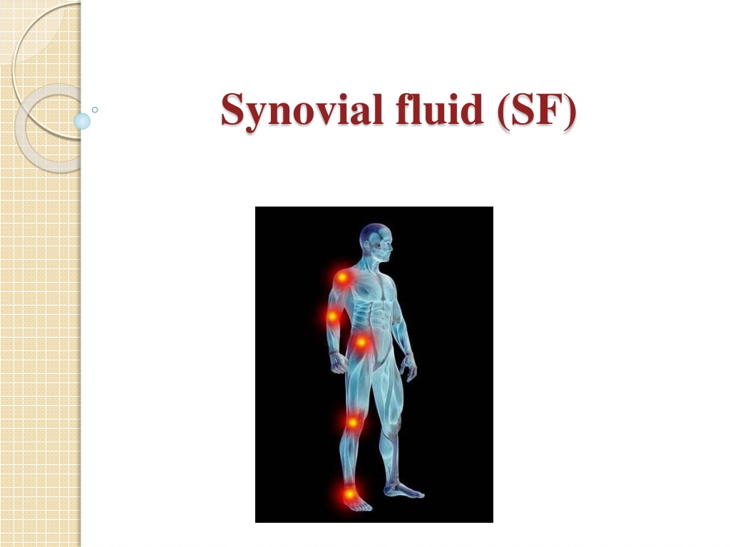


Synovial fluid (SF)
Synovial fluid (SF)
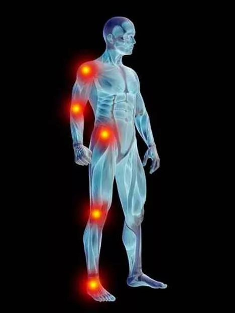

Synovial fluid (SF)
Synovial fluid (joint fluid) (synovia):
Synovial fluid is a plasma dialysate modified by
constituents secreted by the joint tissues
.
-
Joint fluid is called
synovial fluid
because of its
resemblance to egg white.
-
(Syn : like , ovia : egg)
-
It is a viscous, mucinous substance that lubricates
most joints.
-
Human joints are lined with a tissue called
synovium.
-
Synovium produces synovia, also called
synovial
fluid.
-
This fluid capsule cushions diarthrotic joints
allowing the bones to freely articulate.
The inner membrane of synovial joints is called the
synovial membrane
and secretes synovial fluid into
the joint cavity
which is formed by
ultrafiltration
of
plasma across the synovial membrane
This fluid forms a thin layer at the surface of
cartilage.
Normal synovial fluid contains 3-4 mg/ml of
(hyaluronic acid)
a: Hyaluronic acid
Normal synovial fluid contains 3-4 mg/ml of (hyaluronic
acid), a polymer of disaccharides composed of D-glucuronic
acid and D-N-acetyl glucosamine joined by alternating beta-1,4
and beta-1,3 glycosidic bond.
Hyaluronan is synthesized by the synovial membrane and
secreted into the joint cavity thus,
providing the synovial fluid with its characteristic; viscous and
elastic properties
lubricating the surfaces between synovium and cartilage
This high content of hyaluronic acid is a distinctive
feature of the synovial fluid.
It is secreted by synovial cells , which also has a
lubricating role.
B: lubricin
Function of the synovial fluid
Synovial fluid forms a thin layer (roughly 50 µm) at
the surface of cartilage and performing the following
functions:
1) reduction of friction - synovial fluid lubricates the
articulating joints thus reducing friction during joint
movement .
2) shock absorption.
3) The fluid supplies oxygen and nutrients and removes
carbon dioxide and metabolic wastes
4) It also contains phagocytic cells that remove microbes
and the debris that results from normal wear and tear
in the joint.
Collection of the synovial fluid
Synovial
fluid can be collected in a procedure
termed
arthrocentisis
.
Arthrocentisis:
It is the method for obtaining synovial fluids by
using a needle aspiration synovial fluid.
Laboratory testing
• Volume:
The amount of fluid contained in joints is usually small.
The knee joint normally contains up to 4 ml of fluid.
•
Color and clarity:
Synovial fluid: colorless – pale yellow
Apearance: clear.
Other appearances may indicate various disease
states:
yellow/cloudy fluids
usually involve an inflammatory
processes.
A white/cloudy synovial fluid
may contain crystals
red, brown
SF indicates hemorrhage into the joint.
•Viscosity:
Synovial fluid is very viscous due to its high
concentration of polymerized hyaluronate
.
•
Clotting:
- Clotting of synovial fluid can result when
fibrinogen is present.
-
Fibrinog
en may have entered into the synovial
capsule during damage to the synovial membrane
or as a result of a traumatic tap.
•
The mucin clot
:
The
mucin
clot test, also known as
Rope’s test
,
is an estimation of the integrity of the hyaluronic
acid–protein complex (mucin).
Normal synovial fluid forms a tight ropy clot upon
the addition of acetic acid.
A good mucin
clot indicates good integrity of the
hyaluronate . A poor mucin clot, one that breaks up
easily, is associated with destruction or dilution of
hyaluronate.
Chemical examination:
1. Proteins:
The normal range for synovial fluid protein is 1-3 g/dl.
Increased synovial fluid protein levels are seen in
arthritis.
2. Glucose:
Normally, synovial fluid glucose levels are less
than 10 mg/dL lower than serum levels.
-
J
o
i
n
t
d
i
s
o
r
d
e
r
s
t
h
a
t
a
r
e
c
l
a
s
s
i
f
i
e
d
a
s
i
n
f
e
c
t
i
o
u
s
d
e
m
o
n
s
t
r
a
t
e
d
e
c
r
e
a
s
e
s
i
n
s
y
n
o
v
i
a
l
f
l
u
i
d
g
l
u
c
o
s
e
a
n
d
c
a
n
b
e
a
s
m
u
c
h
a
s
2
0
-
1
0
0
m
g
/
d
l
l
e
s
s
t
h
a
n
s
e
r
u
m
l
e
v
e
l
s
.
• Uric acid
:
Synovial fluid uric acid normally ranges from
6 to 8 mg/dL.
-
The presence of uric acid in synovial fluid is
helpful in diagnosis of gout.
•
Lactate dehydrogenase:
. Lactate dehydrogenase (LD) can be elevated in
synovial fluid, while serum levels remain normal.
- Synovial fluid LD levels are usually increased in
(
rheumatoid arthritis )
RA, infectious arthritis,
and gout.
Sputum
Sputum:
It is mucus-like secretion, from lungs, bronchi,
trachea and can be coughed up, spit out or
swallowed.
Contaminated with
Epithelial cells
Nasal secretions
Saliva
Bacteria
-
It is produced by surface epithelial cells and
submucous glands.
-
Mucus layer covers the airways and protects
bronchial epithelium against inhaled noxious
substances.
- T
his mucus-like secretion may become infected,
blood stained, or contain abnormal cells that may
lead to a diagnosis.
-
Irritation of the respiratory system causes both
inflammation of the air passages and a notable
increase in mucus secretion.
Chemical composition:
95% water
5 % solids
including:
- Carbohydrates
- Proteins ( enzymes, immunoglobulins),
- Lipids
- DNA from broken WBC
- Macrophages
- Bronchial epithelial cells
A sputum culture is done to:
Detect and identify bacteria or fungi that are
causing an infection (such as pneumonia or
tuberculosis) of the lungs or the airways leading to
the lungs.
- Symptoms of a lung infection may include
difficulty breathing, pain when breathing, or a
cough that produces bloody or greenish brown
sputum.
-
Normal sputum is colorless and clear.
-
Yellow color if puss is present as in pneumonia.
-
Rusty color may be due to decomposed heamoglobin
-
Red color if there was recent haemorrhage as in
pulmonary infection, or tuberculousis.
Pneumonia
Pneumonia occurs when the lungs become inflamed
and infected.
Because the lungs are infected, pneumonia makes
it difficult to breath.
- It often follows a common cold or the flu.
Pneumonia can be caused by either bacteria, fungi,
parasites or a virus.
The air sacs may fill with fluid .
https://www.youtube.com/watch?v=-
hZ8OGpWBrY&list=PLfk7Z-
Egnx6XQeRf2KKXDrYKtjXy375AH
Synovial fluid, also known as joint fluid or synovia, is a plasma dialysate that lubricates joints and allows for smooth movement. It is produced by the synovium and contains important components like hyaluronic acid and lubricin. This fluid reduces friction, provides shock absorption, supplies nutrients, and removes waste in the joints. Arthrocentisis is a procedure used to collect synovial fluid for diagnostic purposes.

Uploaded on Sep 17, 2024 | 0 Views
Download Presentation

Please find below an Image/Link to download the presentation.
The content on the website is provided AS IS for your information and personal use only. It may not be sold, licensed, or shared on other websites without obtaining consent from the author.If you encounter any issues during the download, it is possible that the publisher has removed the file from their server.
You are allowed to download the files provided on this website for personal or commercial use, subject to the condition that they are used lawfully. All files are the property of their respective owners.
The content on the website is provided AS IS for your information and personal use only. It may not be sold, licensed, or shared on other websites without obtaining consent from the author.
E N D
Presentation Transcript
Synovial fluid (SF) Synovial fluid (joint fluid) (synovia): Synovial fluid is a plasma dialysate modified by constituents secreted by the joint tissues. - Joint fluid is called synovial fluid because of its resemblance to egg white. - (Syn : like , ovia : egg) - It is a viscous, mucinous substance that lubricates most joints.
- Human joints are lined with a tissue called synovium. - Synovium produces synovia, also called synovial fluid. - This fluid capsule cushions diarthrotic joints allowing the bones to freely articulate. The inner membrane of synovial joints is called the synovial membrane and secretes synovial fluid into the joint cavity which is formed by ultrafiltration of plasma across the synovial membrane This fluid forms a thin layer at the surface of cartilage. Normal synovial fluid contains 3-4 mg/ml of (hyaluronic acid)
a: Hyaluronic acid Normal synovial fluid contains 3-4 mg/ml of (hyaluronic acid), a polymer of disaccharides composed of D-glucuronic acid and D-N-acetyl glucosamine joined by alternating beta-1,4 and beta-1,3 glycosidic bond. Hyaluronan is synthesized by the synovial membrane and secreted into the joint cavity thus, providing the synovial fluid with its characteristic; viscous and elastic properties lubricating the surfaces between synovium and cartilage This high content of hyaluronic acid is a distinctive feature of the synovial fluid.
B: lubricin It is secreted by synovial cells , which also has a lubricating role.
Function of the synovial fluid Synovial fluid forms a thin layer (roughly 50 m) at the surface of cartilage and performing the following functions: 1) reduction of friction - synovial fluid lubricates the articulating joints thus reducing friction during joint movement . 2) shock absorption. 3) The fluid supplies oxygen and nutrients and removes carbon dioxide and metabolic wastes 4) It also contains phagocytic cells that remove microbes and the debris that results from normal wear and tear in the joint.
Collection of the synovial fluid Synovialfluid can be collected in a procedure termed arthrocentisis . Arthrocentisis: It is the method for obtaining synovial fluids by using a needle aspiration synovial fluid.
Laboratory testing Volume: The amount of fluid contained in joints is usually small. The knee joint normally contains up to 4 ml of fluid. Color and clarity: Synovial fluid: colorless pale yellow Apearance: clear. Other appearances may indicate various disease states: yellow/cloudy fluids usually involve an inflammatory processes. A white/cloudy synovial fluid may contain crystals red, brown SF indicates hemorrhage into the joint.
Viscosity: Synovial fluid is very viscous due to its high concentration of polymerized hyaluronate. Clotting: - Clotting of synovial fluid can result when fibrinogen is present. - Fibrinogen may have entered into the synovial capsule during damage to the synovial membrane or as a result of a traumatic tap.
The mucin clot : The mucin clot test, also known as Rope s test, is an estimation of the integrity of the hyaluronic acid protein complex (mucin). Normal synovial fluid forms a tight ropy clot upon the addition of acetic acid. A good mucin clot indicates good integrity of the hyaluronate . A poor mucin clot, one that breaks up easily, is associated with destruction or dilution of hyaluronate.
Chemical examination: 1. Proteins: The normal range for synovial fluid protein is 1-3 g/dl. Increased synovial fluid protein levels are seen in arthritis. 2. Glucose: Normally, synovial fluid glucose levels are less than 10 mg/dL lower than serum levels. - Joint disorders that are classified as infectious demonstrate decreases in synovial fluid glucose and can be as much as 20-100 mg/dl less than serum levels.
Uric acid: Synovial fluid uric acid normally ranges from 6 to 8 mg/dL. - The presence of uric acid in synovial fluid is helpful in diagnosis of gout. Lactate dehydrogenase: . Lactate dehydrogenase (LD) can be elevated in synovial fluid, while serum levels remain normal. - Synovial fluid LD levels are usually increased in (rheumatoid arthritis )RA, infectious arthritis, and gout.
Sputum Sputum: It is mucus-like secretion, from lungs, bronchi, trachea and can be coughed up, spit out or swallowed. Contaminated with Epithelial cells Nasal secretions Saliva Bacteria
It is produced by surface epithelial cells and submucous glands. - - Mucus layer covers the airways and protects bronchial epithelium against inhaled noxious substances.
- This mucus-like secretion may become infected, blood stained, or contain abnormal cells that may lead to a diagnosis. - Irritation of the respiratory system causes both inflammation of the air passages and a notable increase in mucus secretion .
Chemical composition: 95% water 5 % solids including: - Carbohydrates - Proteins ( enzymes, immunoglobulins), - Lipids - DNA from broken WBC - Macrophages - Bronchial epithelial cells
A sputum culture is done to: Detect and identify bacteria or fungi that are causing an infection (such as pneumonia or tuberculosis) of the lungs or the airways leading to the lungs. - Symptoms of a lung infection may include difficulty breathing, pain when breathing, or a cough that produces bloody or greenish brown sputum.
- Normal sputum is colorless and clear. - Yellow color if puss is present as in pneumonia. - Rusty color may be due to decomposed heamoglobin - Red color if there was recent haemorrhage as in pulmonary infection, or tuberculousis.
Pneumonia Pneumonia occurs when the lungs become inflamed and infected. Because the lungs are infected, pneumonia makes it difficult to breath. - It often follows a common cold or the flu. Pneumonia can be caused by either bacteria, fungi, parasites or a virus. The air sacs may fill with fluid .
https://www.youtube.com/watch?v=- hZ8OGpWBrY&list=PLfk7Z- Egnx6XQeRf2KKXDrYKtjXy375AH
")
")