Free Science Education Website - Science Prof Online PowerPoint Resources
Science Prof Online (SPO) is a free science education website offering fully-developed Virtual Science Classrooms, science-related PowerPoints, articles, and images. This resource is designed to assist students, educators, and anyone interested in learning about science. The site provides a wide range of educational resources such as practice test questions, lecture PowerPoints, video tutorials, and sample assignments. Stay updated with new materials by following SPO on social media. The PowerPoints are available in various formats for easy access and printing. Explore interactive learning tools and engage with fun learning resources throughout the presentations.

Uploaded on Sep 25, 2024 | 2 Views
Download Presentation

Please find below an Image/Link to download the presentation.
The content on the website is provided AS IS for your information and personal use only. It may not be sold, licensed, or shared on other websites without obtaining consent from the author.If you encounter any issues during the download, it is possible that the publisher has removed the file from their server.
You are allowed to download the files provided on this website for personal or commercial use, subject to the condition that they are used lawfully. All files are the property of their respective owners.
The content on the website is provided AS IS for your information and personal use only. It may not be sold, licensed, or shared on other websites without obtaining consent from the author.
E N D
Presentation Transcript
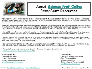
About Science Prof Online PowerPoint Resources Science Prof Online (SPO) is a free science education website that provides fully-developed Virtual Science Classrooms, science-related PowerPoints, articles and images. The site is designed to be a helpful resource for students, educators, and anyone interested in learning about science. The SPO Virtual Classrooms offer many educational resources, including practice test questions, review questions, lecture PowerPoints, video tutorials, sample assignments and course syllabi. New materials are continually being developed, so check back frequently, or follow us on Facebook (Science Prof Online) or Twitter (ScienceProfSPO) for updates. Many SPO PowerPoints are available in a variety of formats, such as fully editable PowerPoint files, as well as uneditable versions in smaller file sizes, such as PowerPoint Shows and Portable Document Format (.pdf), for ease of printing. Images used on this resource, and on the SPO website are, wherever possible, credited and linked to their source. Any words underlined and appearing in blue are links that can be clicked on for more information. PowerPoints must be viewed in slide show mode to use the hyperlinks directly. Several helpful links to fun and interactive learning tools are included throughout the PPT and on the Smart Links slide, near the end of each presentation. You must be in slide show mode to utilize hyperlinks and animations. This digital resource is licensed under Creative Commons Attribution-ShareAlike 3.0: http://creativecommons.org/licenses/by-sa/3.0/ Tami Port, MS Creator of Science Prof Online Chief Executive Nerd Science Prof Online Online Education Resources, LLC info@scienceprofonline.com Alicia Cepaitis, MS Chief Creative Nerd Science Prof Online Online Education Resources, LLC alicia@scienceprofonline.com From the Virtual Microbiology Classroom on ScienceProfOnline.com Image: Compound microscope objectives, T. Port
Laboratory LAB MEETS 1x WEEKLY Be on time. 10 minutes after the start of lab, the doors may be locked. LAB REQUIREMENTS: You must have the following to participate in lab: - Goggles (We have those in lab for you) - Lab coat (We have those in lab for you) - Shoes that completely cover feet, long pants(No shorts or capris), & long hair up. LAB QUIZ & REPORT: Each week there will be an open-book lab quiz available on Moodle. It must be completed before lab. The quiz will cover information from that week s lab (i.e. the upcoming lab). This is how I assess whether or not you are prepared to safely participate in lab. Each Lab Project has an associated Lab Report. See Moodle syllabus for schedule of due dates for homework. WHERE IS THE LAB MANUAL? The links to the Laboratory material are on Moodle. IT IS NOT ALWAYS POSSIBLE TO MAKE UP LABORATORY SESSIONS: From the Virtual Microbiology Classroom on ScienceProfOnline.com Image: : The Muppets Beaker & Dr. Bunsen Honeydew
Laboratory Exercise 1 How to Use a Compound Microscope Basic Microscopy From the Virtual Microbiology Classroom on ScienceProfOnline.com Image: The Far Side by Gary Larson
What am I going to learn from Lab Topic #1? How to make a wet mount slide (Sweeeet)! How to microscopically view wet mounted plant cells & animal cells. How to use and maintain a compound light microscope. How to protect yourself from other people s bodily fluids . From the Virtual Microbiology Classroom on ScienceProfOnline.com Image: Chimp brain in a jar, Gaetan Lee
Anton van Leeuwenhoeks Animalcules (Pronounced Lay-ven-hook) As a draper(merchant who sells cloth and dry goods), he used lenses to examine cloth. This probably led to his interest in lens making. 1632 - 1723 He assembled hundreds of microscopes, some of which magnified objects 270 times. As he looked at things with his microscopes, he discovered micro organisms - organisms so tiny that they were invisible to the naked eye. He called these tiny living organisms animalcules . He first described bacteria, protozoans and many cells of the human body. Everett showing how to use the microscope Images: Leeuwenhoek portrait 1680, Jan Verkolje (I);. Replica of Leeuwenhoek microscope, Jacopo Werther; Man with scope, source unknown From the Virtual Biology Classroom on ScienceProfOnline.com
Compound Light Microscope The Compound Part Simple microscopes have single magnifyinglens (like a magnifying glass). Compound microscopes have two sets of lenses for magnification. Lens closer to the eye = ocular lens (magnifying power of 10x). Lenses closer to the object being viewed = objective lens. (Most light microscopes used in biology have three or four objective lenses). The Light Part Bright-field light microscopes produce a dark image against brighter, backlit background. Provide a 2-D image. Commonly used to view stained cells. Image: Magnifying lamp use to look for part defects, US Navy; Compound light microscope, Moisey From the Virtual Microbiology Classroom on ScienceProfOnline.com
Your Microscope Take Care of Your Scope: It is your responsibility to take care of your scope and learn to use it properly. I randomly check scopes to see if they are put away correctly. If yours is not, I may subtract points from your lab grade. ARM Microscope Log: If you get your scope out and find that it has not been put away properly, make an entry in the microscope log. Getting Scope Out: The scope that you use is numbered to correspond to your seat number, on edge of lab bench in front of you. When transporting your scope, always hold it with one hand under the base, and one hand around the arm. Putting Scope Away: Clean stage if it is oily, and use lens paper to clean lenses. Shortest objective lens (the one with the red band) should be pointing down toward stage. Use course focus to position stage as low as it can go. Always put scope back in numbered parking spot in scope cabinet. BASE ** Now everyone get out their scope ** From the Virtual Microbiology Classroom on ScienceProfOnline.com Image: Compound light microscope, Moisey
Parts of a Compound Light Microscope From the Virtual Microbiology Classroom on ScienceProfOnline.com Image: Compound light microscope, Moisey
Magnification & The Objective Lenses Ocular lens magnifies the specimen 10x. You will always be looking through the ocular and objective lens simultaneously, so multiply ocular magnification x objective power to calculate the Total Magnification (xTM). Rotary nosepiece of your microscope has four objective lenses attached. Shortest lens (red band) should have been pointing down when your scopes were last put away. From the Virtual Microbiology Classroom on ScienceProfOnline.com Image: Microscope objective lenses, T. Port
Scanning Power Objective Lens Red band around it. Magnifies objects 4x. Q: What is the Total Magnification? ____ TM We will only use this lens in today s lab. It is not useful for looking at bacteria. From the Virtual Microbiology Classroom on ScienceProfOnline.com Image: Microscope objective lenses, T. Port
Low Power Objective Lens Has yellow band around it. Magnifies objects 10x. Q: What is the Total Magnification? ____ TM Start with this lens when looking at a bacterial smear. Q: What does the term parfocal mean? From the Virtual Microbiology Classroom on ScienceProfOnline.com Image: Microscope objective lenses, T. Port
High Dry Objective Lens Has blue band around it. Magnifies objects 40x. Q: What is the Total Magnification? ____ TM Switch to this lens after getting your specimen in focus at 100xTM. From the Virtual Microbiology Classroom on ScienceProfOnline.com Image: Microscope objective lenses, T. Port
Microscopy:Electron Microscopes Many microscopic images in your textbook were obtained using electron microscopes. Electron beam wavelengths are shorter than light wavelengths, so better resolving power. Q: What is resolution, in the context of microscopy? Transmission Electron Microscope (TEM): 2-D image Scanning Electron Microscope (SEM): 3-D image Images: Poliovirus, taken with TEM, PHIL #1875, Blood cells, taken with SEM, National Cancer Institute; Transmission electron microscope, Public Health Image Library, image #14236. From the Virtual Microbiology Classroom on ScienceProfOnline.com
How to make a wet mount From the Virtual Microbiology Classroom on ScienceProfOnline.com Image: Wet mount procedure, T. Port
Procedure 1. How to make a wet mount 2. Letter e - What happens to the e when you look at it through the lens? - What happens to your view of the e when your increase magnification? 3. Onion - Wet mount, use stain - Note nucleus and cell wall - View video tutorial 4.Elodea - Wet mount NO stain - Depth of field - 2 layers of cells - Note cell wall, chloroplasts streaming - View video tutorial 5. Cheek cell - Wet mount using NaCl & Methylene blue. - Contrast! - View video tutorial Images: Onion cells, Elodea cells & Cheek cells, SPO General Microbiology Microscopic Images; Elodea plant, Frank Vincentz From the Virtual Microbiology Classroom on ScienceProfOnline.com
Confused? Here are links to fun resources that further explain use of the microscope: Microscopy Laboratory Main Pageon the Virtual Microbiology Classroom of Science Prof Online. Smart Links Compound Microscope Parts and Use videofrom Science ProfOnline. How to Make a Wet Mount of an Elodea Plant Cell video from ScienceProfOnline. How to Make a Wet Mount of an Onion Epithelial Cell video from ScienceProfOnline. How to Make a Wet Mount of a Cheek Cell video from ScienceProfOnline. Play Amoeba, a video game where you are an amoeba that eats and grows. Microscope Mania crossword puzzle. Microscopic Pond Life, an extremely cool collection of videos of a variety of microscopic pond life to the tune of Radiohead s Kid A . From the Virtual Microbiology Classroom on ScienceProfOnline.com (You must be in PPT slideshow view to click on links.)
Are microbes intimidating you? Do yourself a favor. Use the Virtual Microbiology Classroom (VMC) ! The VMC is full of resources to help you succeed, including: practice test questions review questions study guides and learning objectives You can access the VMC by going to the Science Prof Online website www.ScienceProfOnline.com Images: White blood cell, Giant Microbes; Prokaryotic cell, Mariana Ruiz