Female Ovary Histology and Function Overview


HISTOLOGY OF GENITAL ORGANS PART-3

Female reproductive organ
Ovary
Anatomy: Ovaries, Uterine Tubes, Uterus, Vagina, and Vulva
Ovaries – two
1.
Paired glands that provide for the development of oocytes
2.
Production of hormones
3.
Suspended from the dorsal wall to the abdomen by the
mesovarium a. part of the broad ligament
4.
Easily manipulated by rectal palpation
5.
Almond-shaped in most species - Bean-shaped in horse –
Berry-shaped in the sow
6.
Ovulation – release of oocyte occurs over the entire
surface in most species a. in the horse they are confined to
one site – Ovulation Fossa (indentation)
Ovary with follicle
The ovaries have
two functions - "production" and ovulation
and oocytes and the production and secretion of hormones
.
The ovary is attached to the broad ligament by a short fold of
peritoneum, called the
mesovarium
(or ligament of the ovary),
through which vessels and nerves pass to the ovary and enter it
at the hilus of the ovary.
The surface of the ovary is covered by a single layer of cuboidal
epithelium, also called
germinal epithelium
. It is continuous
with the peritoneal mesothelium. Fibrous connective tissue
forms a thin capsule, the
tunica albuginea
, immediately
beneath the epithelium.
Like so many other organs the ovary is divided into
an outer
cortex
and an inner
medulla
. The cortex
consists of a very cellular connective tissue stroma
in which the ovarian follicles are embedded. The
medulla is composed of loose connective tissue,
which contains blood vessels and nerves.
Ovarian Follicles
Ovarian follicles consist of one oocyte and
surrounding
follicular cells
. Follicular
development can be divided into a number of
stages.
The
primary follicle
is are the first morphological stage that
marks the onset of follicular maturation
The previously flattened cell surrounding the oocyte now
form a cuboidal or columnar epithelium surrounding the
oocyte.
Their cytoplasm may have a granular appearance, and they
are for this reason also called
granulosa cells
.
The continued proliferation of these cells will result in the
formation of a stratified epithelium (with a distinct basement
membrane) surrounding the oocyte.
The
zona pellucida
(glycoproteins between interdigitating
processes of oocyte and granulosa cells) becomes visible.
Parenchymal cells of the ovary surrounding the growing
follicle become organised in concentric sheaths, the
theca
folliculi
.
Oocyte surrounded by cells
Secondary follicle are s
mall fluid-filled spaces become visible
between the granulosa cells as the follicle reaches a diameter
of about 400 µm.
These spaces enlarge and fuse to form the
follicular antrum,
which is the defining feature of the secondary follicle
.
The oocyte is now located eccentric in the follicle in
the
cumulus oophorus
, where it is surrounded by granulosa
cells.
The theca folliculi differentiates with the continued growth of
the follicle into a
theca interna
and a
theca externa
.
Vascularization of the theca interna improves, and the
spindle-shaped or polyhedral cells in this layer start to
produce
oestrogens
.
The theca externa retains the characteristics of a highly
cellular connective tissue with smooth muscle cells.
The mature or tertiary or preovulatory or Graafian follicle
increases further in size
The Graafian follicle forms a small "bump" on the surface of
the ovary, the
stigma
(or macula pellucida).
The stigma is characterised by a thinning of the capsule and a
progressive restriction of the blood flow to it. Prior to
ovulation the cumulus oophorus separates from the follicular
wall.
The oocyte is now floating freely in the follicular antrum. It is
still surrounded by granulosa cells which form the
corona
radiata
.
The follicle finally ruptures at the stigma and the oocyte is
released from the ovary.
Follicle along with corpus leuteum
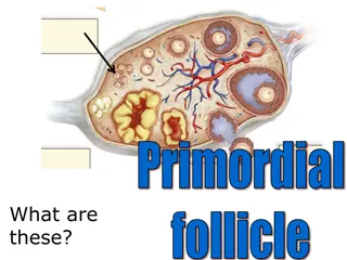
Primordial follicles
are located in the cortex just
beneath tunica albuginea. One layer of flattened
follicular cells surround the oocyte (about 30 µm in
diameter). The nucleus of the oocyte is positioned
eccentric in the cell. It appears very light and
contains a prominent nucleolus.
Most organelles of the oocyte aggregate in the
centre of the cell, where they form the
vitelline
body
Theca cells
The female reproductive organ, the ovary, plays crucial roles in oocyte development, hormone production, and ovulation. Structurally, ovaries have a cortex and medulla, with follicles containing oocytes and surrounding cells. The primary follicle marks the onset of follicular maturation, leading to the formation of a stratified epithelium around the oocyte. The ovary is covered by a single layer of cuboidal epithelium and connected to the broad ligament via the mesovarium.

Download Presentation

Please find below an Image/Link to download the presentation.
The content on the website is provided AS IS for your information and personal use only. It may not be sold, licensed, or shared on other websites without obtaining consent from the author.If you encounter any issues during the download, it is possible that the publisher has removed the file from their server.
You are allowed to download the files provided on this website for personal or commercial use, subject to the condition that they are used lawfully. All files are the property of their respective owners.
The content on the website is provided AS IS for your information and personal use only. It may not be sold, licensed, or shared on other websites without obtaining consent from the author.
E N D
Presentation Transcript
Female reproductive organ Ovary Anatomy: Ovaries, Uterine Tubes, Uterus, Vagina, and Vulva Ovaries two 1. Paired glands that provide for the development of oocytes 2. Production of hormones 3. Suspended from the dorsal wall to the abdomen by the mesovarium a. part of the broad ligament 4. Easily manipulated by rectal palpation 5. Almond-shaped in most species - Bean-shaped in horse Berry-shaped in the sow 6. Ovulation release of oocyte occurs over the entire surface in most species a. in the horse they are confined to one site Ovulation Fossa (indentation)
The ovaries have two functions - "production" and ovulation and oocytes and the production and secretion of hormones. The ovary is attached to the broad ligament by a short fold of peritoneum, called the mesovarium (or ligament of the ovary), through which vessels and nerves pass to the ovary and enter it at the hilus of the ovary. The surface of the ovary is covered by a single layer of cuboidal epithelium, also called germinal epithelium. It is continuous with the peritoneal mesothelium. Fibrous connective tissue forms a thin capsule, the tunica albuginea, immediately beneath the epithelium.
Like so many other organs the ovary is divided into an outer cortex and an inner medulla. The cortex consists of a very cellular connective tissue stroma in which the ovarian follicles are embedded. The medulla is composed of loose connective tissue, which contains blood vessels and nerves. Ovarian Follicles Ovarian follicles consist of one oocyte and surrounding follicular cells. Follicular development can be divided into a number of stages.
The primary follicleis are the first morphological stage that marks the onset of follicular maturation The previously flattened cell surrounding the oocyte now form a cuboidal or columnar epithelium surrounding the oocyte. Their cytoplasm may have a granular appearance, and they are for this reason also called granulosa cells. The continued proliferation of these cells will result in the formation of a stratified epithelium (with a distinct basement membrane) surrounding the oocyte. The zona pellucida (glycoproteins between interdigitating processes of oocyte and granulosa cells) becomes visible. Parenchymal cells of the ovary surrounding the growing follicle become organised in concentric sheaths, the theca folliculi.
Secondary follicle are small fluid-filled spaces become visible between the granulosa cells as the follicle reaches a diameter of about 400 m. These spaces enlarge and fuse to form the follicular antrum, which is the defining feature of the secondary follicle. The oocyte is now located eccentric in the follicle in the cumulus oophorus, where it is surrounded by granulosa cells. The theca folliculi differentiates with the continued growth of the follicle into a theca interna and a theca externa. Vascularization of the theca interna improves, and the spindle-shaped or polyhedral cells in this layer start to produce oestrogens. The theca externa retains the characteristics of a highly cellular connective tissue with smooth muscle cells.
The mature or tertiary or preovulatory or Graafian follicle increases further in size The Graafian follicle forms a small "bump" on the surface of the ovary, the stigma (or macula pellucida). The stigma is characterised by a thinning of the capsule and a progressive restriction of the blood flow to it. Prior to ovulation the cumulus oophorus separates from the follicular wall. The oocyte is now floating freely in the follicular antrum. It is still surrounded by granulosa cells which form the corona radiata. The follicle finally ruptures at the stigma and the oocyte is released from the ovary.
Primordial follicles are located in the cortex just beneath tunica albuginea. One layer of flattened follicular cells surround the oocyte (about 30 m in diameter). The nucleus of the oocyte is positioned eccentric in the cell. It appears very light and contains a prominent nucleolus. Most organelles of the oocyte aggregate in the centre of the cell, where they form the vitelline body