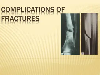
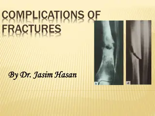
Complications of Fractures: Early and Local Manifestations

 
Complications of fractures
 
 
1-The general complications of fractures (blood loss,
shock, fat embolism, cardiorespiratory failure etc.)
2-Local complications can be divided into :
a-
early 
(thosethat arise during the first few weeks
following injury).
 
b- 
late.
 
Local complications of fractures
 
 Urgent                                                   Less urgent                             Late
Local visceral injury                       Fracture blisters
                                                                                                         Delayed union
Vascular injury                                 Plaster sores
                                                                                                                Malunion
Nerve injury                                  Pressure sores
                                                                                                              Non-union
Compartment                               Nerve entrapment
                                                                                                           Avascular necrosis
syndrome                                      Myositis ossificans
Haemarthrosis                               Ligament injury
                                                                                                          Muscle contracture
Infection                                             Tendon lesions
Gas gangrene                                      Joint stiffness                        joint instability
                                                              Algodystrophy                      Osteoarthritis
 
 
                   EARLY COMPLICATIONS
Early complications may present as part of the
primary
injury or may appear only after a few days or weeks.
1-VISCERAL INJURY
Fractures around the trunk are often complicated
by injuries to underlying viscera, the most
important being penetration of the lung with life-
threatening pneumothorax following rib fractures
and rupture of the bladder or urethra in pelvic
fractures. These injuries require emergency
treatment.


2-VASCULAR INJURY
The fractures most often associated with damage to
a major artery are those around the knee and
elbow, and those of the humeral and femoral
shafts.
The artery may be cut, torn, compressed or
contused,either by the initial injury or
subsequently by jagged bone fragments. The
effects vary from transient diminution of blood
flow to profound ischaemia, tissue death and
peripheral gangrene.
 
 
Clinical features
The patient may complain of paraesthesia or numbnessin the toes or the
fingers. The injured limb is cold and pale, or slightly cyanosed, and the pulse
is weak or absent.
 X-rays will probably show one of the ‘highrisk’fractures listed above. If a
vascular injury is suspected an angiogram  should be performed
immediately; if it is positive, emergency treatment must be started without
further delay.
 
Treatment
   All bandages and splints should be removed. The fracture is re-x-rayed and, if
the position of the bones suggests that the artery is being compressed or
kinked, prompt reduction is necessary.
   The circulation is then reassessed  repeatedly over the next half hour. If there
is no improvement, the vessels must be explored by operation  preferably
with the benefit of preoperative or peroperative angiography.
     A cut vessel can be sutured, or a segment  may be replaced by a vein graft;
if it is thrombosed, endarterectomy may restore the blood flow. If vessel
repair is undertaken, stable fixation is a must and where it is practicable, the
fracture should be fixed internally.
 
 
3- NERVE INJURY
Nerve injury is particularly common with fractures of the humerus or injuries
around the elbow or the knee
A-  Closed nerve injuries
In closed injuries the nerve is seldom severed, and spontaneous recovery
should be awaited – it occurs in90 per cent within 4 months. If recovery
has not occurred by the expected time, and if nerve conduction studies
and EMG fail to show evidence of recovery, the nerve should be explored.
B- Open nerve injuries
With open fractures the nerve injury is more likely to be complete. In these
cases the nerve should be explored at the time of debridement and
repaired at the time or at wound closure.
C- Acute nerve compression
Nerve compression, as distinct from a direct injury, sometimes occurs with
fractures or dislocations around the wrist. Complaints of numbness or
paraesthesiain the distribution of the median or ulnar nerves should be
taken seriously and the patient monitored closely; if there is no
improvement within 48 hours of fracture reduction or splitting of
bandages around the splint, the nerve should be explored and
decompressed.
 
 
                                Common nerve injuries
        Injury                                                                             Nerve
Shoulder dislocation                                                      Axillary
Humeral shaft fracture                                                  Radial
Humeral supracondylar fracture                                Radial or
median
Elbow medial condyle                                                      Ulnar
Monteggia fracture–dislocation                                Posterior-
interosseous
Hip dislocation                                                               Sciatic
Knee dislocation                                                          Peroneal
 
 
        INDICATIONS  FOR  EARLY EXPLORATION
 
Nerve injury associated with open fracture
Nerve injury with fractures that need internal
fixation
Presence of a concomitant vascular injury
Nerve damage diagnosed
 
 
4- COMPARTMENT SYNDROME
Fractures of the arm or leg can give rise to severe
ischaemia, even if there is no damage to a major
vessel.
 
Bleeding, oedema or inflammation (infection)
may increase the pressure within one of the
osseofascial
 
compartments; there is reduced capillary
flow,
 
which results in muscle ischaemia, further
oedema,
still greater pressure and yet more profound ischaemia
a vicious circle that ends, after 12 hours or less, in
necrosis of nerve and muscle within the compartment.
Nerve is capable of regeneration but muscle, once
infarcted, can never recover and is replaced by
inelastic
 
fibrous tissue (Volkmann’s ischaemic
contracture).
A similar cascade of events may be caused by swelling
 
of
a limb inside a tight plaster cast.
 
 
                           Clinical features
High-risk injuries are fractures of the elbow,
forearm bones, proximal third of the tibia, and also
multiple fractures of the hand or foot, crush injuries
and circumferential burns. Other precipitating
factors are operation (usually for internal fixation)
or infection.
The classic features of ischaemia are the five Ps:
    PAIN
    Paraesthesia
     Pallor
     Paralysis
     Pulselessness


However in compartment syndrome the ischaemia  occurs at the
capillary level, so pulses may still be felt
and the skin may not be pale! The earliest of the ‘classic’features are pain
(or a ‘bursting’ sensation), altered sensibility and paresis (or, more usually,
weakness in active muscle contraction).
 
Ischaemic muscle is highly sensitive to stretch. If the limb is unduly
painful, swollen or tense, the muscles (which may be tender) should be
tested by stretching them. 
When the toes or fingers are passively
hyperextended,there is increased pain in the calf or forearm.
 
Confirmation of the diagnosis can be made by measuring the
intracompartmental pressures. So important is the need for early
diagnosis that some surgeons advocate the use of continuous
compartment pressure monitoring for high-risk injuries (e.g. fractures of
the  tibia and fibula) and especially for forearm or leg fracture in patients
who are unconscious. A split catheter is introduced into the compartment
and the pressure is measured close to the level of the fracture. 
A
differential pressure (ΔP) – the difference between diastolic pressure
and compartment pressure – of less than30 mmHg (4.00 kilopascals) is
an  indication for immediate compartment decompression.
 
 
                       Treatment
 
The threatened compartment (or compartments) must be
promptly decompressed. Casts, bandages and dressings
must be completely removed – merely splitting  the plaster
is utterly useless – and the limb should be nursed flat
(elevating the limb causes a further decrease in end
capillary pressure and aggravates the muscle ischaemia).
The ΔP should be carefully monitored; if it falls below 30
mmHg, immediate open fasciotomy is performed. In the
case of the leg, ‘fasciotomy’ means opening all four
compartments through medial and lateral incisions. The
wounds should be left open and inspected 2 days later:
 if thereis muscle necrosis, debridement can be carried out; if
the tissues are healthy, the wounds can be sutured (without
tension) or skin-graft


5-HAEMARTHROSIS
Fractures involving a joint may cause acute
haemarthrosis. The joint is swollen and tense and
the patient resists any attempt at moving it. The
blood should be aspirated before dealing with the
fracture.
6-INFECTION
Open fractures may become infected; closed
fractures hardly ever do unless they are opened by
operation.
Post-traumatic wound infection is now the most
common cause of chronic osteitis.


                                  LATE COMPLICTIONS
1-DELAYED UNION
Causes
A-BIOLOGICAL
Inadequate blood supply 
A badly displaced fracture of a long bone will cause
tearing of both the periosteum and interruption of the intramedullary blood
supply. The fracture edges will become necrotic and dependent on the formation of
an ensheathing callus mass to bridge the break. If the zone of necrosis is extensive,
as might occur in highly comminuted fractures, union may be hampered.
Severe damage to the soft  tissues 
affects fracture healing by: (1) reducing the
effectiveness of muscle splintage; (2) damaging the local blood supply and (3)
diminishing or eliminating the osteogenic input from mesenchymal stem cells
within muscle.
Periosteal stripping 
Over-enthusiastic stripping of periosteum during internal
fixation is an avoidable  cause of delayed union.
 
I
nfection 
Both biology and stability are hampered by active infection: not only is
there bone lysis, necrosis and pus formation, but implants which are used to hold
the fracture tend to loosen
B- BIOMECHANICAL
Imperfect splintage 
Excessive traction (creating a fracture gap) or excessive
movement at the fracture site will delay ossification in the callus. In the forearm
and
 
 
leg a single-bone fracture may be held apart by
anntact fellow bone.
Over-rigid fixation 
Contrary to popular belief,
rigid fixation delays rather than promotes
fracture union.
 
C-PATIENT RELATED
In a less than ideal world, there are patients who
are:
• Immense
• Immoderate
• Immovable
• Impossible.


Clinical features
Fracture tenderness persists and, if the bone is subjectedto stress, pain may be acute.
On x-ray, the fracture line remains visible and there is very little or incomplete callus
formation or periosteal reaction. However, the bone ends are not sclerosed or atrophic.
The appearances suggest that, although the fracture has not united, it eventually will.
 
Treatment
CONSERVATIVE
The two important principles are: (1) to eliminate any possible cause of delayed union
and (2) to promote healing by providing the most appropriate environment.
Immobilization (whether by cast or by internal fixation) should be sufficient to prevent
shear at the fracture site, but fracture loading is an important stimulus to union and can
be enhanced by:  (1) encouraging muscular exercise and (2) by weightbearing in the
cast or brace. The watchword is patience; however, there comes a point with every
fracture where the illeffects of prolonged immobilization outweigh the advantages of
non-operative treatment, or where the risk of implant breakage begins to  loom.
OPERATIVE
Each case should be treated on its merits; however, if union is delayed for more than 6
months and there is no sign of callus formation, internal fixation and bone grafting are
indicated.
 
 
2-NON-UNION
In a minority of cases delayed union gradually turns into
non-union – that is it becomes apparent that the fracture
will never unite without intervention.
Movement can be elicited at the fracture site and pain
diminishes; the fracture gap becomes a type of
pseudoarthrosis.
X-ray 
The fracture is clearly visible but the bone on either
side of it may show either exuberant callus or atrophy. This
contrasting appearance has led to nonunion being divided
into hypertrophic and atrophic types. 
In hypertrophic non-
union
 
the bone ends are enlarged, suggesting that
osteogenesis is still active but not quite capable of bridging
the gap. In 
atrophic
 
non-union
, osteogenesis seems to have
ceased. The bone ends are tapered or rounded with no
suggestionof new bone formation.


  Treatment
CONSERVATIVE
  Non-union is occasionally symptomless, needing no treatment
or, at most, a removable splint. Even if symptoms are present,
operation is not the onlyanswer; with hypertrophic non-
union, 
functional bracing 
may be sufficient to induce union,
but splintage often needs to be prolonged. 
Pulsed
electromagnetic fields 
and low-frequency, 
pulsed ultrasound
can also be used to stimulate union.
OPERATIVE
    1- With hypertrophic non-union and in the
absence of deformity, very rigid fixation alone (internal or
external) may lead to union.
2-With atrophic non-union, fixation alone is not enough. Fibrous
tissue in the fracture gap, as well as the hard, sclerotic bone
ends is excised and bone  grafts are packed around the
fracture  with internal or illizarov fixation


3-MALUNION
When the fragments join in an unsatisfactory position (unacceptable
angulation, rotation or shortening) the fracture is said to be malunited.
Causes are failure to reduce a fracture adequately, failure to hold
reduction while healing proceeds, or gradual collapse of comminuted or
osteoporotic bone.
Clinical features
The deformity is usually obvious, but sometimes the true extent of
malunion is apparent only on x-ray. Rotational deformity of the femur,
tibia, humerus or forearm may be missed unless the limb is compared
with its opposite fellow. Rotational deformity of a metacarpal fracture is
detected by asking the patient to flatten the fingers onto the palm and
seeing whether the normal regular fan-shaped appearance I
reproduced
X-rays are essential to check the position of the fracture while it is
uniting. This is particularly important during the first 3 weeks, when the
situation may change without warning. At this stage it is sometimes
difficult to decide what constitutes ‘malunion’; acceptable norms differ
from one site to another and these are discussed under the individual
fractures
 
 
Treatment
Incipient malunion may call for treatment even before   the fracture has
fully united; the decision on the need
for re-manipulation or correction may be extremely
difficult. A few guidelines are offered:
1. In adults, fractures should be reduced as near to the anatomical
position as possible. Angulation of more than 10–15 degrees in a long
bone or a noticeable rotational deformity may need correction by
remanipulation,
or by osteotomy and fixation.
2. In children, angular deformities near the bone ends (and especially if
the deformity is in the same plane as that of movement of the nearby
joint) will usually remodel with time; rotational deformities will not.
3. In the lower limb, shortening of more than 2.0 cm is seldom acceptable
to the patient and a limb length equalizing procedure may be indicated.
4. The patient’s expectations (often prompted bycosmesis) may be quite
different from the surgeon’s; they are not to be ignored
5. Very little is known of the long-term effects of small angular deformities
on joint function. However, it seems likely that malalignment of more than
15 degrees in any plane may cause asymmetrical loading of the joint
above or beloand the late development of secondary osteoarthritis; this
applies particularly to the large weightbearing joints.
 
 
4- AVASCULAR NECROSIS
Certain regions are notorious for their propensity tovdevelop ischaemia and
bone necrosis after injury , They are: (1) the head of the femur (after fracture of
the femoral neck or dislocation of the hip);
        (2) the proximal part of the scaphoid (after fracture through its waist);
        (3) the lunate (following dislocation) and
        (4) the body of the talus (after fracture of its neck).
Accurately speaking, this is an early complication of bone injury, because
ischaemia occurs during the first few hours following fracture or dislocation.
However, the clinical and radiological effects are not seen until weeks or even
months later.
Clinical features
     There are no symptoms associated with avascular necrosis, but if the fracture fails
to unite or if the bone collapses the patient may complain of pain. X-ray shows
the characteristic increase in x-ray density, which occurs as a consequence of two
factors:
     Disuse osteoporosis in the surrounding parts gives the impression of ‘increased
density’ in the necrotic segment, and
     collapse of trabeculae compacts the bone and increases its density. Where
normal bone meets the necrotic segment a zone of increased radiographic
density may be produced by new bone formation.


Treatment
Treatment usually becomes necessary when joint
function is threatened.
In old people with necrosis of the femoral head
an arthroplasty is the obvious choice;
 in younger people, realignment osteotomy (or, in
some cases, arthrodesis) may be wiser.
 Avascular necrosis in the scaphoid or talus may
need no more than symptomatic treatment, but
arthrodesis of the wrist or ankle is sometimes
needed.
Slide Note
Embed
Share

Fractures can lead to a variety of complications, ranging from general issues like blood loss and shock to local problems such as nerve injury and joint stiffness. Early complications may include visceral or vascular injuries, requiring prompt medical attention. Proper diagnosis and treatment are crucial to prevent long-term consequences associated with fractures.

  • Fractures
  • Complications
  • Injuries
  • Vascular injury
  • Nerve injury

Uploaded on Sep 13, 2024 | 0 Views


Download Presentation

Please find below an Image/Link to download the presentation.

The content on the website is provided AS IS for your information and personal use only. It may not be sold, licensed, or shared on other websites without obtaining consent from the author. Download presentation by click this link. If you encounter any issues during the download, it is possible that the publisher has removed the file from their server.

E N D

Presentation Transcript


  1. Complications of fractures 1-The general complications of fractures (blood loss, shock, fat embolism, cardiorespiratory failure etc.) 2-Local complications can be divided into : a-early (thosethat arise during the first few weeks following injury). b- late.

  2. Local complications of fractures Urgent Less urgent Late Local visceral injury Fracture blisters Delayed union Vascular injury Plaster sores Malunion Nerve injury Pressure sores Non-union Compartment Nerve entrapment Avascular necrosis syndrome Myositis ossificans Haemarthrosis Ligament injury Muscle contracture Infection Tendon lesions Gas gangrene Joint stiffness joint instability Algodystrophy Osteoarthritis

  3. EARLY COMPLICATIONS Early complications may present as part of the primary injury or may appear only after a few days or weeks. 1-VISCERAL INJURY Fractures around the trunk are often complicated by injuries to underlying viscera, the most important being penetration of the lung with life- threatening pneumothorax following rib fractures and rupture of the bladder or urethra in pelvic fractures. These injuries require emergency treatment.

  4. 2-VASCULAR INJURY The fractures most often associated with damage to a major artery are those around the knee and elbow, and those of the humeral and femoral shafts. The artery may be cut, torn, compressed or contused,either by the initial injury or subsequently by jagged bone fragments. The effects vary from transient diminution of blood flow to profound ischaemia, tissue death and peripheral gangrene.

  5. Clinical features The patient may complain of paraesthesia or numbnessin the toes or the fingers. The injured limb is cold and pale, or slightly cyanosed, and the pulse is weak or absent. X-rays will probably show one of the highrisk fractures listed above. If a vascular injury is suspected an angiogram should be performed immediately; if it is positive, emergency treatment must be started without further delay. Treatment All bandages and splints should be removed. The fracture is re-x-rayed and, if the position of the bones suggests that the artery is being compressed or kinked, prompt reduction is necessary. The circulation is then reassessed repeatedly over the next half hour. If there is no improvement, the vessels must be explored by operation preferably with the benefit of preoperative or peroperative angiography. A cut vessel can be sutured, or a segment may be replaced by a vein graft; if it is thrombosed, endarterectomy may restore the blood flow. If vessel repair is undertaken, stable fixation is a must and where it is practicable, the fracture should be fixed internally.

  6. 3- NERVE INJURY Nerve injury is particularly common with fractures of the humerus or injuries around the elbow or the knee A- Closed nerve injuries In closed injuries the nerve is seldom severed, and spontaneous recovery should be awaited it occurs in90 per cent within 4 months. If recovery has not occurred by the expected time, and if nerve conduction studies and EMG fail to show evidence of recovery, the nerve should be explored. B- Open nerve injuries With open fractures the nerve injury is more likely to be complete. In these cases the nerve should be explored at the time of debridement and repaired at the time or at wound closure. C- Acute nerve compression Nerve compression, as distinct from a direct injury, sometimes occurs with fractures or dislocations around the wrist. Complaints of numbness or paraesthesiain the distribution of the median or ulnar nerves should be taken seriously and the patient monitored closely; if there is no improvement within 48 hours of fracture reduction or splitting of bandages around the splint, the nerve should be explored and decompressed.

  7. Common nerve injuries Injury Nerve Shoulder dislocation Axillary Humeral shaft fracture Radial Humeral supracondylar fracture Radial or median Elbow medial condyle Monteggia fracture dislocation Posterior- interosseous Hip dislocation Sciatic Knee dislocation Peroneal Ulnar

  8. INDICATIONS FOR EARLY EXPLORATION Nerve injury associated with open fracture Nerve injury with fractures that need internal fixation Presence of a concomitant vascular injury Nerve damage diagnosed

  9. 4- COMPARTMENT SYNDROME Fractures of the arm or leg can give rise to severe ischaemia, even if there is no damage to a major vessel. Bleeding, oedema or inflammation (infection) may increase the pressure within one of the osseofascial compartments; there is reduced capillary flow, which results in muscle ischaemia, further oedema, still greater pressure and yet more profound ischaemia a vicious circle that ends, after 12 hours or less, in necrosis of nerve and muscle within the compartment. Nerve is capable of regeneration but muscle, once infarcted, can never recover and is replaced by inelastic fibrous tissue (Volkmann s ischaemic contracture). A similar cascade of events may be caused by swelling of a limb inside a tight plaster cast.

  10. Clinical features High-risk injuries are fractures of the elbow, forearm bones, proximal third of the tibia, and also multiple fractures of the hand or foot, crush injuries and circumferential burns. Other precipitating factors are operation (usually for internal fixation) or infection. The classic features of ischaemia are the five Ps: PAIN Paraesthesia Pallor Paralysis Pulselessness

  11. However in compartment syndrome the ischaemia occurs at the capillary level, so pulses may still be felt and the skin may not be pale! The earliest of the classic features are pain (or a bursting sensation), altered sensibility and paresis (or, more usually, weakness in active muscle contraction). Ischaemic muscle is highly sensitive to stretch. If the limb is unduly painful, swollen or tense, the muscles (which may be tender) should be tested by stretching them. When the toes or fingers are passively hyperextended,there is increased pain in the calf or forearm. Confirmation of the diagnosis can be made by measuring the intracompartmental pressures. So important is the need for early diagnosis that some surgeons advocate the use of continuous compartment pressure monitoring for high-risk injuries (e.g. fractures of the tibia and fibula) and especially for forearm or leg fracture in patients who are unconscious. A split catheter is introduced into the compartment and the pressure is measured close to the level of the fracture. A differential pressure ( P) the difference between diastolic pressure and compartment pressure of less than30 mmHg (4.00 kilopascals) is an indication for immediate compartment decompression.

  12. Treatment The threatened compartment (or compartments) must be promptly decompressed. Casts, bandages and dressings must be completely removed merely splitting the plaster is utterly useless and the limb should be nursed flat (elevating the limb causes a further decrease in end capillary pressure and aggravates the muscle ischaemia). The P should be carefully monitored; if it falls below 30 mmHg, immediate open fasciotomy is performed. In the case of the leg, fasciotomy means opening all four compartments through medial and lateral incisions. The wounds should be left open and inspected 2 days later: if thereis muscle necrosis, debridement can be carried out; if the tissues are healthy, the wounds can be sutured (without tension) or skin-graft

  13. 5-HAEMARTHROSIS Fractures involving a joint may cause acute haemarthrosis. The joint is swollen and tense and the patient resists any attempt at moving it. The blood should be aspirated before dealing with the fracture. 6-INFECTION Open fractures may become infected; closed fractures hardly ever do unless they are opened by operation. Post-traumatic wound infection is now the most common cause of chronic osteitis.

  14. LATE COMPLICTIONS 1-DELAYED UNION Causes A-BIOLOGICAL Inadequate blood supply A badly displaced fracture of a long bone will cause tearing of both the periosteum and interruption of the intramedullary blood supply. The fracture edges will become necrotic and dependent on the formation of an ensheathing callus mass to bridge the break. If the zone of necrosis is extensive, as might occur in highly comminuted fractures, union may be hampered. Severe damage to the soft tissues affects fracture healing by: (1) reducing the effectiveness of muscle splintage; (2) damaging the local blood supply and (3) diminishing or eliminating the osteogenic input from mesenchymal stem cells within muscle. Periosteal stripping Over-enthusiastic stripping of periosteum during internal fixation is an avoidable cause of delayed union. Infection Both biology and stability are hampered by active infection: not only is there bone lysis, necrosis and pus formation, but implants which are used to hold the fracture tend to loosen B- BIOMECHANICAL Imperfect splintage Excessive traction (creating a fracture gap) or excessive movement at the fracture site will delay ossification in the callus. In the forearm and

  15. leg a single-bone fracture may be held apart by anntact fellow bone. Over-rigid fixation Contrary to popular belief, rigid fixation delays rather than promotes fracture union. C-PATIENT RELATED In a less than ideal world, there are patients who are: Immense Immoderate Immovable Impossible.

  16. Clinical features Fracture tenderness persists and, if the bone is subjectedto stress, pain may be acute. On x-ray, the fracture line remains visible and there is very little or incomplete callus formation or periosteal reaction. However, the bone ends are not sclerosed or atrophic. The appearances suggest that, although the fracture has not united, it eventually will. Treatment CONSERVATIVE The two important principles are: (1) to eliminate any possible cause of delayed union and (2) to promote healing by providing the most appropriate environment. Immobilization (whether by cast or by internal fixation) should be sufficient to prevent shear at the fracture site, but fracture loading is an important stimulus to union and can be enhanced by: (1) encouraging muscular exercise and (2) by weightbearing in the cast or brace. The watchword is patience; however, there comes a point with every fracture where the illeffects of prolonged immobilization outweigh the advantages of non-operative treatment, or where the risk of implant breakage begins to loom. OPERATIVE Each case should be treated on its merits; however, if union is delayed for more than 6 months and there is no sign of callus formation, internal fixation and bone grafting are indicated.

  17. 2-NON-UNION In a minority of cases delayed union gradually turns into non-union that is it becomes apparent that the fracture will never unite without intervention. Movement can be elicited at the fracture site and pain diminishes; the fracture gap becomes a type of pseudoarthrosis. X-ray The fracture is clearly visible but the bone on either side of it may show either exuberant callus or atrophy. This contrasting appearance has led to nonunion being divided into hypertrophic and atrophic types. In hypertrophic non- union the bone ends are enlarged, suggesting that osteogenesis is still active but not quite capable of bridging the gap. In atrophic non-union, osteogenesis seems to have ceased. The bone ends are tapered or rounded with no suggestionof new bone formation.

  18. Treatment CONSERVATIVE Non-union is occasionally symptomless, needing no treatment or, at most, a removable splint. Even if symptoms are present, operation is not the onlyanswer; with hypertrophic non- union, functional bracing may be sufficient to induce union, but splintage often needs to be prolonged. Pulsed electromagnetic fields and low-frequency, pulsed ultrasound can also be used to stimulate union. OPERATIVE 1- With hypertrophic non-union and in the absence of deformity, very rigid fixation alone (internal or external) may lead to union. 2-With atrophic non-union, fixation alone is not enough. Fibrous tissue in the fracture gap, as well as the hard, sclerotic bone ends is excised and bone grafts are packed around the fracture with internal or illizarov fixation

  19. 3-MALUNION When the fragments join in an unsatisfactory position (unacceptable angulation, rotation or shortening) the fracture is said to be malunited. Causes are failure to reduce a fracture adequately, failure to hold reduction while healing proceeds, or gradual collapse of comminuted or osteoporotic bone. Clinical features The deformity is usually obvious, but sometimes the true extent of malunion is apparent only on x-ray. Rotational deformity of the femur, tibia, humerus or forearm may be missed unless the limb is compared with its opposite fellow. Rotational deformity of a metacarpal fracture is detected by asking the patient to flatten the fingers onto the palm and seeing whether the normal regular fan-shaped appearance I reproduced X-rays are essential to check the position of the fracture while it is uniting. This is particularly important during the first 3 weeks, when the situation may change without warning. At this stage it is sometimes difficult to decide what constitutes malunion ; acceptable norms differ from one site to another and these are discussed under the individual fractures

  20. Treatment Incipient malunion may call for treatment even before the fracture has fully united; the decision on the need for re-manipulation or correction may be extremely difficult. A few guidelines are offered: 1. In adults, fractures should be reduced as near to the anatomical position as possible. Angulation of more than 10 15 degrees in a long bone or a noticeable rotational deformity may need correction by remanipulation, or by osteotomy and fixation. 2. In children, angular deformities near the bone ends (and especially if the deformity is in the same plane as that of movement of the nearby joint) will usually remodel with time; rotational deformities will not. 3. In the lower limb, shortening of more than 2.0 cm is seldom acceptable to the patient and a limb length equalizing procedure may be indicated. 4. The patient s expectations (often prompted bycosmesis) may be quite different from the surgeon s; they are not to be ignored 5. Very little is known of the long-term effects of small angular deformities on joint function. However, it seems likely that malalignment of more than 15 degrees in any plane may cause asymmetrical loading of the joint above or beloand the late development of secondary osteoarthritis; this applies particularly to the large weightbearing joints.

  21. 4- AVASCULAR NECROSIS Certain regions are notorious for their propensity tovdevelop ischaemia and bone necrosis after injury , They are: (1) the head of the femur (after fracture of the femoral neck or dislocation of the hip); (2) the proximal part of the scaphoid (after fracture through its waist); (3) the lunate (following dislocation) and (4) the body of the talus (after fracture of its neck). Accurately speaking, this is an early complication of bone injury, because ischaemia occurs during the first few hours following fracture or dislocation. However, the clinical and radiological effects are not seen until weeks or even months later. Clinical features There are no symptoms associated with avascular necrosis, but if the fracture fails to unite or if the bone collapses the patient may complain of pain. X-ray shows the characteristic increase in x-ray density, which occurs as a consequence of two factors: Disuse osteoporosis in the surrounding parts gives the impression of increased density in the necrotic segment, and collapse of trabeculae compacts the bone and increases its density. Where normal bone meets the necrotic segment a zone of increased radiographic density may be produced by new bone formation.

  22. Treatment Treatment usually becomes necessary when joint function is threatened. In old people with necrosis of the femoral head an arthroplasty is the obvious choice; in younger people, realignment osteotomy (or, in some cases, arthrodesis) may be wiser. Avascular necrosis in the scaphoid or talus may need no more than symptomatic treatment, but arthrodesis of the wrist or ankle is sometimes needed.

More Related Content

giItT1WQy@!-/#giItT1WQy@!-/#giItT1WQy@!-/#giItT1WQy@!-/#giItT1WQy@!-/#giItT1WQy@!-/#