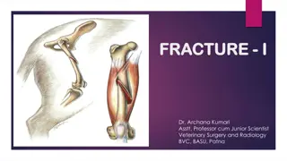
Long Bone Implants and Osteosynthesis Techniques
Fractures in long bones can be categorized based on their form, aiding in prognosis and management. Different types of fractures include comminuted, transverse, oblique, crushing, intra-articular, detachment, and selected fractures. Long bone implants play a crucial role in osteosynthesis, with materials like wire, screws, and plaques being commonly used. Various metals such as cobalt-nickel alloys, stainless medical steel, and titanium are utilized in the construction of these implants. This article also delves into the specific uses and properties of wire, screws, and plaques in the context of internal osteosynthesis for fractures.

Download Presentation

Please find below an Image/Link to download the presentation.
The content on the website is provided AS IS for your information and personal use only. It may not be sold, licensed, or shared on other websites without obtaining consent from the author.If you encounter any issues during the download, it is possible that the publisher has removed the file from their server.
You are allowed to download the files provided on this website for personal or commercial use, subject to the condition that they are used lawfully. All files are the property of their respective owners.
The content on the website is provided AS IS for your information and personal use only. It may not be sold, licensed, or shared on other websites without obtaining consent from the author.
E N D
Presentation Transcript
Fractures are categorized according to their form. Grouping is useful in determining prognosis and management: A comminuted fracture: This is when the bone continues to have its normal shape and the fracture looks like a crack on the x-ray. Transverse fracture: It is perpendicular to the long axis of the bone. In many cases, once reassigned, it remains firmly in place. Oblique fracture: It is oblique to the long axis of the bone. It is unstable and leaves the position of restoration. Crushing fracture: It is the fracture where the bone, due to the exercise of great force, has been broken into several pieces. Intra-articular fracture: It is the fracture where there is damage to the articular surface. It may cause arthritis in the joint in the long run. Detachment fracture: It is the fracture where a small part of the bone has been detached from its position, but without the bone losing its supporting capacity. Selected fracture: These are the fractures where skin injury is present. Depending on the degree of exposure of the bone to the wound, they are divided into three categories. The third category is where the bone is exposed outside the skin. In complicated fractures there is a risk of microbial inflammation and osteomyelitis.
Long bone implants Osteosynthesis Materials The main osteosynthesis materials are: 1. Wire 2. Screws 3. Plaques 4. Angular heels 5. Intramedullary cells 6. External osteosynthesis devices The metals from which they are made are: Cobalt-nickel alloys Stainless medical steel Titanium
Long bone implants Wire The wire is used to hold small pieces of bone In combination with Kirchner type needles, it can be used as a traction tape for the internal osteosynthesis of fractures (olecranon patella)
Long bone implants Screw The screw is a very useful material in the osteosynthesis of fractures It is possible to use it alone (inter-destructive compression), or by stabilizing other materials such as plates & stones There are many types of bone screws, but there are two main types: Spongy Cortical
Long bone implants Plaques There are various types of plates, most of which are used to achieve a completely stable osteosynthesis, but & plates for the application of biological osteosynthesis, which provides relative stability
Long bone implants The main types of plaque Dynamic Compression Plate (D.C.P. Dynamic Compression Plate) Dynamic compression plate of partial (limited) contact (L.C.-D.C.P. Limited Contact - Dynamic Compression Plate) Tubular plates Reconstruction Plates Special plates (Type Y, Type T, Type Cobra, etc.)
Long bone implants Intramedullary Helios Intramedullary arthrodesis is indicated in the treatment of fractures mainly of the diaphysis of long luminal bones They are applied either open or closed with the help of a fluoroscopic machine Intramedullary Helios There are several types of intramedullary cords The main ones are: Unsecured helos with glyphs Unsecured helos without glyphing Insurable helos with glyphing Insurable helos without glyphs
Long bone implants Angular Suns They consist of plate and iron They are indicated in the osteosynthesis of fractures of the central & distal end of the femur (transtrochanteric & supracondylar femur fractures) The main categories of these threads are: Fixed Angle Angular Heels Variable angle angled heels Angled fixed angle screws with sliding screw
External Osteosynthesis Device, which is placed outside the body & which stabilizes fracture ends using pins, which connect the bone to the device External osteosynthesis is a safe method for fracture stabilization External osteosynthesis systems, depending on the type of their frame, are divided into: One sided Bilaterally Circularly External Osteosynthesis Advantages: It does not impair the bone perfusion in the area of the fracture No major surgical trauma is required for placement It is useful for stabilizing open fractures It can be adjusted without requiring new surgery. It is considered a good choice in cases of increased risk of inflammation It is a fairly safe method in cases of bone infection Disadvantages: Development of inflammation of the skin at the points of entry of the needles Stiffness (femur fractures) It is relatively bulky and often not tolerated by the patient
http://www.klinikiagiosloukas.gr/issues.php?i=13&a=164 The method invented and applied by the world-famous orthopedic surgeon G. Ilizarov for the lengthening and correction of bone and joint deformities has successfully toured the world and offered quality of life to thousands of patients. https://www.newsbeas t.gr/health/arthro/2067 784/katagmata-knimis
(Ilizarov) http://www.klinikiagiosloukas.gr/issues.php?i=13&a=164 For the first time in world medical practice, Ilizarov put into practice the method of growing the tissues of the organism. In other words, he observed and proved that after osteotomy and with the removal of parts of the bone, new bone is created. At the same time, vessels, nerves and soft tissue are created (expansive histogenesis method). At the same time, he created the external osteosynthesis of the bones which consists of needles, rings and rods. By applying the method, people gained the unique ability to increase their body height, correct post-traumatic deformities and heal from hereditary and acquired skeletal diseases.
. , . . Ilizarov. 2014;19(2): 171-179. Gavriil Abramovich Ilizarov (15/06/1921- 24/07/1992) was a Russian doctor Ilizarov device Cyclic systems of external ossification of various types
. , . . Ilizarov. 2014;19(2): 171-179. In particular, it consists of: rings of various types (full, half rings, 5/8, with curved ends), arches, connecting plates (straight, twisted, curved, with threaded ends), connecting rods (threaded, with holes, telescopic, Z-shaped), simple and oiled needles, nails, special half pins, male and female supports and especially for half pins, screws (slotted, connecting, Russian type), nuts, washers (slotted, toothed, conical, spherical), threaded sockets, flexible links, simple and dynamic needle tensioners, surgical chisels, hammers, keys, angle meters, cutters, pliers, bone clamps, surgical drills and surgical burs.
. ,. . Ilizarov. 2014;19(2): 171-179. Results The advantages of the Ilizarov system are as follows: great stability, percutaneous approach and closed reduction of the fracture, minimizing the risk of infections, reduced blood loss and need for transfusions, possibility of correcting the deformation of the axis during the healing of the fracture, loading of the affected leg from the first postoperative day, removal of this at outpatient level. The disadvantages of this are as follows: soft tissue infection through the needles, loosening of needles, sutures and screws, mood disorders and mild depressive symptoms. Conclusion In summary, we must note that the Ilizarov method and device is an effective alternative proposal for dealing with intractable orthopedic problems. This is based on the biomechanical principle of axial compressive loading and micromotion in the osteogenesis zone, stimulating bone bridging of the fracture, with the result that this method is successful in cases where other methods have failed. But the Ilizarov device is a complex system of many mechanical parts. Its application is therefore quite difficult and requires a significant learning curve from the orthopedic surgeon, in order to cope with the difficulties of the surgical operations.
Taylor Spatial Frame (a) Mechanical parts of IEF, (b-e) clinical application of IEF, (f) Taylor Spatial Frame, (g) Ortho-SUV Frame, (h-j) clinical application of HEF Ilizarov external fixator (IEF) ( ) Hexapod external fixator ( F) ( ) Taylor Spatial Frame ( )
(a) Deformity form of foot and ankle deformity. (b) Correction pattern using an external fixator.
15-12-22 10-11-22 26-01-23 10-11-22 (0,5 /24 ) 31-01-22. 43 . (3 - 1 /24 , Ilizarov) Inequality coverage 43 mm. The gap is covered with denser bone Bone formation Incision in the tibia to treat inequality (3 weeks after surgery-removal of 1mm/24 hours, using the Ilizarov technique) From 10-11-22 the bone was removed with (0.5 mm/24 hours) until 31-01-22.
1. . , . . Ilizarov. 2014;19(2): 171-179. 2. http://www.klinikiagiosloukas.gr/issues.php?i=13&a=164 3. Wikipedia 4. Wikipedia 5. Configuration synthesis and structural design of lower-mobility parallel external fixators based on corrective degree-of- freedom classification


")


 Deformity form of foot and ankle deformity.")
 Deformity form of foot and ankle deformity.")
 Deformity form of foot and ankle deformity.")