Pathological Fractures

Pathological fractures occur in weakened bones due to underlying diseases or conditions, often manifesting with minimal trauma. Common causes include neoplastic lesions, infections, metabolic imbalances, and developmental disorders. Recognizing these fractures is crucial for timely intervention and appropriate treatment to prevent complications and improve outcomes.

Uploaded on Mar 26, 2024 | 2 Views
Download Presentation

Please find below an Image/Link to download the presentation.
The content on the website is provided AS IS for your information and personal use only. It may not be sold, licensed, or shared on other websites without obtaining consent from the author.If you encounter any issues during the download, it is possible that the publisher has removed the file from their server.
You are allowed to download the files provided on this website for personal or commercial use, subject to the condition that they are used lawfully. All files are the property of their respective owners.
The content on the website is provided AS IS for your information and personal use only. It may not be sold, licensed, or shared on other websites without obtaining consent from the author.
E N D
Presentation Transcript
CASE You are an FY2 working in A&E and have been asked to review an 48 year old female who has presented with hip pain following a fall. What would you to do first?
CASE She tripped over her rug and fell onto the floor and was unable to tolerate any movement afterwards. She remembers everything before, during and after the fall as she did not lose consciousness Just before she tripped, she was walking in her slippers from her kitchen to the living room. When asked about her drug history she says she can t remember what they are called but she takes pills to lower her blood pressure and a pain killer for ongoing hip pain PMHx includes breast cancer
CASE What would you like to do next?
CASE On examination you notice the following: RR 20 BP 110/80 HR 90 Temp 37 degrees Shortened and externally rotated right leg, the right hip is very tender to touch and has significant bruising in the area
CASE Based on examination findings and the history what is your top differential? What is the underlying cause?
Pathological Fractures What are they Pathological fractures are fractures that occur in a bone that has been weakened by disease. They can be spontaneous or follow on from a minor trauma/low energy injury that wouldn t normally result in a fracture
Pathological Fractures - Causes Neoplastic Primary bone tumours Secondary or Metastatic bone tumours Infections Osteomyelitis Hydadtid disease of bone Tumour like lesions Solitary Bone Cysts (SBC) Aneurysmal Bone Cyst (ABC) Fibrous dysplasia Non-ossifying fibroma Metabolic and Hormonal Imbalances Osteomalacia + Riockets Cushing s syndrome Hyperparathyroidism Developmental disorders Osteogenesis imperfecta Osteopetrosis Defect of bone remodelling Pagets disease
Pathological Fractures Main causes/Risk Factors Commonly seen in >50s Carcinomas that spread to bone: Breast, Lung, Thyroid, Renal, Prostate Common sites for mets: Spine, Pelvis, Ribs, Skull, Proximal femur, Proximal humerus
Pathological Fractures Age Underlying cause At birth Osteogenesis imperfecta 0 5 years Osteomyeltits 5 20 years Osteomyelitis, Primary bone malignancies 20 50 years Malignancy, Osteomalacia >50 years Osteomalacia, Multiple myeloma, Secondaries
Pathological Fractures How do they present? Most common location for fractures: Subtrochanteric femur, humeral head and metaphyseal junction, vertebral body Often present with pain around the location of the fracture B symptoms (if malignancy is the underlying cause or co-morbid) Bone pain
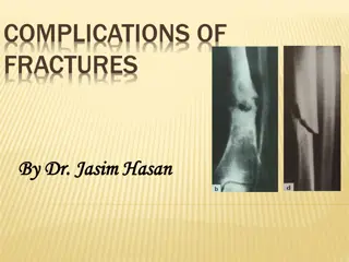
Pathological Fractures vs High Energy Fractures Pathological Fractures Occur at low energy (fall from standing height etc.) Typically patients have no history of trauma Transverse fracture pattern seen on X-rays Presence of risk factors High Energy Fractures History of trauma Spiral or Comminuted fracture pattern seen on X-rays
Pathological Fractures vs High Energy Fractures Pathological Fractures High Energy Fractures Displaced spiral fracture of the distal tibial shaft comminuted subtrochanteric fracture of the proximal femur Displaced transverse fracture of the distal diaphysis of the left femur. The fracture an underlying bony lytic lesion.
Pathological Fractures Suspect a pathological fracture if: The fracture occurs spontaneously w/out Trauma Pain is present at the site before the fracture They have had multiple recent fractures The patient is older than 45 There is a history of malignancy
Pathological Fractures: Differentials Osteomalacia Osteoporosis Osteopetrosis Pagets disease Stress fracture Benign fracture Infection
Pathological Fractures: Investigations Investigation X-ray of limb including joint above and below the lesion is how a definitive diagnosis is made. X-ray should include two views Lytic lesions -> Lung, thyroid and renal Majority of breast and prostate CA related lesions are blastic Consider a CT scan CTAP: look for primary lesion in all patients >50 w/single bony lesion Bone scan: used to identify other skeletal lesions Would see myeloma and thyroid carcinoma as cold Bloods: Serum Ca, FBC, U+Es, PSA, LFTs Urinalysis -> renal CA Bone biopsy should be taken if primary carcinoma is not identified to rule out a primary bone lesion.
Pathological Fractures: Management Non-operative management Bisphosphonate therapy -> for lytic, blastic and mixed lesions (SE: Osteonecrosis of the jaw) Denosumab -> for bone mets from solid tumors and multiple myeloma (SE: Osteonecrosis of the jaw) Radiation therapy -> palliation of pain and local tumor control Chemotherapy and Hormone Therapy
Pathological Fractures: Bisphosphonate Fractures Also known as atypical femoral fractures or Bisphosphonate-related proximal femoral fractures. They are usually associated with the long term term use of bisphosphonates. In order to reduce the risk of atypical fractures and osteonecrosis of the jaw a Bisphosphonate holiday of approximately 1-2 years after. Drug holidays are usually considered after 3 years from Zoledronic acid and 5 years after Alendrontae and Risedronate depending on their individual risk.
Pathological Fractures: Management Operative management Prophylactic stabilization is considered in the following circumstances: Involvement of the cortex Increased pain Pure lysis The pathological fracture would occur in a weight bearing area Stabilization of complete fracture -> indicated if stabilization would improve quality of life, non-surgical treatment has failed and if the patient is in significant pain
Pathological Fractures: Mirels Classification Mirels classification is a system used to assess the risk of a patient developing a pathological fracture in the long bones It takes into account the site, location on the long bone, lesion type Score 1 2 3 Site Upper limb Lower limb Peritrochanteric Pain Mild Moderate Functional Lesion Blastic Mixed Lytic Size (width of bone) <1/3 1/3 2/3 >2/3 Score <7 no surgery Score >7 prophylactic fixation