Applications of CNNs in Skin Cancer Diagnosis
This study delves into the utilization of Convolutional Neural Networks (CNNs) for diagnosing skin cancer, particularly melanoma. It explores the challenges in distinguishing melanoma from benign and atypical conditions at a cellular level, emphasizing the importance of accurate mitosis detection. The preprocessing techniques, dataset composition, and comparison of CNN models such as Efficient Spatial Pyramid of Dilated Convolutions (ESPNet) and Densely Connected Convolutional Networks (DenseNet161) are discussed. The effectiveness of these models in enhancing diagnostic accuracy is highlighted, showcasing the potential for advanced machine learning algorithms in improving skin cancer diagnosis procedures.
Download Presentation

Please find below an Image/Link to download the presentation.
The content on the website is provided AS IS for your information and personal use only. It may not be sold, licensed, or shared on other websites without obtaining consent from the author.If you encounter any issues during the download, it is possible that the publisher has removed the file from their server.
You are allowed to download the files provided on this website for personal or commercial use, subject to the condition that they are used lawfully. All files are the property of their respective owners.
The content on the website is provided AS IS for your information and personal use only. It may not be sold, licensed, or shared on other websites without obtaining consent from the author.
E N D
Presentation Transcript
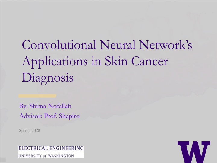
Convolutional Neural Networks Applications in Skin Cancer Diagnosis By: Shima Nofallah Advisor: Prof. Shapiro Spring 2020
What is Melanoma? > Melanoma is the most aggressive type of skin cancer. > Pathologists look at a skin biopsy slide and determine if its overall structure is normal, abnormal, or malignant. > Diagnostic errors are much more frequently than in other tissues and can lead to under- and over-diagnosis of cancer. Figure 1. An example of Invasive Melanoma Stage T1b with Hematoxylin and Eosin stain
What is Melanoma? Structural entities: Dermis Epidermis Melanocyte nests Cellular entities: Nuclei Melanocyte Mitosis Diagnoses: Benign Atypia Melanoma in situ Invasive T1a Invasive T1b
Mitosis Detection > Distinguishing mitoses from normal nuclei is a challenge. Figure 5. Examples of a sampled Mitosis (Left) and a sampled Nuclei (Right).
Preprocessing > Data augmentation: : Rotations of 45, 90, 135 or 225 degrees. Mirroring horizontal and vertical. > The final dataset: 4364 mitosis samples. 12640 non-mitosis samples. > Dataset randomly split: Training: 60% Validation: 20% Testing: 20%
Method and Model > In recent years, with the development of fast and accessible GPUs, Convolutional Neural Networks (CNNs) have dominated computer vision research due to their impressive performance, and mitosis detection is not an exception. > We ran our experiment two separate times on two well-designed CNNs and compared their results: 1. Efficient Spatial Pyramid of Dilated Convolutions (ESPNet) 2. Densely Connected Convolutional Networks (DenseNet161)
Method and Model > ESPNet A fast and efficient convolutional neural network for high resolution inputs. Figure 7. (Left) Comparing standard convolution and ESP. (Right) Block diagram of ESP module.
Method and Model > DenseNet: Whereas traditional convolutional networks with L layers have L connections one between each layer and its subsequent layer the DenseNet network has L(L+1)/2 direct connections. Figure 8. DenseNet architectures for ImageNet.
Method and Model > Hyperparameters Adam optimizers. learning rate decay schedule with step size = 5 and ? = 0.1. 20 epochs. cross-entropy loss function. > Evaluation Metrics Accuracy = (TP+TN)/(TP+FP+FN+TN) Precision = TP / (TP + FP) Recall = TP / (TP + FN) F1 score = 2 (Precision Recall) Precision + Recall Sensitivity = TP / (TP + FN) Specificity = TN / (TN + FP)
Results > Evaluation results of ESPNet and DenseNet161 on Melanoma
Dataset > Our dataset comes from 240 H&E stained slides of skin biopsy images, acquired by the University of Washington School of Medicine in the MPATH study (R01 CA151306). > Our dataset contains five different diagnoses: 1. Benign 2. Atypia 4. Invasive Melanoma T1a 3. Melanoma in Situ 5. Invasive Melanoma T1b.
Benign vs. Invasive (1 vs. 5) Normalized input Gradient Layer 5 heatmap True Positive (Invasive)
Benign vs. Invasive (1 vs. 5) Normalized input Gradient Layer 5 heatmap True Negative (Benign)
Benign & Atypia vs. Invasive Atypia Invasive Invasive
Latest Results Class 1 vs. 5 Class 1 &2 vs. 5 Train: 1: 31 slices, 5: 72 slices Test: 1: 26 slices, 5: 74 slices Train: 1 & 2: 74 slices, 5: 72 slices Test: 1 & 2 : 75 slices, 5: 74 slices Accuracy F1 Precision Recall Total 1 5 1 5 1 5 1 vs. 5 0.78 0.54 0.86 0.59 0.83 0.5 0.88 Total 1&2 5 1&2 5 1&2 5 1&2 vs. 5 0.819 0.83 0.81 0.80 0.84 0.85 0.78


