Anatomy and Physiology of the Ear by Dr. Farid Alzhrani
This detailed presentation covers the anatomy of the ear, including the external ear structures, nerve supply, tympanic membrane layers, and divisions of the middle ear. Explore the intricacies of the auricle, external auditory canal, and the tympanic membrane, elucidated with informative images.

Download Presentation

Please find below an Image/Link to download the presentation.
The content on the website is provided AS IS for your information and personal use only. It may not be sold, licensed, or shared on other websites without obtaining consent from the author.If you encounter any issues during the download, it is possible that the publisher has removed the file from their server.
You are allowed to download the files provided on this website for personal or commercial use, subject to the condition that they are used lawfully. All files are the property of their respective owners.
The content on the website is provided AS IS for your information and personal use only. It may not be sold, licensed, or shared on other websites without obtaining consent from the author.
E N D
Presentation Transcript
( Ear I ) Anatomy and Physiology of the ear Dr. Farid Alzhrani Assistant professor Consultant of Otolaryngology, head and neck surgery King Abdulaziz University Hospital
A) External Ear Auricle External Canal
Nerve Supply of External Ear Cervical II & III ( greater auricular and lesser occipital) V cranial nerve (auriculotemporal) X cranial nerve (auricular or Arnold s) Fibres from VII cranial nerve
TYMPANIC MEMBRANE It forms the partition between the external auditory canal and the middle ear. Parts : Pars Tensa. 1. Pars Flaccida. 2.
TYMPANIC MEMBRANE The Tympanic membrane consist of three layers: Outer layer, of epithelial (ectodermal) origin, 1. The middle layer or lamina propria, of mesodermal origin, 2. The inner layer, of endodermal origin, comprising the middle ear mucosa. 3.
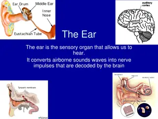
ANATOMY OF THE EAR External Ear a) Middle Ear b) Inner Ear c)
B) Middle Ear http://download.e-bookshelf.de/download/0000/6688/46/L-X-0000668846-0001409245.XHTML/images/c02f009.jpg Eustachian (Pharyngo- 1. tympanic) Tube Tympanum (Middle Ear 2. Cavity) Mastoid Antrum and Air 3. Cells
Middle Ear Eustachian (Pharyngo-Tympanic) Tube Connect ear nasopharynx. the middle with cavity Lies adjacent to the ICA
Middle Ear Eustachian (Pharyngo-Tympanic) Tube
Middle Ear Eustachian (Pharyngo-Tympanic) Tube
Middle Ear Eustachian (Pharyngo-Tympanic) Tube
Middle Ear Eustachian (Pharyngo-Tympanic) Tube
Middle Ear Tympanic cavity (Middle ear cavity)
Middle Ear Tympanic cavity (Middle ear cavity)
Middle Ear Tympanic cavity (Middle ear cavity) Middle ear cavity divided into three parts
Middle Ear Tympanic cavity (Middle ear cavity) LINING OF THE MIDDLE EAR : Mucous membrane of the middle ear space consists of stratified cuboidal epithelium, which changes to pseudostratified ciliated epithelium around the mouth of the eustachian tube.
Middle Ear Tympanic cavity (Middle ear cavity) Motor nerve supply of the middle ear muscles : Sensory nerve supply of the middle ear mucosa : 1. Stapedius muscle supplied by the stapedial branch of the facial nerve. 2. Tensor tympani muscle supplied by the mandibular division of the trigeminal nerve. Tympanic branch of the glossopharyngeal nerve. 1. Auriculotemporal branch of the trigeminal nerve. 2.
Middle Ear Mastoid antrum and air cells Air-containing cells of the mastoid process are continuous with the air in the middle ear.
Middle Ear Mastoid antrum and air cells Pneumatization is complete between the sixth and twelfth years of life. Normal tubal function is a prerequisite for biologically active, healthy middle ear mucosa, and thus for the normal process of pneumatization.
ANATOMY OF THE EAR External Ear a) Middle Ear Cleft b) Inner Ear c)
C) Inner Ear Consists of : Labyrinth. 1. Bony Labyrinth Membranous Labyrinth 2. Internal Auditory Canal.
Inner Ear Bony Labyrinth Its parts : 1. Bony Cochlea 2. Vestibule 3. Bony semicircular canals
Inner Ear Bony Labyrinth Its contents : 1. Perilymph 2. Membranous labyrinth
Inner Ear Membranous Labyrinth Its parts : Cochlear duct 1. Saccule and utricle 2. Membranous semicircular ducts 3.
Inner Ear Membranous Labyrinth Its contents : Endolymph 1. Sensory epithelium 2. Cochlea: organ of Corti Utricle & saccule: maculae Semicircular canals: cristae
Inner Ear Internal Auditory Canal Contains: Cochleovetibular nerve Facial nerve
Inner Ear Internal Auditory Canal
FUNCIONS OF THE EXTERNAL EAR Protection of the middle ear Curvature Cerumen Auditory functions: Sound conduction Increase sound pressure by the resonance function
FUNCTIONS OF THE EUSTACHIAN TUBE Protection Ventilation Drainage
FUNCIONS OF THE MIDDLE EAR Conduction of sound Protection to the inner ear stapedial reflex
FUNCIONS OF THE INNER EAR Hearing Function: Transduction of sound to action potentials Vestibular Function: Participate in maintaining body balance
")

 External Ear")


 Middle Ear")


 Inner Ear")