Anatomy and Physiology of the Larynx: Overview and Cartilages
Exploring the anatomy and physiology of the larynx, this content discusses the location, movements, and cartilages of the larynx. Details on the unpaired (thyroid, cricoid, epiglottis) and paired (arytenoid, corniculate, cunieform) cartilages are provided, along with their functions and characteristics. Additionally, the functions and structures of the epiglottis, arytenoid cartilages, and other associated structures are outlined for a comprehensive understanding.
Download Presentation

Please find below an Image/Link to download the presentation.
The content on the website is provided AS IS for your information and personal use only. It may not be sold, licensed, or shared on other websites without obtaining consent from the author.If you encounter any issues during the download, it is possible that the publisher has removed the file from their server.
You are allowed to download the files provided on this website for personal or commercial use, subject to the condition that they are used lawfully. All files are the property of their respective owners.
The content on the website is provided AS IS for your information and personal use only. It may not be sold, licensed, or shared on other websites without obtaining consent from the author.
E N D
Presentation Transcript
Anatomy and Physiology of the larynx Dr. Rafid Y. Al-maidi , ENT lecturer Basra Medical College.
The larynx lies in the front of the hypopharynx opposite the third to sixth cervical vertebrae. It moves vertically and in anteroposterior direction during swallowing and phonation . It can also be passively moved from side to side producing a characteristic grating sensation called laryngeal crepitus . In an adult , the larynx ends at the lower border of C6 vertebrae.
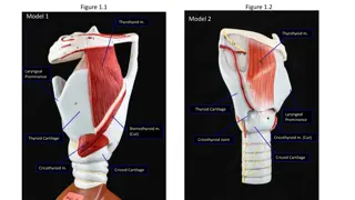
Laryngeal cartilages : Larynx has three unpaired and three paired cartilages . Unpaired : thyroid ,cricoid, epiglottis. Paired : arytenoid , corniculate , cunieform. 1- thyroid : it is the largest cartilage . Its two alae meet anteriorly forming an angle of 90 degree in males and 120 degree in females. Vocal cords are attached to middle of thyroid angle. 2- cricoid cartilage: it is only cartilage forming a complete ring. Its posterior part is expand to from a lamina while anteriorly it is narrow forming an arch.
3- epiglottis: it is leaf like, yellow, elastic cartilage,forming anterior wall of laryngeal inlet. It is attached to body of the hyoid bone by hyoepiglottic ligament, which is divided it into suprahyoid and infrahyoid epiglottis. A stalk-like process of epiglottis (petiole) attaches the epiglottis to the thyroid angle just above the attachment of the vocal cords. Anterior surface of epiglottis is separated from thyro -hyoid membrane and upper part of thyroid cartilage by potential space filled with fat ---the pre-epiglottic space The pre-epiglottic space may be invaded in carcinoma of supraglottic larynx or base of the tongue .
Posterior surface of epiglottis is concavoconvex concave above but convex below forming a bulge called tubercle of epiglottis ,which obstructs view of anterior commissure when examining larynx by indirect laryngoscopy . Epiglottis is not essential for swallowing and can be amputated in carcinoma with little aspiration. 4- Aryteniod cartilages: they are paired.each arytenoid cartilage is pyramidal in shape. It has a base which articulate with cricoid cartilage , muscular process , directed laterally to give attachment to intrinsic laryngeal muscles a vocal process directed anteriorly ,giving attachments to vocal cords and an apex which supports the corniculate cartilage. 5- Corniculate cartilage(of Santorini): they are paired , each articulates with apex of arytenoid cartilage as if forming its horn.
6- Cuneiform cartilage : ( of Wrisberg):they are rod shaped each is situated in aryepiglottic fold in front of corniculate cartilage and provides passive supports to the fold Thyroid , cricoid , most of the arytenoid cartilages are hyaline cartilages whereas epiglottis ,corniculate ,cuneiform and tip of arytenoid near the corniculate cartilage are elastic fibrocartilage . Hyaline cartilage can undergo ossification it begins at age of 25 years in thyroid , a little later the cricoid and arytenoid ,and is complete by 65 years of age . Calcification seen in these cartilages can be confused with foreign bodies of oesophagus or larynx on x-rays.
Laryngeal joints: Cricoarytenoid joint: it is a synovial joint surrounded by capsular ligament. It is formed between the base of arytenoid and a facet on the upper border of cricoid lamina. Cricothyroid joint : it is also a synovial joint . Each is formed by the inferior cronua of thyroid cartilage with a facet on the cricoid cartilage.
Muscles of larynx: They are of two types : Intrinsic : which attach laryngeal cartilages to each other. Extrinsic : which attach larynx to the surrounding structures. Intrinsic muscles : they may act on vocal cords or laryngeal inlet . A- acting on vocal cords - abductors : posterior cricoarytenoid muscle. - adductors : lateral cricoarytenoid muscle. interarytenoid muscle thyroarytenoid muscle(external part) - tensors : cricothyroid , vocalis ( internal part of thyroarytenoid ).
B-acting on laryngeal inlet : -opening of laryngeal inlet : thyroepiglottic (part of thyro- -arytenoid) - closure of laryngeal inlet: interarytenoid (oblique part) aryepiglottic ( posterior oblique part of interarytenoids) Extrinsic muscles: they connect the larynx to the neighbouring structures and are divided into elevators or depressors of larynx. a)elevators: stylopharyngeus , salpingopharyngeus, thyrohyoid,palatopharyngeus b)Depressors: sternohyoid , sternothyroid and omohyoid .
Cavity of the larynx: Laryngeal cavity starts at the laryngeal inlet where it communicates with the pharynx and ends at the lower border of cricoid cartilage where it is continuous with the lumen of trachea. Inlet of larynx: it is an oblique opening bounded anteriorly by free margin of epiglottis , on the sides , by aryepiglottic folds and posteriorly by interarytenoid fold. Vestibule : it extends from laryngeal inlet to the vestibular folds. Its anterior wall is formed by posterior surface of epiglottis;sides by aryepiglottic folds and posterior wall by mucous membrane over the anterior surface of arytenoids. Subglottic space: it extends from vocal cord to the lower border of cricoid cartilage .
Vestibular folds(false vocal cords): two in number , each is a fold of mucous membrane extending anteroposteriorly across the laryngeal cavity . It contains vestibular ligament , a few fibers of muscle (thyroarytenoidus ) and mucous glands. Vocal folds(true vocal cords): they are two pearly white sharp bands extending from the middle of thyroid angle to the vocal process of arytenoids. Glottis:(rima glottidis): it is elongated space between vocal cords anteriorly , and vocal processes and base of arytenoids posteriorly. Ventricle (sinus of larynx): it is a deep elliptical space between vestibular and vocal folds, also extending a short distance above and lateral to vestibular fold.
Mucous Membrane of the Larynx: Epithelium : it is ciliated columnar type except over the vocal cords and upper part of the vestibule where it is stratified squamous type. Mucous glands: are distributed all over the MM and particularly numerous on the posterior surface of epiglottis , posterior part of the aryepiglottic folds and in the saccules . There are no mucous glands in the vocal folds. Lymphatic drainage : 1- Supraglottic larynx above VC is drained to upper deep cervical LN . 2- Infraglottic below VC to lower deep cervical LN. No lymphatics in VC .
Physiology of Larynx: 1- Protection of the lower airways. 2- Phonation. 3- Respiration. 4- Fixation of the chest.
