Acute Otitis Media in Primary Care


Barbara Adams and Mike Pointon

Aims and objectives
Know how to assess and manage common ENT
problems in primary care
Know about watchful waiting and use of delayed
prescriptions
Know how and when to refer to ENT secondary care
for non-urgent referrals
Know about ENT emergencies and how to refer
Acute otitis media (AOM) definitions
AOM: Infection in middle ear, characterised by presence of
middle ear effusion associated with acute onset of signs
and symptoms of middle ear inflammation
Recurrent AOM: ≥3 episodes in 6m or ≥4 in 1y with absence
of middle ear disease between episodes
Persistent AOM (treatment failure): symptoms persist after
initial management (no antibiotics, delayed antibiotics or
immediate antibiotic prescribing strategy) or symptoms
worsening
AOM: causes & complications
Bacterial infection: most common- strep pneumoniae, h
influenzae (only 10% due to type B and preventable by HIB
vaccine), moraxella catarrhalis
Viral infection: most common- respiratory syncytial virus
and rhinovirus
Complications: hearing loss; chronic perforation and
otorrhoea, CSOM, cholesteatoma, intracranial
complications
AOM: diagnosis
Presents with earache (!)
In younger children-non specific symptoms,
e.g rubbing ear, fever, irritability, crying, poor
feeding, restlessness at night, cough, or
rhinorrhoea
Mastoiditis
AOM
AOM
AOM Differential diagnoses
Other URTI: may be mild redness of TM, self limiting
Otitis media with effusion (OME)/ glue ear: fluid in middle ear
without signs of acute inflammation of TM
CSOM: persistent inflammation and TM perforation with
exudate >2-6w. May lead to . . . . . .
Acute mastoiditis (rare)- swelling, tenderness and redness over
mastoid bone, pinna pushed forward
Bullous myringitis (rare)- haemorrhagic bullae on TM caused by
Mycoplasma pneumoniae (90% spontaneous resolution)
Management of AOM: when to refer or admit?
Advise a no antibiotic or delayed antibiotic strategy for most people
with suspected AOM but:
consider antibiotics in children < 3m,
bilateral AOM
systemically unwell
high risk of complications e.g. immunosuppression, CF.
For all antibiotic prescribing strategies: inform patient average duration
of illness for untreated AOM is 4 days.
Admit: According to “Feverish illness in Children” NICE Guidance
Adults and children with suspected complications e.g. meningitis,
mastoiditis, or facial paralysis
Amoxicillin or Erythromycin
Follow up of AOM
Routine follow up not usually required
Follow up if:
symptoms worse or not settling within 4 days
otorrhoea persists >2w
perforation
if hearing loss persists in absence of pain or fever, ie OME
Recurrent AOM: Second line co-amoxiclav
http://guidance.nice.org.uk/CG47
Feverish illness in children
http://guidance.nice.org.uk/CG69
Respiratory Tract Infections
Otitis media with effusion (OME) / Glue ear
Definition: non-purulent collection of fluid in middle ear
(must be > 2/52 after recent AOM to be classed as Glue ear)
Causes:
Eustachian tube dysfunction
> 50% due to AOM especially in < 3 yrs
Other: low grade bacterial/viral infections; gastric reflux;
nasal allergies; adenoids or nasal polyps; CF; Down’s
Pressure changes e.g. with flying or scuba diving (adults)
Symptoms:
hearing loss
absence of earache or systemic upset
can present with problems of speech/language development,
behaviour or social interaction
Other causes of hearing loss (or perceived loss)-
Foreign body in EAC
perforated TM
SNHL
listening problems inc ADHD and learning difficulty
Initial management of OME
Ask about developmental delay or language difficulties
Hearing test
Drugs not recommended as OME usually self limiting
but consider ICS if there is associated allergic rhinitis
Early intervention with grommets
gives no benefit for long-term
hearing, language and behaviour and
increases risk of TM abnormalities.
Subgroup with hearing loss > 25DB
may benefit from early grommet
insertion.
OME general advice:
good prognosis, self-limiting and >90% get resolution within 6m;
limited proven benefit from drugs
OME in adults is unusual in adults and need referral to ENT
(unilateral could mean nasopharyngeal ca)
Grommets – general points:
usually stop functioning after 10m
approx 50% require reinsertion within 5y
conductive deafness after extrusion improves slowly
Complications are otorrhoea, may need specialist input.
most activities unaffected, i.e. can fly and swim but avoid
immersion; re hearing loss should face child when speaking
Adenoidectomy: is usually second line treatment for OME but no UK
national guideline; conflicting evidence.
No evidence for Tonsillectomy in OME
Chronic Suppurative Otitis Media (CSOM)
Symptoms
persistent painless otorrhoea >2w
May be preceded by AOM, trauma and grommets
Differentials
OE, FB, wax
Assessment
Exclude intracranial involvement, facial paralysis or
mastoiditis- needs admission
otherwise routine referral
Otitis externa (OE)
Inflammation of EAC
Localised OE: folliculitis that can progress to a furuncle
Diffuse OE: more widespread inflammation e.g. swimmers
ear
OE defined as: acute if episode<3w; chronic if >3m
Malignant OE: extends to mastoid and temporal bones
resulting in osteitis. Typically in elderly diabetics. Suspect
if pain seems disproportionate to clinical findings
Localised OE
Causes: usually infected hair root by staph aureus
Symptoms: severe ear pain (compared to size of lesion); relief if
furuncle bursts; hearing loss if EAC very swollen
Signs: tiny red swelling in EAC (early); later has white or yellow
pus-filled centre which can completely occlude EAC
Management: analgesia; hot compress; antibiotic only if severe
infection or high risk patient - flucloxacillin or erythromycin
Refer: if needs I+D, no response to antibiotic or cellulitis
spreading outside EAC
Acute diffuse OE
Causes:
bacterial infection- pseudomonas or staph aureus
seborrhoeic dermatitis
fungal infection- usually candida
contact dermatitis - meds (sudden onset) or hearing aids/earplugs (insidious onset)
Symptoms: any combination of ear pain, itch, discharge and hearing
loss
Signs:
EAC and/or external ear are red, swollen or eczematous
serous/purulent discharge
inflamed TM – may be difficult to visualise
pain on moving ear or jaw
Investigations: rarely useful but if treatment fails, send swab for
bacterial and fungal culture
Management: Use topical ear preparation for 7
days;
2% acetic acid for mild cases
antibiotic plus steroid e.g. Locorten-Vioform
Gentisone HC (NB not if perforation)
If wax/debris obstructing EAC or extensive swelling
or cellulitis
Pope wick
Dry mopping (children)
Microsuction (ENT PCC)
Advise re prevention of OE: keep ears clean and dry;
treat underlying eczema/psoriasis
Failure of topical meds:
review diagnosis/compliance
consider PO fluclox or erythromycin
?fungal (spores in EAC)
Swab and refer
Chronic OE
Causes:
Secondary fungal infection- due to prolonged use of topical antibacterials or
steroids
Seborrhoeic dermatitis; contact dermatitis
Sometimes no cause can be found for OE
Symptoms:
mild discomfort; pain usually mild
Signs:
lack of ear wax; dry, hypertrophic skin leading to canal stenosis; pain on exam
Assess risk /precipitating factors; severity of symptoms; signs of fungal
infection- whitish cotton-like strands in EAC, black or white balls of
aspergillus. Look for signs of dermatitis, evidence of allergy (ear plugs etc) or
focus of fungal infection elsewhere, e.g. Skin, nails, vagina- can cause 2’
inflammation EAC
Investigations:
only take swab for C+S if treatment fails as interpretation can be difficult:
sensitivities are determined for systemic use and much higher concentrations
can be achieved by topical use; organisms may be contaminants, usually fungal
overgrowth after using antibacterial drops due to suppressed normal bacterial
flora
Chronic OE
Management:
advise general measures as for acute diffuse OE
Treatment depends on cause - often requires more than one
strategy:
if fungal infection- top antifungal, refer if poor response
seborrhoeic dermatitis- antifungal and steroid combined
If no cause evident- 7d course top steroid +/- acetic acid spray. If
good response, may need to continue steroid but reduce
potency/dose.
If cannot be withdrawn after 2-3m, refer ENT. If poor response, try
trial of top antifungal
Refer ENT if contact sensitivity (re patch testing); if EAC occluded;
if malignant OE suspected.
Foreign Bodies
Management depends on what it is:
Batteries – immediate referral to ENT
Inert FB – e.g. retained grommet, beads, foam - not so
urgent
Organic – e.g. food, insects. May cause infection therefore
should be dealt with sooner. For insects – drown in olive oil
first.
Some FBs may resolve with syringing, but if not refer to
PCC
Do not attempt to remove under direct visualisation as
more likely to cause harm
Anterior or Posterior – hx gives clues
> 90% from Little’s Area
Age gives clue – more likely posterior in Elderly
Cause: Idiopathic, trauma (nose picking), dry mucosa,
hypertension, coagulopathy, NSAID, Warfarin, tumour
CAN BE FATAL!!!
First Aid: Compression & Ice
Epistaxis
Avoid blowing their nose (1/52)
Avoid hot drinks (1/52)
Naseptin cream 1/52
Admit: If cannot control, elderly, warfarinised, low
platelets, recurrent excessive bleeding
PCC: If not settling with conservative rx
AgNO3 cautery – can be done in GP
Packing, Electrocautery, Surgery (SPA ligation, ECA
ligation, embolisation)
Cautery: What you need:
A good lightsource
Nasal speculum (or large aural speculum)
Lignocaine (with adrenalin)
Cotton wool
Cautery sticks
Causative factors – allergic, viral, bacterial, fungal, autoimmune.
Acute <12wks, Chronic >12wks, Recurrent (>4/yr)
15% population. 6 million lost working days / yr in the UK
Presents as “My cold won’t go away” – persistant symptoms of URTI,
without improvement after 10-14 days or worsening after 5 days
Major:
Nasal congestion/obstruction
Purulent discharge
Loss of smell
Facial pain / ear pain or fullness
Rhinosinusitis
Minor:
Tenderness over sinus area
Fever
Headache
Halitosis
Fatigue / Lethargy
Post nasal drip
What to exclude on examination:
Periorbital swelling, extraocular muscle dysfunction, decreased VA
or proptosis
Foreign bodies
Concomitant otitis media (in children)
CNS complications
Polypoid changes or deviated septum
What to expect on examination:
Erythema / swelling of nasal mucosa
Mucopurulent secretions
Tenderness over sinuses
Differentials
Allergic rhinitis (seasonal or perennial)
Usually just nasal symptoms and usually persistent
Nasal FB – unilateral blockage or discharge
Sinonasal tumour – chronic, unilateral blockage, discharge
(bloody)
Other causes of facial pain
Tension Headache
TMJ dysfunction or bruxism
Neuropathic
Dental pain (hot/cold drinks, chewing)
Investigations
Xrays / Bloods / Swabs = not required, only indicated if > 12
wks and failure to respond to Rx – will probably refer at that
stage (rigid endoscopy / coronal CT / allergy testing)
Consider emergency admission to hospital if symptoms are
accompanied by:
Systemic illness
Swelling or cellulitis in face
Signs of CNS involvement
Orbital involvement
Consider urgent ENT referral if:
Persistant unilateral symptoms such as (suspecting sinonasal
tumour):
Bloodstained discharge
Non-tender facial pain
Facial swelling
Unilateral polyps
Consider routine referral to ENT if:
More than 3-4 episodes per year lasting > 10 days with no symptoms
between episodes
Management of acute rhinosinusitis
(guidelines on map of medicine)
Viral is 200 times more common than bacterial
Viral URTI usually precedes bacterial
Bacterial usually has more severe and prolonged
symptoms
Strep pneumoniae, H. influenzae, Moraxella Catarrhalis
First line :
Amoxicillin
Doxycycline, erythromycin, clarithromycin (pen allergic)
Second Line:
Co-amoxiclav
Azithromycin (pen allergic)
Management of chronic rhinosinusitis (referral toolkit)
Commonest in children aged 1-4
Rare in adults
Potential risk to airway
Suspect if persistant unilateral symptoms of blockage
or foul smelling discharge
Unless very easy to get at, and very compliant child,
best not attempted in GP (sometimes only get one
shot!)
Nasal Foreign Bodies
Best viewed from above – looking
at deviation of nasal bones –
difficult if swollen
Exclude septal haematoma
Requires immediate drainage to
prevent abscess or permanent
saddle nose deformity
Otherwise refer to PCC for
manipulation 7-10 days post
injury. For old injuries routine
ENT referral
Nasal Fracture
Nasal blockage will almost always be accompanied by snoring
Have OSA in the back of your mind
Defined as the presence of at least five obstructive events per
hour during sleep
Features
Impaired alertness
Cognitive impairment
Excessive sleepiness (Epworth scale)
Morning headaches
Choking or SOB feeling at night
Nocturia
Unrefreshing sleep
Sleep quality of partners affected (“does he stop breathing at
night?”)
Refer to Respiratory in the first instance
Consider OSA
Sore throat: causes
Common infections:
rhinovirus; coronovirus, parainfluenza virus; common cold (25%
sore throats)
GABHS causes 15-30% sore throats in children and 10% in adults
Herpes simplex virus type 1 (more rarely type 2) = 2%
Epstein Barr virus: infectious mononucleosis (glandular
fever)- <1%
. Suspect IM if sore throat persists >2w - do FBC
and IM screen.
Non-
infectious causes
Physical irritation
Hayfever
Stevens Johnson syndrome
Kawasaki disease
Oral mucositis 2’ chemo /radiotherapy
Sore throat: complications
Complications of streptococcal
pharyngitis are rare:
Suppurative complications:
OM
acute sinusitis
peritonsilar cellulitis / peritonsillar
abscess (quinsy)
Pharyngeal abscess
Retropharyngeal abscess, more
common in children
Non suppurative complications are rare:
rheumatic fever
post-streptococcal glomerulonephritis
R sided quinsy showing
displacement of uvula to L
Sore throat: when to refer
Admit if stridor or respiratory difficulty
Trismus, drooling, dysphagia.
Dehydration /unable to take fluids
Severe suppurative complications, ie if abnormal throat
swelling/suspected abscess
Systemically unwell and at risk of immunosuppression
Suspect Kawasaki disease
Profoundly unwell and cause unknown
Sore throat: management in primary care
Reassure sore throat usually self limiting and symptoms resolve within
3d in 40% cases, 1w in 85% (even if due to streptococcal infection)
Advise see healthcare professional if symptoms do not improve, and
urgently if breathing difficulties, stridor, drooling, muffled voice,
severe pain, dysphagia or unable to take fluids or systemically ill
Symptoms of infectious mononucleosis usually resolve within 1-2w,
mild cases within days. But lethargy continues for some time and rarely
may continue for months or years. Advise re contact sport.
Advise regular paracetamol, ibuprofen, fluids ++ but avoid hot drinks;
saline mouthwashes; discuss role of antibiotics
Consider delayed prescription or immediate antibiotics – use Centor
scoring - Antibiotic regime: Prescribe phenoxymethylpenicillin for 10d;
or erythromycin or clarithromycin for 5d. Avoid amoxicillin (EBV)
Indications for tonsillectomy for recurrent acute
sore throat
Sore throats are due to acute tonsillitis
Episodes of sore throat are disabling and prevent normal
functioning
Seven or more well documented, clinically significant,
adequately treated sore throats in the preceding year
or
Five or more such episodes in each of the preceding two
years
or
Three or more such episodes in each of the preceding three
years
SIGN 2010, Management of sore throat and indications for tonsillectomy
http://www.sign.ac.uk/pdf/qrg117.pdf
Vertigo
Vertigo:
‘is a symptom and refers to a perception of spinning or rotation of
the person or their surroundings in the absence of physical
movement’
Peripheral vertigo = labyrinthine cause
Benign paroxysmal positional vertigo (BPPV)
Vestibular neuronitis:
Meniere’s disease:
Central vertigo = cerebellar cause
Common
Migraine
Uncommon
stroke and TIA
cerebellar tumour
acoustic neuroma
MS
Assessment of vertigo
Most balance problems that present in primary care are not
rotatory vertigo, but unsteadiness. A full time GP is likely
to see 10-20 people with vertigo in 1y
To determine vertigo rather than dizziness, ask:
“do you feel light-headed or do you see the world spin around
you as if you had just got off a roundabout”
about timing, duration, onset, frequency and severity of
symptoms
aggravating factors, e.g. head movement
effect on daily activities
associated symptoms:
hearing loss, tinnitus (unilateral/bilateral), headache,
diplopia, dysarthria /dysphagia, ataxia
,
nausea, vomiting
Assessment of vertigo: medical history
Recent URTI or ear infection suggests vestibular
neuronitis or labyrinthitis
Migraine: inc likelihood of migrainous vertigo
Head trauma/ recent labyrinthitis: BPPV
Trauma to ear: perilymph fistula
Anxiety or depression can worsen symptoms or cause
feelings of lightheadedness (e.g. from hyperventilation)
Acute alcohol intoxication can cause vertigo
Examination
ENT – incl. Weber and Rinnes tests
Full Neuro incl cerebellar testing + gait. Particularly
looking for nystagmus
Assessment of vertigo: specific tests
Romberg’s test:
identifies peripheral or central cause of vertigo (but not
sensitive for differentiating between them)
Ask patient to stand up straight, feet together, arms
outstretched with eyes closed. If patient unable to keep
balance- the test is positive (usually fall to side of lesion)
A positive test suggests problem with proprioception or
vestibular function.
Hallpike manoeuvre:
to confirm diagnosis of BPPV
Hallpike manoeuvre - demonstration
Be cautious with patients with neck or back pathology or carotid
stenosis as manouvre involves turning and extending neck
http://northerndoctor.com/2010/09/27/dizziness-dix-hallpike-and-the-epley-
manoeuvre/
Ask patient to:
report any vertigo during test
keep eyes open and stare at examiner’s nose
sit upright on couch, head turned 45’ to one side
lie them down rapidly until head extended 30’ over end of bed, one ear
downward If neck problems- can be done without neck extension
observe eyes closely for 30 sec for nystagmus- note type and direction
support head in position and sit up
Repeat with other side
test is positive for BPPV if vertigo and nystagmus (torsional and beating
towards ground) are present and nystagmus shows latency, fatigue and
adaptation
Features of central causes of vertigo
severe or prolonged
new onset headache
focal neurological deficits
central type nystagmus (vertical)
excess nausea and vomiting
prolonged severe imbalance (inability to stand up even
with eyes open)
Features of peripheral causes of vertigo
BPPV:
vertigo induced by moving head position
episodes last for seconds
Vestibular neuronitis and labyrinthitis:
vertigo persists for days and improves with time
no hearing loss or tinnitus with vestibular neuronitis
in labyrinthitis, sudden hearing loss with vertigo and tinnitus may
be present
Meniere’s disease:
ages 20-50y men> women
vertigo, not provoked by position change
episodes last 30 min to several hours
symptoms of tinnitus, hearing loss and fullness in ear
may be clusters of attacks and long remissions
Medication used in vertigo
prochlorperazine
cyclizine
cinnarizine
promethazine
Tinnitus
Unwanted perception of sound within head, in absence of
sound from external environment
Can be described as ringing, hissing, buzzing, roaring or
humming. Classified as-
Subjective tinnitus:
sound only heard by patient; assoc with abnormalities of auditory
system
Objective tinnitus:
sound heard by patient and examiner; caused by physical
abnormality that produces sound near or within ear
Disorders associated with subjective tinnitus
Two thirds people with tinnitus have disorder causing
hearing loss; one third have idiopathic tinnitus
Most commonly assoc with disorders causing sensorineural
hearing loss (SNHL):
age related
noise induced
Meniere’s disease
Less commonly assoc with disorders causing conductive
hearing loss:
impacted wax
otosclerosis (rare)
Uncommonly, subjective tinnitus is associated with:
Ototoxic drugs
Cytotoxic drugs (e.g. Cisplatin, methotrexate)
Aminoglycosides (gentamicin)
macrolides, quinine, aspirin, NSAIDs and loop diuretics
Ear infections:
(OM, OME, CSOM)
Neurological disorders:
acoustic neuroma; schwannoma, MS
Metabolic disorders:
Hypothyroidism; diabetes
Psychological disorders:
anxiety and depression
Trauma
Disorders associated with objective tinnitus
Objective tinnitus is very rare
Due to:
Vascular disorders:
AVMs; vascular tumours;
High output states:
anaemia; thyrotoxicosis; Paget’s disease
Management of tinnitus in primary care
Assess underlying cause
Refer to ENT:
All patients with objective tinnitus
Patients with subjective tinnitus, following hearing test,
who have associated SNHL
Tinnitus associated with conductive hearing loss when
treatable causes not identified or managed in primary
care
Tinnitus secondary to head or neck injury
Tinnitus of uncertain cause
Tinnitus that is causing distress despite primary care
management
Foreign Bodies
Feeling of food (most commonly) stuck in throat /
oesophagus
If complete dysphagia of acute onset, then very high
chance of a FB obstruction
If delayed onset of FB sensation after eating, and mild
symptoms, could simply be abrasion, symptoms will go
in 48 hrs.
Refer if not resolved
Oesophageal food bolus: coke or pineapple juice,
buscopan (IM) or GTN (SL) can help
Lower motor neurone
(involving forehead)
Motor supply to the scalp,
facial muscles & stapedius
Taste to anterior 2/3 of the
tongue
Possible causes:
Traumatic
facial lacerations, blunt trauma ( BOS fracture), newborn paralysis
Neoplastic
parotid tumors, tumors of the external canal and middle ear,
metastatic lesions, SCC, cholesteatoma, acoustic neuroma
Infectious
herpes zoster oticus (Ramsey-Hunt syndrome), AOM, CSOM,
malignant otitis externa
Idiopathic
Bell's palsy although traditionally defined as idiopathic it is thought to
be associated with herpes simplex virus type 1
Characteristics of a peripheral facial paralysis include:
Motor
unable to wrinkle forehead
unable to raise eyebrow
unable to wrinkle nasolabial fold
unable to purse lips or show teeth
inability to completely close eye
(classified using House-Brackmann scale)
Decreased taste sensation
Hyperacusis
Reduction of lacrimation
Need full head & neck examination
If Ramsey-Hunt will give aciclovir
All will get steroids (40mg
prednisolone daily)
Eye taping at night and lacrilube if
cannot close eye
Referral to PCC
Will get hearing test on the day and
subsequent follow up
+/- Ophthalmology referral
Prognosis depends on cause
Sialolithiasis (calculi)
Sialadenitis (inflammation)
Acute
Chronic
Recurrent
Tumours
Other
Examination
Inspect the enlarged gland and all others
Tender – Sialadenitis / Sialolithiasis
Non-Tender – Tumour
More than one gland affected – autoimmune or viral (e.g.
Mumps)
Overlying inflammation might point towards infection
Test facial nerve
Inspect the oral cavity (bimanual)
May be able to palpate a stone
May be able to express pus from the duct
80-95% in SMG, 5-20% in Parotid
Intermittent pain and swelling at meal times.
Acidic or spicy foods cause worse symptoms
Swelling appears before, and persists after the pain
Most common in 3
rd
– 6
th
decades
Very rarely cause complete salivary obstruction
Palpation of SMG
SMG duct (Wharton’s)
openings
Stone inside duct opening
Opening of Parotid Duct (Stensen’s)
Adjacent to maxillary 2
nd
molar
Management
Sour foods (sialogogues) to stimulate saliva flow
Massaging the affected gland to promote saliva flow
Artificial saliva products and/or frequent small drinks
Antibiotics may be required for episodes of acute
inflammation (see Sialadenitis)
Refer if not settling
Most commonly affects the Parotid (Parotitis)
Elderly, dehydrated, debilitated
Pain & fever
Tender swelling with redness, may be purulent
discharge from the duct
Management
Rehydration
Staph aureus is most common organism
Flucloxacillin
Co-amoxiclav
Refer for admission if:
Fails to improve after 5/7 ABx
Facial nerve involvement
Requiring IV fluids
Prophylaxis
Adequate fluid intake
Avoidance of anticholinergics
Good oral hygiene (gargles etc)
Stimulation of salivation e.g. gum chewing
Chronic
Usually from partial duct obstruction
Refer
Recurrent
Consider swabbing any duct discharge
Refer
Usually more insidious onset
Usually painless
Going to be referring under 2ww rules for neck lump
Autoimmune – Sjogren’s
Metabolic – Myxoedema, DM, Cushing’s, Bulimia,
Alcoholism, Cirrhosis, Gout
Drug induced – OCP, Coproxamol
Viral – Mumps
(only 5 slides to go . . . . . . )
Same day – SHO
Primary Care Clinic - SHO
2 week wait – faxed
Routine referrals –
Voice/Balance/General/Thyroid/Oncology –
written/C&B
Audiology – written/C&B
Microsuction – written/C&B
ENT SHO through switchboard or bleep 585
Ward 15 (Adults) or Ward 17/18 (Children)
Located in Head & Neck outpatients YDH
Accessed through SHO
AM & PM Mon, Tues, Thu, Fri
Usually will get appt within a week, sooner if clinical
need.
SHO led with support from Staff Grades / SpR
Have access to audiometry on the day
Otitis Externa
Nasal Fracture
Epistaxis
VII n palsy
Recent parotid swelling (stones/infection)
Sudden SNHL
Foreign bodies
Submandibular swellings usually go via max facs to
exclude dental abscess
NICE Guidance CG27 June 2005
Refer urgently patients with:
an unexplained lump in the neck, of recent onset, or a previously
undiagnosed lump that has changed over a period of 3 to 6 weeks
an unexplained persistent swelling in the parotid or submandibular
gland
an unexplained persistent sore or painful throat
unilateral unexplained pain in the head and neck area for more than 4
weeks, associated with otalgia (ear ache) but a normal otoscopy
unexplained ulceration of the oral mucosa or mass persisting for more
than 3 weeks
unexplained red and white patches (including suspected lichen planus)
of the oral mucosa that are painful or swollen or bleeding
For patients with persistent symptoms or signs related to the oral cavity
in whom a definitive diagnosis of a benign lesion cannot be made, refer
or follow up until the symptoms and signs disappear. If the symptoms
and signs have not disappeared after 6 weeks, make an urgent referral.
Hoarseness > 3/52
CXR
ENT if NAD
Refer urgently patients with a thyroid swelling associated with any of the following:
a solitary nodule increasing in size
a history of neck irradiation
a family history of an endocrine tumour
unexplained hoarseness or voice changes
cervical lymphadenopathy
very young (pre-pubertal) patient
patient aged 65 years and older
-
Do not delay referral with Ix (e.g. TFTs / USS)
-
Request thyroid function tests in patients with a thyroid swelling without stridor or
any of the features listed above. Refer patients with hyper-/hypo-thyroidism and an
associated goitre, non-urgently, to an endocrinologist. Patients with goitre and
normal thyroid function tests without any of the features listed above should be
referred non-urgently
http://guidance.nice.org.uk/CG27
Learn about assessing and managing common ENT problems in primary care, watchful waiting, and referral guidelines. Explore the definitions, causes, complications, and diagnosis of Acute Otitis Media (AOM) along with its differential diagnoses and management strategies. Enhance your knowledge in recognizing AOM symptoms, differentiating from other URTIs, and understanding potential complications like mastoiditis.

Download Presentation

Please find below an Image/Link to download the presentation.
The content on the website is provided AS IS for your information and personal use only. It may not be sold, licensed, or shared on other websites without obtaining consent from the author.If you encounter any issues during the download, it is possible that the publisher has removed the file from their server.
You are allowed to download the files provided on this website for personal or commercial use, subject to the condition that they are used lawfully. All files are the property of their respective owners.
The content on the website is provided AS IS for your information and personal use only. It may not be sold, licensed, or shared on other websites without obtaining consent from the author.
E N D
Presentation Transcript
Aims and objectives Know how to assess and manage common ENT problems in primary care Know about watchful waiting and use of delayed prescriptions Know how and when to refer to ENT secondary care for non-urgent referrals Know about ENT emergencies and how to refer
Acute otitis media (AOM) definitions AOM: Infection in middle ear, characterised by presence of middle ear effusion associated with acute onset of signs and symptoms of middle ear inflammation Recurrent AOM: 3 episodes in 6m or 4 in 1y with absence of middle ear disease between episodes Persistent AOM (treatment failure): symptoms persist after initial management (no antibiotics, delayed antibiotics or immediate antibiotic prescribing strategy) or symptoms worsening
AOM: causes & complications Bacterial infection: most common- strep pneumoniae, h influenzae (only 10% due to type B and preventable by HIB vaccine), moraxella catarrhalis Viral infection: most common- respiratory syncytial virus and rhinovirus Complications: hearing loss; chronic perforation and otorrhoea, CSOM, cholesteatoma, intracranial complications
Mastoiditis AOM: diagnosis Presents with earache (!) In younger children-non specific symptoms, e.g rubbing ear, fever, irritability, crying, poor feeding, restlessness at night, cough, or rhinorrhoea AOM AOM
AOM Differential diagnoses Other URTI: may be mild redness of TM, self limiting Otitis media with effusion (OME)/ glue ear: fluid in middle ear without signs of acute inflammation of TM CSOM: persistent inflammation and TM perforation with exudate >2-6w. May lead to . . . . . . Acute mastoiditis (rare)- swelling, tenderness and redness over mastoid bone, pinna pushed forward Bullous myringitis (rare)- haemorrhagic bullae on TM caused by Mycoplasma pneumoniae (90% spontaneous resolution)
Management of AOM: when to refer or admit? Advise a no antibiotic or delayed antibiotic strategy for most people with suspected AOM but: consider antibiotics in children < 3m, bilateral AOM systemically unwell high risk of complications e.g. immunosuppression, CF. For all antibiotic prescribing strategies: inform patient average duration of illness for untreated AOM is 4 days. Admit: According to Feverish illness in Children NICE Guidance Adults and children with suspected complications e.g. meningitis, mastoiditis, or facial paralysis Amoxicillin or Erythromycin
Follow up of AOM Routine follow up not usually required Follow up if: symptoms worse or not settling within 4 days otorrhoea persists >2w perforation if hearing loss persists in absence of pain or fever, ie OME Recurrent AOM: Second line co-amoxiclav http://guidance.nice.org.uk/CG47 Feverish illness in children http://guidance.nice.org.uk/CG69 Respiratory Tract Infections
Otitis media with effusion (OME) / Glue ear Definition: non-purulent collection of fluid in middle ear (must be > 2/52 after recent AOM to be classed as Glue ear) Causes: Eustachian tube dysfunction > 50% due to AOM especially in < 3 yrs Other: low grade bacterial/viral infections; gastric reflux; nasal allergies; adenoids or nasal polyps; CF; Down s Pressure changes e.g. with flying or scuba diving (adults) Symptoms: hearing loss absence of earache or systemic upset can present with problems of speech/language development, behaviour or social interaction
Other causes of hearing loss (or perceived loss)- Foreign body in EAC perforated TM SNHL listening problems inc ADHD and learning difficulty Initial management of OME Ask about developmental delay or language difficulties Hearing test Drugs not recommended as OME usually self limiting but consider ICS if there is associated allergic rhinitis
Hearing loss > 25 DB and/or Speech & Language delay Hearing Loss < 25 DB Rpt audiogram at 3/12 Refer Early intervention with grommets gives no benefit for long-term hearing, language and behaviour and increases risk of TM abnormalities. Subgroup with hearing loss > 25DB may benefit from early grommet insertion. If persistent OME refer
OME general advice: good prognosis, self-limiting and >90% get resolution within 6m; limited proven benefit from drugs OME in adults is unusual in adults and need referral to ENT (unilateral could mean nasopharyngeal ca) Grommets general points: usually stop functioning after 10m approx 50% require reinsertion within 5y conductive deafness after extrusion improves slowly Complications are otorrhoea, may need specialist input. most activities unaffected, i.e. can fly and swim but avoid immersion; re hearing loss should face child when speaking Adenoidectomy: is usually second line treatment for OME but no UK national guideline; conflicting evidence. No evidence for Tonsillectomy in OME
Chronic Suppurative Otitis Media (CSOM) Symptoms persistent painless otorrhoea >2w May be preceded by AOM, trauma and grommets Differentials OE, FB, wax Assessment Exclude intracranial involvement, facial paralysis or mastoiditis- needs admission otherwise routine referral
Otitis externa (OE) Inflammation of EAC Localised OE: folliculitis that can progress to a furuncle Diffuse OE: more widespread inflammation e.g. swimmers ear OE defined as: acute if episode<3w; chronic if >3m Malignant OE: extends to mastoid and temporal bones resulting in osteitis. Typically in elderly diabetics. Suspect if pain seems disproportionate to clinical findings
Localised OE Causes: usually infected hair root by staph aureus Symptoms: severe ear pain (compared to size of lesion); relief if furuncle bursts; hearing loss if EAC very swollen Signs: tiny red swelling in EAC (early); later has white or yellow pus-filled centre which can completely occlude EAC Management: analgesia; hot compress; antibiotic only if severe infection or high risk patient - flucloxacillin or erythromycin Refer: if needs I+D, no response to antibiotic or cellulitis spreading outside EAC
Acute diffuse OE Causes: bacterial infection- pseudomonas or staph aureus seborrhoeic dermatitis fungal infection- usually candida contact dermatitis - meds (sudden onset) or hearing aids/earplugs (insidious onset) Symptoms: any combination of ear pain, itch, discharge and hearing loss Signs: EAC and/or external ear are red, swollen or eczematous serous/purulent discharge inflamed TM may be difficult to visualise pain on moving ear or jaw Investigations: rarely useful but if treatment fails, send swab for bacterial and fungal culture
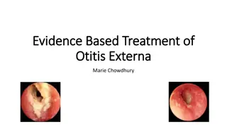
Management: Use topical ear preparation for 7 days; 2% acetic acid for mild cases antibiotic plus steroid e.g. Locorten-Vioform Gentisone HC (NB not if perforation) If wax/debris obstructing EAC or extensive swelling or cellulitis Pope wick Dry mopping (children) Microsuction (ENT PCC) File:OtitisExterna10.JPG Advise re prevention of OE: keep ears clean and dry; treat underlying eczema/psoriasis Failure of topical meds: review diagnosis/compliance consider PO fluclox or erythromycin ?fungal (spores in EAC) Swab and refer
Chronic OE Causes: Secondary fungal infection- due to prolonged use of topical antibacterials or steroids Seborrhoeic dermatitis; contact dermatitis Sometimes no cause can be found for OE Symptoms: mild discomfort; pain usually mild Signs: lack of ear wax; dry, hypertrophic skin leading to canal stenosis; pain on exam Assess risk /precipitating factors; severity of symptoms; signs of fungal infection- whitish cotton-like strands in EAC, black or white balls of aspergillus. Look for signs of dermatitis, evidence of allergy (ear plugs etc) or focus of fungal infection elsewhere, e.g. Skin, nails, vagina- can cause 2 inflammation EAC Investigations: only take swab for C+S if treatment fails as interpretation can be difficult: sensitivities are determined for systemic use and much higher concentrations can be achieved by topical use; organisms may be contaminants, usually fungal overgrowth after using antibacterial drops due to suppressed normal bacterial flora
Chronic OE Management: advise general measures as for acute diffuse OE Treatment depends on cause - often requires more than one strategy: if fungal infection- top antifungal, refer if poor response seborrhoeic dermatitis- antifungal and steroid combined If no cause evident- 7d course top steroid +/- acetic acid spray. If good response, may need to continue steroid but reduce potency/dose. If cannot be withdrawn after 2-3m, refer ENT. If poor response, try trial of top antifungal Refer ENT if contact sensitivity (re patch testing); if EAC occluded; if malignant OE suspected.
Foreign Bodies Management depends on what it is: Batteries immediate referral to ENT Inert FB e.g. retained grommet, beads, foam - not so urgent Organic e.g. food, insects. May cause infection therefore should be dealt with sooner. For insects drown in olive oil first. Some FBs may resolve with syringing, but if not refer to PCC Do not attempt to remove under direct visualisation as more likely to cause harm
Epistaxis Anterior or Posterior hx gives clues > 90% from Little s Area Age gives clue more likely posterior in Elderly Cause: Idiopathic, trauma (nose picking), dry mucosa, hypertension, coagulopathy, NSAID, Warfarin, tumour CAN BE FATAL!!! First Aid: Compression & Ice
Avoid blowing their nose (1/52) Avoid hot drinks (1/52) Naseptin cream 1/52 Admit: If cannot control, elderly, warfarinised, low platelets, recurrent excessive bleeding PCC: If not settling with conservative rx AgNO3 cautery can be done in GP Packing, Electrocautery, Surgery (SPA ligation, ECA ligation, embolisation)
Cautery: What you need: A good lightsource Nasal speculum (or large aural speculum) Lignocaine (with adrenalin) Cotton wool Cautery sticks
Rhinosinusitis Causative factors allergic, viral, bacterial, fungal, autoimmune. Acute <12wks, Chronic >12wks, Recurrent (>4/yr) 15% population. 6 million lost working days / yr in the UK Presents as My cold won t go away persistant symptoms of URTI, without improvement after 10-14 days or worsening after 5 days Major: Nasal congestion/obstruction Purulent discharge Loss of smell Facial pain / ear pain or fullness
Minor: Tenderness over sinus area Fever Headache Halitosis Fatigue / Lethargy Post nasal drip What to exclude on examination: Periorbital swelling, extraocular muscle dysfunction, decreased VA or proptosis Foreign bodies Concomitant otitis media (in children) CNS complications Polypoid changes or deviated septum What to expect on examination: Erythema / swelling of nasal mucosa Mucopurulent secretions Tenderness over sinuses
Differentials Allergic rhinitis (seasonal or perennial) Usually just nasal symptoms and usually persistent Nasal FB unilateral blockage or discharge Sinonasal tumour chronic, unilateral blockage, discharge (bloody) Other causes of facial pain Tension Headache TMJ dysfunction or bruxism Neuropathic Dental pain (hot/cold drinks, chewing) Investigations Xrays / Bloods / Swabs = not required, only indicated if > 12 wks and failure to respond to Rx will probably refer at that stage (rigid endoscopy / coronal CT / allergy testing)
Consider emergency admission to hospital if symptoms are accompanied by: Systemic illness Swelling or cellulitis in face Signs of CNS involvement Orbital involvement Consider urgent ENT referral if: Persistant unilateral symptoms such as (suspecting sinonasal tumour): Bloodstained discharge Non-tender facial pain Facial swelling Unilateral polyps Consider routine referral to ENT if: More than 3-4 episodes per year lasting > 10 days with no symptoms between episodes
Management of acute rhinosinusitis (guidelines on map of medicine) Viral is 200 times more common than bacterial Viral URTI usually precedes bacterial Bacterial usually has more severe and prolonged symptoms Strep pneumoniae, H. influenzae, Moraxella Catarrhalis First line : Amoxicillin Doxycycline, erythromycin, clarithromycin (pen allergic) Second Line: Co-amoxiclav Azithromycin (pen allergic)
More than 7 days Fewer than 7 days Consider antibiotics Advice on self-care measures -paracetamol or ibuprofen -intranasal decongestant (1 week max) +/- oral decongestant (limited evidence) -Saline douching -Warm face packs (5-10 mins, tds may help drainage) -Maintaining hydration & rest -(topical steroids if polypoid change) Follow up for complications, compliance, expect improvement after 72 hrs with first line Abx Follow up for complications & compliance Consider change of ABx Recurrent acute episodes Less than 6/52 between episodes More than 6/52 between episodes Use second line antibiotics Use first line antibiotics
Management of chronic rhinosinusitis (referral toolkit) Initial drug therapy for 2-3 months duration of topical nasal steroid spray (nasonex/avamys) +/- antihistamine If symptoms of allergic aetiology perform skin prick or immunoglobulin assay Give PIL http://www.patient.co.uk/health/Sinusitis-Chronic.htm Advice re smoking (ENT would usually advocate daily saline douching) If initial treatment fails: Commence topical nasal steroid drop for 4 weeks (returning to steroid spray afterwards) Consider oral prednisolone 25mg od for 2 weeks Broad spectrum antibiotics only if purulent nasal discharge If no response to above treatment then refer
Nasal Foreign Bodies Commonest in children aged 1-4 Rare in adults Potential risk to airway Suspect if persistant unilateral symptoms of blockage or foul smelling discharge Unless very easy to get at, and very compliant child, best not attempted in GP (sometimes only get one shot!)
Nasal Fracture Best viewed from above looking at deviation of nasal bones difficult if swollen Exclude septal haematoma Requires immediate drainage to prevent abscess or permanent saddle nose deformity Otherwise refer to PCC for manipulation 7-10 days post injury. For old injuries routine ENT referral
Consider OSA Nasal blockage will almost always be accompanied by snoring Have OSA in the back of your mind Defined as the presence of at least five obstructive events per hour during sleep Features Impaired alertness Cognitive impairment Excessive sleepiness (Epworth scale) Morning headaches Choking or SOB feeling at night Nocturia Unrefreshing sleep Sleep quality of partners affected ( does he stop breathing at night? ) Refer to Respiratory in the first instance
Sore throat: causes Common infections: rhinovirus; coronovirus, parainfluenza virus; common cold (25% sore throats) GABHS causes 15-30% sore throats in children and 10% in adults Herpes simplex virus type 1 (more rarely type 2) = 2% Epstein Barr virus: infectious mononucleosis (glandular fever)- <1%. Suspect IM if sore throat persists >2w - do FBC and IM screen. Non-infectious causes Physical irritation Hayfever Stevens Johnson syndrome Kawasaki disease Oral mucositis 2 chemo /radiotherapy
Sore throat: complications Complications of streptococcal pharyngitis are rare: Suppurative complications: OM acute sinusitis peritonsilar cellulitis / peritonsillar abscess (quinsy) Pharyngeal abscess Retropharyngeal abscess, more common in children Non suppurative complications are rare: R sided quinsy showing displacement of uvula to L rheumatic fever post-streptococcal glomerulonephritis
Sore throat: when to refer Admit if stridor or respiratory difficulty Trismus, drooling, dysphagia. Dehydration /unable to take fluids Severe suppurative complications, ie if abnormal throat swelling/suspected abscess Systemically unwell and at risk of immunosuppression Suspect Kawasaki disease Profoundly unwell and cause unknown
Sore throat: management in primary care Reassure sore throat usually self limiting and symptoms resolve within 3d in 40% cases, 1w in 85% (even if due to streptococcal infection) Advise see healthcare professional if symptoms do not improve, and urgently if breathing difficulties, stridor, drooling, muffled voice, severe pain, dysphagia or unable to take fluids or systemically ill Symptoms of infectious mononucleosis usually resolve within 1-2w, mild cases within days. But lethargy continues for some time and rarely may continue for months or years. Advise re contact sport. Advise regular paracetamol, ibuprofen, fluids ++ but avoid hot drinks; saline mouthwashes; discuss role of antibiotics Consider delayed prescription or immediate antibiotics use Centor scoring - Antibiotic regime: Prescribe phenoxymethylpenicillin for 10d; or erythromycin or clarithromycin for 5d. Avoid amoxicillin (EBV)
Indications for tonsillectomy for recurrent acute sore throat Sore throats are due to acute tonsillitis Episodes of sore throat are disabling and prevent normal functioning Seven or more well documented, clinically significant, adequately treated sore throats in the preceding year or Five or more such episodes in each of the preceding two years or Three or more such episodes in each of the preceding three years SIGN 2010, Management of sore throat and indications for tonsillectomy http://www.sign.ac.uk/pdf/qrg117.pdf
Vertigo Vertigo: is a symptom and refers to a perception of spinning or rotation of the person or their surroundings in the absence of physical movement Peripheral vertigo = labyrinthine cause Benign paroxysmal positional vertigo (BPPV) Vestibular neuronitis: Meniere s disease: Central vertigo = cerebellar cause Common Migraine Uncommon stroke and TIA cerebellar tumour acoustic neuroma MS
Assessment of vertigo Most balance problems that present in primary care are not rotatory vertigo, but unsteadiness. A full time GP is likely to see 10-20 people with vertigo in 1y To determine vertigo rather than dizziness, ask: do you feel light-headed or do you see the world spin around you as if you had just got off a roundabout about timing, duration, onset, frequency and severity of symptoms aggravating factors, e.g. head movement effect on daily activities associated symptoms: hearing loss, tinnitus (unilateral/bilateral), headache, diplopia, dysarthria /dysphagia, ataxia, nausea, vomiting
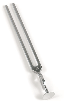
Assessment of vertigo: medical history Recent URTI or ear infection suggests vestibular neuronitis or labyrinthitis Migraine: inc likelihood of migrainous vertigo Head trauma/ recent labyrinthitis: BPPV Trauma to ear: perilymph fistula Anxiety or depression can worsen symptoms or cause feelings of lightheadedness (e.g. from hyperventilation) Acute alcohol intoxication can cause vertigo Examination ENT incl. Weber and Rinnes tests Full Neuro incl cerebellar testing + gait. Particularly looking for nystagmus
Assessment of vertigo: specific tests Romberg s test: identifies peripheral or central cause of vertigo (but not sensitive for differentiating between them) Ask patient to stand up straight, feet together, arms outstretched with eyes closed. If patient unable to keep balance- the test is positive (usually fall to side of lesion) A positive test suggests problem with proprioception or vestibular function. Hallpike manoeuvre: to confirm diagnosis of BPPV
Hallpike manoeuvre - demonstration Be cautious with patients with neck or back pathology or carotid stenosis as manouvre involves turning and extending neck http://northerndoctor.com/2010/09/27/dizziness-dix-hallpike-and-the-epley- manoeuvre/ Ask patient to: report any vertigo during test keep eyes open and stare at examiner s nose sit upright on couch, head turned 45 to one side lie them down rapidly until head extended 30 over end of bed, one ear downward If neck problems- can be done without neck extension observe eyes closely for 30 sec for nystagmus- note type and direction support head in position and sit up Repeat with other side test is positive for BPPV if vertigo and nystagmus (torsional and beating towards ground) are present and nystagmus shows latency, fatigue and adaptation
Features of central causes of vertigo severe or prolonged new onset headache focal neurological deficits central type nystagmus (vertical) excess nausea and vomiting prolonged severe imbalance (inability to stand up even with eyes open)
Features of peripheral causes of vertigo BPPV: vertigo induced by moving head position episodes last for seconds Vestibular neuronitis and labyrinthitis: vertigo persists for days and improves with time no hearing loss or tinnitus with vestibular neuronitis in labyrinthitis, sudden hearing loss with vertigo and tinnitus may be present Meniere s disease: ages 20-50y men> women vertigo, not provoked by position change episodes last 30 min to several hours symptoms of tinnitus, hearing loss and fullness in ear may be clusters of attacks and long remissions


 definitions")


 / Glue ear")

-")


")
")


")
")


")