Exploring Fission Near 198Pb with AT-TPC at FRIB: Insights from Curtis Hunt
Delve into the intriguing world of fission studies near 198Pb using the AT-TPC at FRIB. Supported by the DOE Office of Science, this research probes nuclear structure, fission properties, and fusion-fission reactions. By employing innovative techniques like the Heavy Isotope Tagger and active target
1 views • 39 slides
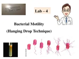
Bacterial Motility Testing Using Hanging Drop Technique
The Hanging Drop Technique is a method used to determine the motility of bacteria by suspending them in a drop of fluid on a slide. This procedure involves preparing a hanging drop slide, inoculating the drop with bacteria, sealing it with petroleum jelly, and observing the organisms under a microsc
2 views • 17 slides

Forensic Analysis Instruments: Microscope vs. Borescope
Instruments like the stereomicroscope, comparison microscope, and borescope are used in forensic analysis for individual examination, side-by-side comparison, superimposition, and barrel analysis. The comparison microscope is utilized for analyzing fired cartridge cases, while the borescope helps in
0 views • 8 slides
Comparison Microscope and Borescope
Instruments like Stereomicroscope, Comparison Microscope, and Borescope are used for individual examination, side-by-side comparison, superimposition, and analysis of barrels in forensic investigations. Comparison microscopes are used for examining fired cartridge cases but have limitations such as
1 views • 8 slides
Introduction to Hanging Drop Technique in Microbiology
The hanging drop technique is a method used in microbiology to observe living microorganisms suspended in a fluid under a microscope. This technique allows for the examination of motility and morphology of bacteria, providing valuable information for research and analysis. By creating a wet mount us
0 views • 17 slides
Microbiology Study: Hanging Drop Technique in Department of Microbiology, College of Medicine
In the Department of Microbiology at the College of Medicine, the Hanging Drop Technique is utilized to study live microorganisms. This technique involves suspending microorganisms in fluid on a hollow ground slide, allowing for observation of their morphology under a microscope. By creating a wet m
0 views • 8 slides
Isolation of AM Fungi by Wet Sieving and Sucrose Gradient Methods
Wet sieving is a popular technique to isolate different sizes of spores from soil samples. Developed by Gerdemann and Nicolson in 1963, this method involves passing an aqueous suspension through different sieves to collect spores of varying sizes. The process includes agitating the soil-water mixtur
0 views • 14 slides
Evolution of Microscopes: From Reading Stones to Phase-Contrast Microscopy
The history of microscopes dates back to the 11th century with the invention of the reading stone, leading to the creation of wearable eyeglasses in the 13th century. The first microscope was developed in the late 16th century by Zacharias Jansen, paving the way for the compound microscope and the t
0 views • 17 slides
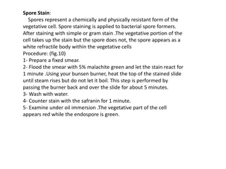
Bacterial Staining Techniques Overview
Spore staining, acid-fast staining, and negative staining are essential techniques in microbiology for differentiating bacterial structures based on staining properties. Spore staining highlights spore formers, acid-fast staining differentiates acid-fast bacteria, and negative staining detects capsu
5 views • 6 slides

Understanding WBC Differential Count in Blood Analysis
WBC (white blood cell) count is essential in assessing a patient's health. A differential count helps determine the percentage of different types of white blood cells. This analysis can provide important insights into various health conditions like infections, allergies, and systemic illnesses. The
1 views • 9 slides
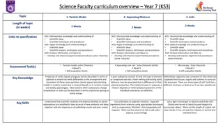
Year 7 Science Curriculum Overview: Particle Model, Separating Mixtures, Cells
Understand scientific concepts, techniques, and procedures through topics such as particle model, separating mixtures, and cells. Learn to apply knowledge, conduct scientific inquiries, and analyze information to develop experimental procedures. Key skills include techniques for separating mixtures
1 views • 29 slides
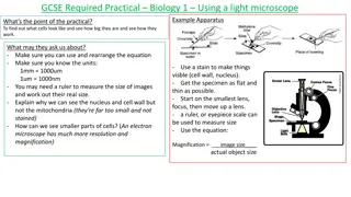
GCSE Required Practical Biology Experiments Overview
Explore GCSE Required Practical Biology experiments including using a light microscope, investigating osmosis, conducting food tests, and studying amylase enzyme activity. Understand the objectives, apparatus, results, and potential questions that may be asked in each experiment. Gain insights into
0 views • 5 slides

Techniques for Monitoring Asbestos Fiber Concentration in Ambient and Indoor Air using SEM
Explore the methods and results of an international proficiency testing (PT) scheme for measuring asbestos fibers in indoor and ambient air. The use of Scanning Electron Microscope (SEM) based techniques, including ISO 14966 and VDI 3492 guidelines, is discussed. The importance of PT schemes in ensu
0 views • 27 slides
Mastering Microscope Focusing Techniques
Understand the essential parts of a light microscope, the magnification process, general procedures for handling a microscope, focusing techniques for specimens, and the relationship between magnification and field of view. Learn how to avoid damaging the microscope and achieve optimal results durin
0 views • 18 slides
Microbiology Lab: The Hanging Drop Preparation
Learn how to prepare living microorganisms for microscopic study using the hanging drop technique. This method involves suspending microorganisms in a fluid and creating a hanging drop on a hollow ground slide, allowing for observation of morphology and motility. Follow step-by-step procedures to ma
0 views • 17 slides

Understanding Wilms Tumor in WAGR Syndrome
WAGR syndrome is a rare genetic condition associated with Wilms Tumor, aniridia, and developmental delays. Wilms Tumor is a form of kidney cancer mostly affecting children and is linked to genetic disorders like WAGR syndrome. The risk of Wilms Tumor is higher in children with WAGR syndrome, with po
1 views • 14 slides
Proper Use of a Microscope: Essential Steps and Tips
Learn the correct way to use a microscope with detailed instructions like carrying it with care, preparing slides, adjusting lighting, focusing, and maintaining cleanliness. Following these steps ensures optimal performance and clear images while using a microscope. Remember to handle the lenses car
0 views • 4 slides
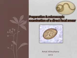
Direct Fecal Smear Preparation and Microscopic Examination Procedure
Direct fecal smears are useful for quick screenings to detect intestinal parasites. This article outlines the preparation and microscopic examination procedure for direct fecal smears. It covers advantages, disadvantages, fecal collection methods, and steps for preparing and examining fecal smears u
0 views • 16 slides

Understanding Specimen Preparation in Electron Microscopy
Living things cannot survive in an electron microscope due to the high temperature generated by the electron beam, vacuum inside the microscope, and need for specimen preparation steps like fixation, dehydration, freezing, cutting, and mounting. Fixation involves stabilizing tissue with chemicals, d
3 views • 10 slides
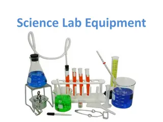
Essential Science Lab Equipment Overview
Explore a range of essential science lab equipment including a Triple Beam Balance for measuring mass, Goggles for eye protection, Mortar and Pestle for crushing and mixing substances, Bunsen Burner for heating and sterilization, Beaker for liquids, Test Tube Clamp for handling test tubes, Test Tube
0 views • 23 slides

Impact of the Renaissance on Medicine in Early Modern Britain
In the early modern period in Britain from 1450 to 1800, the Renaissance brought about significant changes to medicine. This era marked a rebirth in Europe with increased interest in classical architecture, art, and scientific advancements. The impact of the Renaissance on medicine included the chal
9 views • 8 slides
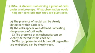
Identifying Plant Cells Under Microscope
Observing well-defined cells with clear presence of cell walls would help conclude that the cells are plant cells, distinguishable from animal cells by the absence of cell walls. The detection of nuclei within each cell is a common characteristic observed in both plant and animal cells.
1 views • 274 slides
Disposable Glucose Nanosensor Development Project
This project aims to develop a disposable glucose nanosensor and testing method to detect a range of concentrations. The goal is to create a low-cost and easy-to-fabricate nanosensor by designing an effective fabrication process. Steps involve cleaning the microscope slide, depositing titanium and a
0 views • 14 slides
Understanding The Light Microscope: A Comprehensive Guide
Delve into the world of microscopy with Dr. Mohammed Hussein's Lab Manual No. 1, covering the fundamentals of the light microscope, its components, lenses, magnification power, resolving power, and detailed instructions on how to focus your microscope effectively for optimal results. Explore the his
0 views • 19 slides
Understanding the Proper Use and Care of a Compound Microscope
Explore the proper use and care of a compound microscope, including the different parts of the microscope, magnification calculations, general procedures for handling the microscope, and more. Learn about objectives, eyepiece magnification, total magnification, and essential maintenance tips to ensu
0 views • 20 slides

Innovative Gadgets from AliExpress: Unbox & Explore New Tech Marvels
Uncover the latest innovative gadgets from AliExpress in a thrilling unboxing experience. From a mini microscope with impressive magnification to a revolutionary rivet adapter for power drills, discover cutting-edge devices like a flame torch for everyday use, staple-less paper binding solution, and
0 views • 6 slides
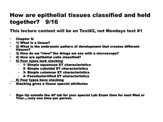
Understanding Epithelial Tissues: Classification and Cellular Characteristics
Epithelial tissues are classified based on their structure and organization, with various types exhibiting distinct features like stacking or lack of stacking. These tissues are held together by special attributes, contributing to their functions within organs and systems. By examining tissues under
0 views • 14 slides
Understanding the Compound Light Microscope and Its Applications
Explore the fundamental concepts of microscopy through Professor Diane Hilker's detailed lectures on the use and care of the compound light microscope. Dive into topics like magnification, resolution, and the practical application of microscopes in scientific research. Enhance your understanding of
0 views • 17 slides
Understanding Microscope Functionality and Different Types
Learn about the essential parts and functions of microscopes, including magnification, resolution, and different types such as bright-field, dark-field, phase-contrast, dissecting, and inverted microscopes. Discover how parfocal microscopes maintain focus and the roles of ocular lenses, nose pieces,
0 views • 28 slides
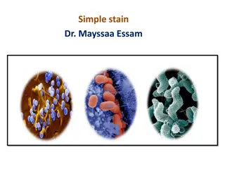
Understanding Bacterial Morphology and Simple Stain Techniques
Explore the characteristics of bacteria based on their shape and structure, grouped into spherical, rod-shaped, and helical types. Learn about bacterial smear preparation principles and the simple stain procedure for observing bacterial cells under a microscope. Discover various bacterial species an
0 views • 13 slides
Understanding Streptococci: Classification and Characteristics
Streptococci are Gram-positive cocci commonly found in chains or pairs. This article covers their classification based on hemolysis patterns and Lancefield grouping, along with general characteristics and principles of differentiation tests. Learn about the infections caused by different Streptococc
0 views • 39 slides
Microscope Focusing and Bacteria Morphology Guide
Learn how to focus a microscope for simple staining, understand the morphology of bacteria such as cocci, bacillus, spirals, and pleomorphic shapes, and discover the process of preparing a slide smear with detailed steps and images.
0 views • 11 slides
Exploring Microscopic Life Through a Microscope
Journey into the microscopic world by examining Elodea leaves and Paramecium under a microscope. Discover the intricate details of these tiny organisms, observe their movement, and learn about their structures. Engage in hands-on activities to explore the fascinating realm of microscopic life.
0 views • 16 slides
Understanding Microscopes: Light vs. Electron Microscopes
Learn about the differences between light microscopes (LM) and electron microscopes (EM), including their magnification power, resolving power, and key parts. Explore the types of electron microscopes such as Transmission Electron Microscope (TEM) and Scanning Electron Microscope (SEM) for advanced
0 views • 8 slides
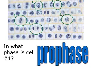
Microscope Parts and Cell Phases - Educational Overview
Discover the different phases of cell division through informative images highlighting prophase, interphase, metaphase, telophase, and anaphase. Learn the names of essential microscope parts such as the arm, body, coarse adjustment, fine adjustment, eyepiece, and stage. Test your knowledge by identi
0 views • 27 slides
Introduction to Microscopy: Care, Terms, and Operation
Explore the world of microscopy with proper care tips, essential terms like magnification and resolution, and detailed operation steps for using a microscope effectively. Learn about the components of a microscope and the process of preparing and viewing specimens at different magnification levels.
0 views • 27 slides
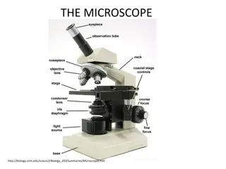
Understanding the Components of a Light Microscope
A light microscope is an essential instrument for examining small objects. It utilizes visible light and magnifying lenses to reveal details not visible to the naked eye. Learn about the different types of light microscopes and their components, such as the ocular lens, nosepiece, objective lenses,
0 views • 11 slides
Understanding the Microscope in Nursing Education
Explore the significance of the microscope in the field of nursing education through this lecture by Assistant Lecture Zahraa Mahmoud Hussain at the University of Basrah College of Nursing. Learn about the types of microscopes, their common uses in medical and life sciences, and the essential parts
0 views • 7 slides
Tagger Microscope Quadrupole Commissioning Study Overview
Tagger microscope quadrupole commissioning study for GlueX collaboration meeting led by Richard Jones from the University of Connecticut in May 2016. The study focuses on electron collimation, momentum conservation, tagmagnification, and performance optimization. Questions address collimated stripe
0 views • 14 slides
Exploring the Components of a Light Microscope in Basic Sciences at University of Basrah College of Dentistry
In this lab session led by Dr. Hanaa Ali Hussein, students learn about the essential parts of a light microscope, including the ocular lens, body tube, prism, arm, resolving nose piece, objective lens, mechanical stage, adjustment knobs, illuminator, base, condenser lens, iris diaphragm, and light f
0 views • 10 slides