Understanding Autoimmunity and Immunological Tolerance

Autoimmunity is a condition where the body's immune cells mistakenly attack its own tissues, leading to damage. Immunological tolerance helps prevent this by mechanisms like central and peripheral tolerance. Central tolerance involves deleting self-reactive immune cells during maturation in key organs like the thymus and bone marrow. Breaches in tolerance mechanisms can lead to autoimmune diseases.

Download Presentation

Please find below an Image/Link to download the presentation.
The content on the website is provided AS IS for your information and personal use only. It may not be sold, licensed, or shared on other websites without obtaining consent from the author. Download presentation by click this link. If you encounter any issues during the download, it is possible that the publisher has removed the file from their server.
E N D
Presentation Transcript
Autoimmunity Autoimmunity Dr. Mohit Bhatia Assistant Professor Department of Microbiology AIIMS, Rishikesh
Learning objectives By the end of this session student should be able to understand: Central and peripheral tolerance Theories of autoimmunity Autoimmune diseases
AUTOIMMUNITY AUTOIMMUNITY Condition in which the body s own immunologically competent cells or antibodies act against its self-antigens resulting in structural or functional damage. Paul Ehrlich had first introduced the concept of autoimmunity; he termed this condition as horror autotoxicus .
AUTOIMMUNITY AUTOIMMUNITY Normally immune system does not react to its own antigens due to a protective mechanism called tolerance. Any breach in tolerance mechanisms predispose to several autoimmune diseases.
IMMUNOLOGICAL TOLERANCE IMMUNOLOGICAL TOLERANCE State in which an individual is incapable of developing an immune response against his own tissue antigens. Mediated by two broad mechanisms: oCentral tolerance oPeripheral tolerance
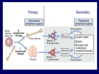
Central tolerance Central tolerance Refers to the deletion of self-reactive T and B lymphocytes during their maturation in central lymphoid organs (i.e., in the thymus for T cells and in the bone marrow for B cells). In thymus: oDuring the T cell development in thymus any self-antigens are encountered processed and presented by thymic antigen presenting cells (APCs) in association with self-MHC. oAny developing T cell that expresses a receptor for such self- antigen is negatively selected (i.e. deleted by apoptosis).
Central Central tolerance tolerance In bone marrow: Receptor editing - process by which many of the B cells reactivate the machinery of antigen receptor gene rearrangement (mainly genes coding for light chains), so that a different (edited) B cell receptor will be produced which no longer recognizes the self-antigen. Negative selection- If receptor editing fails, they undergo apoptosis.
Peripheral tolerance Peripheral tolerance Back-up mechanisms that occur in the peripheral tissues to counteract the self-reactive T cells that escape central tolerance.
Peripheral tolerance Peripheral tolerance - - Mechanisms Mechanisms Ignorance- Self-reactive T cells might never encounter the self-antigen which they recognize. Anergy: oDefined as unresponsiveness to antigenic stimulus. oThe self-reactive T cells interact with the APCs presenting the self antigen, but the co-stimulatory signal is blocked. oThe B7 molecules on APC bind to CTLA-4 molecules on T cells instead of CD28 molecules.
Peripheral tolerance Peripheral tolerance Phenotypic skewing: oSelf-reactive T cells interacting with APCs presented with self-antigens, undergo full activation. oSecrete non-pathogenic cytokines and chemokine receptors profile.
Peripheral tolerance Peripheral tolerance Apoptosis by AICD: oActivation-induced cell death oActivation of T cells induces upregulation of Fas ligand which subsequently interacts with the death receptor Fas leading to apoptosis.
Peripheral tolerance Peripheral tolerance Regulatory T cells (Treg cells): oTreg cells can down regulate the self-reactive T cells through secreting certain cytokines (e.g., IL-10 and transforming growth factor [TGF- ]) or killing by direct cell to cell contact. Dendritic cells (DCs): oImmature DCs and tolerogenic DCs capture the self-antigen for processing. oDown regulate the expression of molecules of co-stimulatory ligands such as B7 molecules or act indirectly by induction of regulatory T cells. Sequestration of self-antigen: Certain self-antigens can evade immune recognition by sequestration in immunologically privileged sites, e.g. corneal proteins, testicular antigens and antigens from brain.
MECHANISMS OF AUTOIMMUNITY MECHANISMS OF AUTOIMMUNITY Breakdown of T-Cell Anergy: In the presence of tissue necrosis and local inflammation express co-stimulatory molecules (B7) . oMultiple sclerosis, rheumatoid arthritis and psoriasis Failure of AICD- Failure of the auto reactive activated T cells to undergo activation induced cell death (AICD) oSLE (systemic lupus erythematosus)
MECHANISMS OF AUTOIMMUNITY MECHANISMS OF AUTOIMMUNITY Loss of Treg cells. Providing T cell help to stimulate self-reacting B cells: oAntibody response to self-antigens occurs only when potentially self-reactive B cells receive help from T cells.
MECHANISMS OF AUTOIMMUNITY MECHANISMS OF AUTOIMMUNITY Release of Sequestered Antigens: oSequestered antigens -never been exposed to the tolerance mechanisms during development of immune system. oInjury to the organs leads to release of such sequestered antigens which are very well capable of mounting an immune response. oSpermatozoa and ocular antigens release can cause post vasectomy orchitis and post-traumatic uveitis.
MECHANISMS OF AUTOIMMUNITY MECHANISMS OF AUTOIMMUNITY Molecular Mimicry: oSome microorganisms share antigenic determinants (epitopes) with self-antigens. oImmune response against such microbes would produce antibodies that can cross-react with self-antigen. oExample: Acute rheumatic fever and multiple sclerosis (molecular mimicry involving T-cell epitopes).
MECHANISMS OF AUTOIMMUNITY MECHANISMS OF AUTOIMMUNITY Polyclonal Lymphocyte Activation oPolyclonal T cell activation - Superantigens released from microbes (e.g. Staphylococcus aureus), polyclonally activate the T cells directly by binding to antigen non-specific V region of T cell receptors. oPolyclonal B cell activation can be induced by products of various microbes such as Epstein Barr virus, HIV, etc.
MECHANISMS OF AUTOIMMUNITY MECHANISMS OF AUTOIMMUNITY Bystander activation: oNonspecific activation of bystander self-reactive TH1 cells. oLeads to cytokine influx which causes an increased infiltration of various non-specific T cells at the site of infection. Epitope spreading Molecular sequestration (Cryptic self epitopes)
AUTOIMMUNE DISEASES AUTOIMMUNE DISEASES Single Organ or Cell Type Autoimmune Diseases Self-antigen present on Disease Autoimmune anemias Autoimmune hemolytic anemia Type of immune response & Important features RBC membrane proteins Auto-antibodies to RBC antigens triggers complement mediated lysis or antibody-mediated opsonization of the RBCs Drugs such as penicillin or methyldopa interact with RBCs so that the cells become antigenic Auto-antibodies to intrinsic factor block the uptake of vitamin B 12; leads to megaloblastic anemia Auto-antibodies against platelet membrane antigens leads to platelet count Drug induced hemolytic anemia Pernicious anemia Drugs alter the red cell membrane antigens Intrinsic factor (a membrane-bound protein on gastric parietal cells) Idiopathic Thrombocytopenic Purpura Platelet membrane proteins (glycoproteins IIb-IIIa or Ib-IX)
AUTOIMMUNE DISEASES AUTOIMMUNE DISEASES Single Organ or Cell Type Autoimmune Diseases Self-antigen present on Renal and lung basement membranes lungs followed by complement mediated injury leads to progressive kidney damage and pulmonary haemorrhage Disease Type of immune response & Important features Goodpasture syndrome Auto-antibodies bind to basement-membrane antigens on kidney glomeruli and the alveoli of the Myasthenia gravis Acetylcholine receptors Blocking type of auto-antibody directed against Ach receptors present on motor nerve endings, leads to progressive weakening of the skeletal muscles Graves disease Thyroid-stimulating hormone (TSH) receptor Anti TSH- auto-antibody (stimulates thyroid follicles, leads to hyperthyroid state)
AUTOIMMUNE DISEASES AUTOIMMUNE DISEASES Disease Self-antigen present on Type of immune response & Important features Hashimoto s thyroiditis Thyroid proteins and Auto-antibodies and TDTH cells targeted against thyroid antigen leads to suppression of thyroid gland. Seen in middle aged females Hypothyroid state is produced ( production of thyroid hormones) Streptococcal antigen- antibody complexes are deposited on glomerular basement membrane TDTH cells and auto-antibodies directed against pancreatic beta cells cause production of insulin cells Post-streptococcal glomerulonephritis Insulin-dependent diabetes mellitus Kidney Beta cells present in islets of Langerhans of pancreas
AUTOIMMUNE DISEASES AUTOIMMUNE DISEASES Single Organ or Cell Type Autoimmune Diseases Systemic Autoimmune Diseases Self-antigen present on Auto-antibodies are produced against various tissue antigens such as DNA, nuclear protein, RBC and platelet membranes. Disease Systemic lupus erythematosus Type of immune response & Important features Age & sex- Women (20-40 years of age) are commonly affected; female to male ratio is-10:1. Immune complexes (self Ag- auto Ab) are formed; which are deposited in various organs Major symptoms- Fever, butterfly rash over the cheeks, arthritis, pleurisy, and kidney dysfunction
AUTOIMMUNE DISEASES AUTOIMMUNE DISEASES Single Organ or Cell Type Autoimmune Diseases Systemic Autoimmune Diseases Self-antigen present on Here, a group of auto-antibodies against the host IgG antibodies are produced called RA factor. It is an IgM antibody directed against the Fc region of IgG. ACPA (Anti citrullinated peptide antibodies) are also produced Disease Rheumatoid arthritis Type of immune response & Important features Age & sex- Women (40-60 years of age) affected Auto-antibodies bind to circulating IgG, forming IgM-IgG complexes that are deposited in the joints and can activate the complement cascade. Major symptoms- Main feature-Arthritis (chronic inflammation of the joints, begins at synovium; most common joints involved are-small joints of the hands, feet and cervical spine) Other features-hematologic, cardiovascular, and respiratory systems are also frequently affected
AUTOIMMUNE DISEASES AUTOIMMUNE DISEASES Single Organ or Cell Type Autoimmune Diseases Systemic Autoimmune Diseases Disease Self-antigen present on Type of immune response & Important features Sj gren syndrome Ribonucleoprotein (RNP) antigens SS-A (Ro) and SS-B (La) present on salivary gland, lacrimal gland, liver, kidney, thyroid Auto-antibodies to the RNP antigens SS-A (Ro) and SS-B (La); leads to immune-mediated destruction of the lacrimal and salivary glands resulting in dry eyes (keratoconjunctivitis sicca) and dry mouth (xerostomia)
AUTOIMMUNE DISEASES AUTOIMMUNE DISEASES Single Organ or Cell Type Autoimmune Diseases Systemic Autoimmune Diseases Self-antigen present on Nuclear antigens such as DNA topoisomerase and centromere present in heart, lungs, GIT, kidney, etc Disease Scleroderma (Systemic Sclerosis) Type of immune response & Important features Helper T cell (mainly) and auto-antibody mediated. Excessive fibrosis of the skin, throughout the body Two types- 1.Diffuse scleroderma- Auto-antibodies against DNA topoisomerase I (anti-Scl 70) is elevated 2.Limited scleroderma- Anticentromere antibody, characterized by CREST syndrome-calcinosis, Raynaud phenomenon, esophageal dysmotility, sclerodactyly, and telangiectasia
AUTOIMMUNE DISEASES AUTOIMMUNE DISEASES Single Organ or Cell Type Autoimmune Diseases Systemic Autoimmune Diseases Self-antigen present on Sacroiliac joints & other vertebrae Several types- Ankylosing spondylitis Reiter Syndrome Psoriatic Arthritis Spondylitis with Inflammatory Bowel Disease Reactive arthritis Disease Seronegative Spondyloarthropathies Type of immune response & Important features Common characteristics- They present as rheumatoid arthritis like features, but differ from it by- Association with HLA-B27 Pathologic changes begin in the ligamentous attachments to the bone rather than in the synovium Involvement of the sacroiliac joints, and/or arthritis in other peripheral joints Absence of RFs (hence the name "seronegative") Auto-Ab and immune complex mediated
AUTOIMMUNE DISEASES AUTOIMMUNE DISEASES Single Organ or Cell Type Autoimmune Diseases Systemic Autoimmune Diseases Disease Self-antigen present on Type of immune response & Important features Multiple sclerosis Brain (white matter) Self-reactive T cells produce characteristic inflammatory lesions in brain that destroys the myelin sheath of nerve fibres; leads to numerous neurologic dysfunctions
LABORATORY DIAGNOSIS OF AUTOIMMUNE DISEASES LABORATORY DIAGNOSIS OF AUTOIMMUNE DISEASES Autoimmune diseases Laboratory diagnosis oCoombs test - red cells are incubated with an anti human IgG antiserum IgG auto-antibodies are present on the red cells, the cells are agglutinated by the antiserum Autoimmune hemolytic anemias Goodpasture syndrome Biopsies from patients are stained with fluorescent-labeled anti- IgG and anti-C3b reveal linear deposits of IgG and C3b along the basement membranes.
LABORATORY DIAGNOSIS OF AUTOIMMUNE DISEASES LABORATORY DIAGNOSIS OF AUTOIMMUNE DISEASES Autoimmune diseases Laboratory diagnosis SLE (Systemic lupus erythematosus) Detection of autoantibodies by indirect immunofluorescence assay (most widely used) and ELISA based techniques. ANA (antinuclear antibody)- Positive in >90% cases (screening test). Anti-double stranded DNA (dsDNA)-Highly specific (Confirmation). Anti-Sm antibodies Lupus band test- Direct immunofluorescence test - detect deposits of immunoglobulins and complement proteins in the patient's skin. LE cell test- No longer used because the LE cells are only found in 50 75% of SLE cases.
LABORATORY DIAGNOSIS OF AUTOIMMUNE DISEASES LABORATORY DIAGNOSIS OF AUTOIMMUNE DISEASES Autoimmune diseases Laboratory diagnosis Scleroderma Anti-Scl 70 antibody is raised, detected by indirect immunofluorescence assay Sj gren s syndrome Detection of SS-A (or anti-Ro) and SS-B (or anti-La) antibodies by indirect immunofluorescence assay.
LABORATORY DIAGNOSIS OF AUTOIMMUNE DISEASES LABORATORY DIAGNOSIS OF AUTOIMMUNE DISEASES Autoimmune diseases Laboratory diagnosis Rheumatoid arthritis RA factor (by latex agglutination test)- RA factor is an IgM autoantibody directed against Fc portion of IgG, good sensitivity. False positive - seen in other autoimmune diseases. ACPA (Anti-citrullinated peptide antibodies) is an auto-antibody to citrullin protein. It is positive only in 67% of cases; but is highly specific. Rose-Waaler test to detect RA factor is of historical importance, no longer used now.