Understanding Cellular Events in Inflammation and Repair

Explore the lecture on Cellular Events in Inflammation by Dr. Maha Arafah, focusing on the sequence of vascular changes in acute inflammation, phases of changes in vascular caliber and flow, and objectives covering leukocyte extravasation, chemotaxis, phagocytosis, and microbial killing. Reference material from Robbins Basic Pathology 9th edition.

Download Presentation

Please find below an Image/Link to download the presentation.
The content on the website is provided AS IS for your information and personal use only. It may not be sold, licensed, or shared on other websites without obtaining consent from the author. Download presentation by click this link. If you encounter any issues during the download, it is possible that the publisher has removed the file from their server.
E N D
Presentation Transcript
INFLAMMATION AND REPAIR Lecture 2 Cellular Events in Inflammation (Foundation Block, Pathology) Maha Arafah Associate Professor Department of Pathology Dr. Maha Arafah King Saud University Email: marafah@hotmail.com 2017 1
5. Describe the sequence of vascular changes in acute inflammation Events of acute Inflammation Acute inflammation has three main events: (1) Hemodynamic changes (alterations in vascular caliber that lead to an increase in blood flow) vascular (2) Increased vascular permeability (structural changes in the microvasculature that permit plasma proteins and leukocytes to leave the circulation) cellular (3) Emigration of the leukocytes from the microcirculation (their accumulation in the focus of injury, and their activation to eliminate the offending agent) 4
5. Describe the sequence of vascular changes in acute inflammation Phases of changes in Vascular Caliber and Flow 1. Transient vasoconstriction of arterioles It disappears within 3-5 seconds in mild injuries 2. Vasodilatation: It involves the arterioles results in opening of new microvasculature beds in the area leading to increasing blood flow Histamine effect 3. Slowing of the circulation due to increased permeability of the microvasculature, this leads to outpouring of protein-rich fluid in the extravascular tissues. 4. Stasis: slow circulation due to dilated small vessels packed with red cells 5
Objectives 1. Describe the steps involved in extravasation of leukocytes from the blood to the tissues. 2. Know the steps at which selectins and integrins act. 3. Describe the meaning and utility of chemotaxis. Understand the role that chemokines play in inflammation. 4. Describe the steps involved in phagocytosis and the role of IgG and C3b as opsonins and receptors. 5. List the mechanisms of microbial killing. 6. Know various defects in leukocyte function.
Reference book and the relevant page numbers.. Robbins Basic Pathology 9thedition, pages 34-41
Acute Inflammation CELLULAR EVENTS: A critical function of inflammation is to deliver leukocytes to the site of injury LEUKOCYTE EXTRAVASATION and to activate the leukocytes to perform their normal functions in host defense. WHAT ARE THESE FUNCTION? Leukocytes ingest offending agents, kill bacteria and other microbes, and get rid of necrotic tissue and foreign substances. They may induce tissue damage and prolong inflammation, since the leukocyte products that destroy microbes and necrotic tissues can also injure normal host tissues.
1. Describe the steps involved in extravasation of leukocytes from the blood to the tissues.
1. Describe the steps involved in extravasation of leukocytes from the blood to the tissues. Recruitment of leukocytes A multistep process involving attachment of circulating leukocytes to endothelial cells and their migration through the endothelium (extravasation) 3 steps: 1. In the lumen: i. Margination ii. rolling iii. adhesion to endothelium Vascular endothelium normally does not bind circulating cells 2. Transmigration across the endothelium (also called diapedesis) 3. Migration in interstitial tissues toward a chemotactic stimulus
1. Describe the steps involved in extravasation of leukocytes from the blood to the tissues. Resident tissue macrophages, mast cells, and endothelial cells respond to injury by secreting the cytokines TNF, IL-1, histamine and chemokines
1. Describe the steps involved in extravasation of leukocytes from the blood to the tissues. Leukocytes Rolling Within a Venule Margination Because blood flow slows early in inflammation (stasis), the endothelium can be lined by neutrophils (pavementation) Margination is the first step of leukocytes action during acute inflammation cells
Steps involved in extravasation of leukocytes from the blood to the tissues Adhesion Migration across the endothelium Diapedesis
2. Know the steps at which selectins and integrins act. Adhesion Molecules and Receptors 1. Selectins, consist of: 1. E-selectin: confined to endothelium induced by TNF&IL-1 2. P-selectin: present in endothelium and platelets from Weibel-Palade bodies 3. L-selectin: expressed on most leukocyte and endothelium Resident tissue macrophages, mast cells, and endothelial cells respond to injury by secreting the cytokines TNF, IL-1, histamine and chemokines which stimulate selectin E-selectin & P-selectin bind to Sialyl-Lewis X glycoprotein and slow the leukocytes
2. Know the steps at which selectins and integrins act. Adhesion Molecules and Receptors 2. Integrins rolling are transmembrane heterodimeric glycoproteins, made up of and chains, expressed on leukocytes and bind to ligands on endothelial cells Integrins are up regulated on leukocytes by C5a & LTB4 resulting in firm adhesion with vessel wall
2. Know the steps at which selectins and integrins act. Leukocyte Adhesion Deficiency Two types: LAD type 1 is a deficiency of 2- integrin LAD type 2 is mutations in fucosyl transferase required for synthesis of sialylated oligosaccharide These normally binds selectins. Clinical findings: Delayed separation of umbilical cord Increased circulating neutrophils (leukocytosis due to loss of the marginating pool) Recurrent bacterial infection that lack pus formation Poor wound healing
2. Know the steps at which selectins and integrins act. Adhesion Molecules and Receptors 3. The immunoglobulin family molecules : ICAM-1 (intercellular adhesion molecule 1) VCAM-1 (vascular cell adhesion molecule 1) IL-1 and TNF activate intercellular adhesion molecule (ICAM) and vascular cell adhesion molecule (VCAM) on venular endothelial cells. adhesion to endothelium
2. Know the steps at which selectins and integrins act. Adhesion Molecules and Receptors 4. Mucin-like glycoproteins: PECAM-1 - these glycoproteins are found in the extracellular matrix and on cell surfaces. Neutrophils moving along the venular endothelium dissolve the venular basement membrane (release type IV collagenase) exposed by previous histamine-mediated endothelial cell contraction and enter the interstitial tissue. Neutrophils, monocytes, lymphocytes, eosinophils, and basophils all use the same pathway to migrate from the blood into tissues.
2. Know the steps at which selectins and integrins act. Leukocyte Adhesion and Transmigration Migration of the leukocytes through the endothelium is called: Transmigration BV lumen or Diapedesis Diapedesis occurs predominantly in the postcapillary venules Extravascular
Extravasation of leukocytes from the blood to the tissues Leukocyte Adhesion and Transmigration The type of emigrating leukocyte varies with the age of the inflammatory response In most forms of acute inflammation: neutrophils predominate in the inflammatory infiltrate during the first 6 to 24 hours, then are replaced by monocytes in 24 to 48 hours
Extravasation of leukocytes from the blood to the tissues WHY?
Extravasation of leukocytes from the blood to the tissues Leukocyte Adhesion and Transmigration neutrophils are more numerous in the blood, they respond more rapidly to chemokines, but are short-lived; they undergo apoptosis and disappear after 24 to 48 hours, whereas monocytes survive longer.
Extravasation of leukocytes from the blood to the tissues Leukocyte Adhesion and Transmigration The type of emigrating leukocyte varies with the type of stimulus: In viral infections, lymphocytes may be the first cells to arrive In some hypersensitivity reactions and parasitic infection, eosinophil may be the main cell type Chronic inflammation: lymphocytes, plasma cells and macrophages are present
3. Describe the meaning and utility of chemotaxis. Understand the role that chemokines play in inflammation. Chemotaxis After extravasation, leukocytes emigrate in tissues toward the site of injury by a process called chemotaxis, defined as locomotion oriented along a chemical gradient !!!! Chemoattractants Neutrophils are attracted by bacterial products, IL-8, C5a & LTB4 Chemokines act on the adherent leukocytes and stimulate the cells to migrate toward the site of injury or infection
3. Describe the meaning and utility of chemotaxis. Understand the role that chemokines play in inflammation. Chemotaxis Chemoattractants Exogenous and endogenous substances Exogenous agents are bacterial products. Endogenous chemoattractants include several chemical mediators: (1) components of the complement system, particularly C5a (2) products of the lipoxygenase pathway, mainly leukotriene B4 (LTB4) (3) cytokines, particularly those of the chemokine family (e.g., IL-8).
All these chemotactic agents bind to specific seven-transmembrane G-protein-coupled receptors on the surface of leukocytes
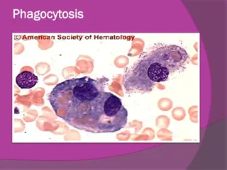
4. Describe the steps involved in phagocytosis and the role of IgG and C3b as opsonins and receptors. Leukocyte Activation Phagocytosis Intracellular destruction Liberation of substances that destroy extracellular microbes and dead tissues Production of mediators
4. Describe the steps involved in phagocytosis and the role of IgG and C3b as opsonins and receptors. Phagocytosis Phagocytosis involves three distinct but interrelated steps (1) Recognition and Attachment of the particle to be ingested by the leukocyte (2) its Engulfment, with subsequent formation of a phagocytic vacuole (3) killing or Degradation of the ingested material.
4. Describe the steps involved in phagocytosis and the role of IgG and C3b as opsonins and receptors. Phagocytosis by neutrophils Immune phagocytosis is much more efficient than nonspecific phagocytosis
4. Describe the steps involved in phagocytosis and the role of IgG and C3b as opsonins and receptors. Leukocyte activation (1) Recognition and Attachment (Opsonization) Is the process of coating a particle, such as a microbe, to target it for phagocytosis The substances that do this are opsonins. These substances include: antibodies (IgG) complement proteins (C3) And others: lectins (mannose-binding lectin (MBL), collectins,fibronectin, fibrinogen, and C-reactive protein These can coat microbes and are recognized by receptors on phagocytes (Fc and C3b receptors).
4. Describe the steps involved in phagocytosis and the role of IgG and C3b as opsonins and receptors.
4. Describe the steps involved in phagocytosis and the role of IgG and C3b as opsonins and receptors. Phagocytosis 2. Engulfment During engulfment, extensions of the cytoplasm (pseudopods) flow around the particle to be engulfed, eventually resulting in complete enclosure of the particle within a phagosome The phagocytic vacuole then fuses with a lysosomal granule, resulting in phagolysosome
Defects in Leukocyte Function Ch diak-Higashi syndrome Protein involved in organelle membrane fusion (no phagolysosomes) Protein trafficking defect ( microtubule defect) Autosomal recessive Clinical feature: Increased risk of pyogenic infection Neutropenia (defect in generation from BM) Giant granule formation (granules formed cannot move in cytoplasm) Defective primary hemostasis ( platelet granule are not secreted) Albinism Peripheral neuropathy
4. Describe the steps involved in phagocytosis and the role of IgG and C3b as opsonins and receptors.
5 . List the mechanisms of microbial killing. Phagocytosis Killing and Degradation 2 mechanisms for Microbial killing: 1. Oxygen-dependent mechanisms 2. Oxygen-independent mechanisms
5 . List the mechanisms of microbial killing. 1. Oxygen-Dependent Mechanisms The H2O2-MPO-halide system is the most efficient bactericidal system in neutrophils O2 SOD H2O2 _ oxidase O2 HOCL MPO Chronic granulomatous disease Decreased oxidative burst.
Oxygen-Dependent Mechanisms Chronic granulomatous disease Decreased oxidative burst. 2 types: A. X-linked: NADPH oxidase (membrane component) B. Autosomal recessive: a. NADPH oxidase (cytoplasmic components) b. Myeloperoxidase deficiency: (absent MPO-H2O2 system) pt. have increased risk of candida infection Infection and granuloma formation with catalase positive organisms e.g. S aureus, Norcardia and Aspergillus
Oxygen-Dependent Mechanisms Comparison of Chronic Granulomatous Disease and Myeloperoxidase Deficiency
5 . List the mechanisms of microbial killing. 2. Oxygen-independent mechanisms through the action of substances in leukocyte granules. These include: Bactericidal permeability increasing protein (BPI) Lysozyme Lactoferrin Major basic protein Defensins Can potentiate further inflammation by damaging tissues These harmful proteases are controlled by a system of anti-proteases in the serum Neutrophil granules contain other enzymes, such as elastase, that also contribute to microbial killing
Neutrophil extracellular traps (NETs) A, Healthy neutrophils with nuclei stained red and cytoplasm green. B, Release of nuclear material from neutrophils (note that two have lost their nuclei), forming extracellular traps. C, An electron micrograph of bacteria (staphylococci) trapped in NETs. NETs provide an additional mechanism of killing microbes that does not involve phagocytosis
Defects in Leukocyte Function
6. Know various defects in leukocyte function. Defects in Leukocyte Function Genetic 1. Leukocyte adhesion deficiency 1and 2 2. Ch diak-Higashi syndrome 3. Chronic granulomatous disease: A. X-linked: NADPH oxidase (membrane component) B. Autosomal recessive: a. NADPH oxidase (cytoplasmic components) b. Myeloperoxidase deficiency
6. Know various defects in leukocyte function. Defects in Leukocyte Function Acquired Thermal injury, diabetes, malignancy, sepsis, immunodeficiencies Chemotaxis Hemodialysis, diabetes mellitus Adhesion Leukemia, anemia, sepsis, diabetes, neonates, malnutrition Phagocytosis and microbicidal activity
TAKE HOME MESSAGES: 1. Several steps are involved in extravasation of leukocytes from the blood to the tissues. 2. Phagocytosis is important step to get rid of necrotic material and bacteria. 3. Various defects in leukocyte function are present. These could be genetic defects or acquired.


")