Methods for Determination of Microbial Growth

Quantitative determination of microbial growth is crucial for various purposes, with two commonly used methods being the standard plate count and spectrophotometric measurement. The standard plate count method estimates living microbial cell density, while spectrophotometric measurement relies on turbidity of microbial cells. For mold growth, the viable plate count method is preferred due to the uneven distribution of mycelia in liquid culture. The viable plate count method involves counting distinct growing colonies on agar plates, with specific colony number recommendations for accurate results. Dilution steps are essential to ensure accurate counting of microbial cells present in liquid culture. On the other hand, turbidimetric determination is based on the relationship between transmitted light and cell density in liquid culture. Both methods play a vital role in assessing microbial growth.
- Microbial Growth
- Standard Plate Count
- Spectrophotometric Measurement
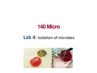
- Viable Plate Count
- Turbidimetric Determination

Download Presentation

Please find below an Image/Link to download the presentation.
The content on the website is provided AS IS for your information and personal use only. It may not be sold, licensed, or shared on other websites without obtaining consent from the author. Download presentation by click this link. If you encounter any issues during the download, it is possible that the publisher has removed the file from their server.
E N D
Presentation Transcript
Lab10 Estimation or Determination of Microbial growth The quantitative determination of microbial growth (Bacteria, molds, yeasts) is required for many aims. Two methods are most widely used for determination of microbial numbers are the standard or viable plate count method and spectrophotometric (turbidity) measurement method. The standard plate count method is an indirect estimation of the cells density, (only the living microbial cells). The spectrophotometric measurement is based on turbidity of the microbial cells alive or dead. This method is not convenient for estimation of the molds growth, because the formation of unhemogenuously distribution of mycelia or hyphal cells in liquid growth culture. The growth in this case could be determined by viable plate count method. The increment in the cells number or in the turbidity in adefinite periods indicate that the growth is occurred . A) viable plate count method. This method is based on the number of a distinct growing colonies on the agar plate .The favorite colonies number is suggested between 25-250 or 30-300 colonies. Fewer than 25 colonies are not acceptable for statistical reasons , and more than 250 or 300 colonies are also not accepted because the colonies are too closed or overlapped to each other .In this case it can not be distinguished as a distinct colony-forming units (
CFUs).Depending upon the assumption that each viable cell is separated for all others and will develop into a single colony (CFU)thus the number of colonic represents the number of cells, that can grow under growth conditions .For the above reasons the sample of the liquid growth culture must be diluted with sterile saline or phosphate buffer until the suspected cells number is enough to count. Materials Procedures 1- Label the bottom of the six Petri-dishes with following dilutions: 10-4, 10-5, 10-6, 10-7 to 10-9. 2- Label the four bottles of saline or phosphate as follows 10-2, 10-4, 10-6, 10-8 or the tubes 10-1, 10-2, 10-3 up to 10-10. 3- Under aseptic conditions transfer 1 ml of liquid culture to the 99 ml saline the result is the first dilution step equal to 1:100 or 10-2 .Then shake the bottle rapidly to serve the distribution of the bacterial cells and break up any cells clumps that may be present. 4- Immediately after shaking transfer 1 ml from 10-2 bottle to the second 99 ml saline bottle to gain 10-4 dilution. 5- Shake well and transfer to the third 99 ml saline bottle the dilution represents 10-6 Reap the process once more to gain 10-8 dilution see the
Turbidimetric determination This method is basically depended on the reciprocal relation between the amount of transmitted light and the suspension density of the bacterial cells in liquid culture. The transmission of light in the visible spectrophotometer through the distilled water is 100% ,when the sample contains any suspended particles , these particles reflects the light beams .The reflected beams could be measured on the scale of the spectral at a definite wave length. Therefore the amount of transmitted light decreases as the cell population (density) increases. Materials 24 48 hr. tryptic soy broth culture of E.coli 3- ml sterile tryptic soy broth tubes (6 tubs) 3-5 ml graduated pipettes with pipette. Empty sterile tubes. Spectrophotometer. (See picture 28) Procedures To make twofold dilution for standard curve (or serial decimal dilution) table 5 tubes the 1st is empty, the tubes No. 2,3,4,5 each one filled with 3-ml sterile tryptic soy broth. From the original broth culture transfer with sterile pipette 3-ml to 1st tube and 3- ml to the tube NO.2 mix well and transfer 3 ml from tube No.2 to the tube No.3.
Figure (22) twofold serial dilution. Picture (23) Spectrophotometer
Figure (4 Growth curve and indirect determination of generation time

.Depending upon the assumption that each viable cell is")
 viable plate count method.")

 twofold serial dilution. Picture (23)")