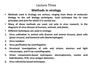
Understanding General Virology: Key Concepts and Detection Methods
Viruses are intracellular parasites lacking metabolic proteins, relying on host cells for survival. Their structure includes a protein coat, nucleic acid, and may have an envelope. Host cell receptors play a crucial role in viral infection. Detection methods include electron microscopy, viral culture, and antigen detection.

Download Presentation

Please find below an Image/Link to download the presentation.
The content on the website is provided AS IS for your information and personal use only. It may not be sold, licensed, or shared on other websites without obtaining consent from the author. Download presentation by click this link. If you encounter any issues during the download, it is possible that the publisher has removed the file from their server.
E N D
Presentation Transcript
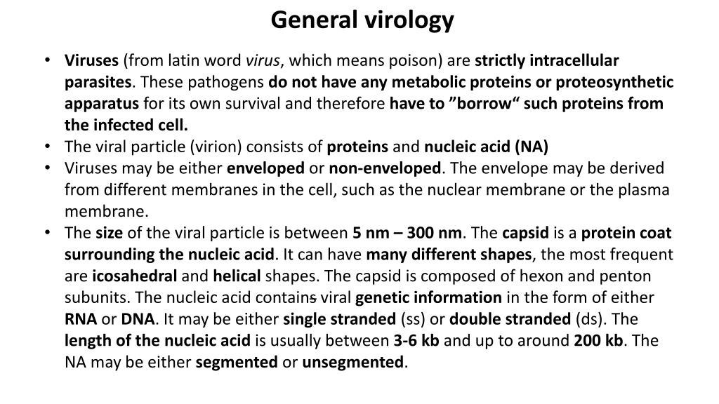
General virology Viruses (from latin word virus, which means poison) are strictly intracellular parasites. These pathogens do not have any metabolic proteins or proteosynthetic apparatus for its own survival and therefore have to borrow such proteins from the infected cell. The viral particle (virion) consists of proteins and nucleic acid (NA) Viruses may be either enveloped or non-enveloped. The envelope may be derived from different membranes in the cell, such as the nuclear membrane or the plasma membrane. The size of the viral particle is between 5 nm 300 nm. The capsid is a protein coat surrounding the nucleic acid. It can have many different shapes, the most frequent are icosahedral and helical shapes. The capsid is composed of hexon and penton subunits. The nucleic acid contains viral genetic information in the form of either RNA or DNA. It may be either single stranded (ss) or double stranded (ds). The length of the nucleic acid is usually between 3-6 kb and up to around 200 kb. The NA may be either segmented or unsegmented.
Host cells must have receptors to which viral proteins can bind to, enabling the virus to begin the first step of infection. An example is the CD20 receptor on B cells, which the Epstein-Barr virus (EBV) can recognize and bind to. Such receptors make the cell permissive to infection. Infection is then characterized by - doubling time, which is the time it takes for the number of viral particles to double. Different structure of viral capsids
Taxonomy Note: RNA only Pircorna and Reo are non-enveloped
Electron microscopic evidence (direct detection) Virions can be detected using electron microscopy A B C Electron microscopic photographs of different viruses. Filovirus (A), e.g. Ebola, Marburg, retrovirus (B), e.g. HIV, enveloped icosahedron herpesvirus (C)
Virus detection by other methods culture (direct detection) Colorless plagues are seen below as dots of lysed tissue culture cells caused by the cytopathic effect of the virus. The number of the plaques - plaque forming unit (PFU). Viral culture - plaques in the cell culture (on the left, in Petri dishes). Picture (A) shows the detail of the cell monolayer, picture (B) show details of the destroyed cells in the plaque.
Antigen detection (direct detection) In immunochromatography the presence of an antigen is determined by the principle of antigen-antibody complex formation. This complex will then be shown as a colorful line. A specimen is added into a well (1). The antigen binds to the labeled specific antibody for that antigen (2). This complex moves together with some free antibodies by capillary action (from the left to the right in the figure below) to form a colorful line in position of immobilized antibodies against the antigen (3). Note: reaction control - unconjugated antibodies (4) moves toward a goat anti-rabbit antibody to create a colored complex (5), indicating that the test has worked properly.
Antigen detection (direct detection) Detection of viral particles using immunofluorescence is depicted in figure below Note: Examples of fluorescentmicroscopic detection of viral proteins. This case shows positivity of cytomegalovirus pp65 (CMV) in human cells
PCR - polymerase change reaction (direct detection) PCR amplifies a specific target DNA or RNA. RNA first needs to be reversely transcribed by reverse transcriptase to create a DNA copy (cDNA). Specific amplified products (amplicons) are detected by gel electrophoresis or, in the case of RQ-PCR (Real-time Quantitative PCR - RQ-PCR) using DNA probes that emit fluorescence, which can be quantitatively measured by a graph. Principle of PCR
Detection of specific amplicon using gel electrophoresis and RQ-PCR Principle of RQ-PCR: In comparison to conventional PCR, RQ-PRCR uses fluorescein labeled DNA probes. If the probes are complementary to the tested target they hybridize to the target (template). When newly synthesized double stranded DNA gets close to the hybridized probe, the exonuclease activity of polymerase cleaves fluorescein from the probe and fluorescence is measured (C).
Serology (indirect detection) Principle and results of Enzyme-linked ImmunoSorbent Assay (ELISA) are depicted in the figure below. This procedure is used to detect specific patient antibodies. Enzyme-linked immunosorbent Assay (ELISA) is frequently used for the detection of viral particles, or antibodies (viral antigen is coated to the plate instead of first antibody) (A). Red/pink spots in panel B present the low/high positivity.
Serology (indirect detection) Principles (on the left) and results (on the right) of complement-fixation reaction (CFR) are depicted in fig. below This procedure is used to detect specific patient antibodies. A positive result, specific antibodies (Ab) bind to antigen (Ag - depicted as colorless circles in the figure) creating a complex of antibodies and antigens (Ab-Ab). Complement binds to the Ab-Ag complex and cannot lyse red blood cells (depicted as full circles in the figure), which subsequently sediment at the bottom of the tube. B negative result, where complement lyses the red blood cells creating a pinkish colour from the reaction (view from side and above). C titres of both the reactions are 1:64 (both reactions in duplicates)


")

")
")
")

")
")