Understanding Leishmania: Parasites and Diseases Overview
Discover the three main forms of Leishmaniassis caused by different species, the clinical types of cutaneous leishmaniasis, and the life cycle of Leishmania parasites. Learn about the transmission, manifestations, and types of this disease, including cutaneous and visceral leishmaniasis.

Download Presentation

Please find below an Image/Link to download the presentation.
The content on the website is provided AS IS for your information and personal use only. It may not be sold, licensed, or shared on other websites without obtaining consent from the author. Download presentation by click this link. If you encounter any issues during the download, it is possible that the publisher has removed the file from their server.
E N D
Presentation Transcript
LECTURE: Leishmania Editing File Important Doctor s notes Extra explanation Only F or only M " " .
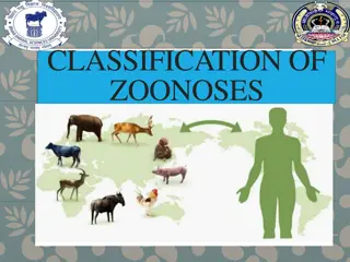
Leishmania Leishmania Parasites and Diseases Parasites and Diseases There are three 3 main form of Lishmaniassis each caused by a different species : SPECIES Disease Leishmania tropica* Leishmania major* Leishmania aethiopica Leishmania mexicana Cutaneous leishmaniasis Affects the skin only Different stages of Haemoflagellate protozoa Leishmania braziliensis Mucocutaneous leishmaniasis In the areas containing mucous membrane Leishmania donovani* Leishmania infantum* Leishmania chagasi Visceral leishmaniasis Internal * Endemic in Saudi Arabia Promastigotes of Leishmania Amastigote of Leishmania
The life cycle of Leishmania: The life cycle of Leishmania: Lishmania spp survive within the macrophages in the human body as intracellular parasites cell mediated immunity determines the host response to infection and clinical manifestations of the disease. The disease is more severe in immunocompromised patients. Route of transmission : via the bite of infected blood sucking Sandflies.
1 1- - Clinical types of cutaneous leishmaniasis Clinical types of cutaneous leishmaniasis COMMON TYPES : known as (oriental sore) : Oriental sore is classical self-limited ulcer. Leishmania major: Leishmania tropica: human and Zoonotic cutaneous leishmaniasis(dogs,rodents) : wet lesions with severe reaction. From human to human or from human to animal or from animal to human Anthroponotic (human only) cutaneous leishmaniasis: Dry lesions with minimal ulceration. From human to human only This starts as a painless papule at the site of Sand fly bite ,generally the face ,which enlarges ,The lesion ulcerates after a few months with an indurated margin. In some other cases the ulcer may spread with an inflammatory zone around , these known as (wet-type-lesion) which heal slowly. Which takes more time than dry lesions In some cases the ulcer remains dry and heals readily (dry-type-lesion) lesion of cutaneous lishmaniasis:
1 1- - Clinical types of cutaneous leishmaniasis Clinical types of cutaneous leishmaniasis Un COMMON TYPES : Diffuse cutaneous leishmaniasis (DCL): Leishmaniasis recidiva (lupoid leishmaniasis): -Caused by L. aethiopica, diffuse nodular non-ulcerating lesions, seen in a part of Africa, people with low immunity to Leishmania antigens. -Diffuse cutaneous (DCL) , and consists of nodules and a thickening of the skin, generally without any ulceration . Severe immunological reaction to leishmania antigen leading to persistent dry skin lesions. Diffuse cutaneous leishmaniasis(DCL) Leishmaniasis recidiva
2 2- - Mucocutaneous leishmaniasis Mucocutaneous leishmaniasis In the nasopharyngeal area The lesion starts as a pustular swelling in the mouth or on the nostrils. The lesion may become ulcerative after many months and then extend into the naso- pharyngeal mucous membrane. Secondary infection is very common with destruction of the nasal cartilage and the facial bone. Caused by: L. braziliensis.
For cutaneous & muco For cutaneous & muco- -cutaneous leishmaniasis : cutaneous leishmaniasis : Diagnosis: The parasite can be isolated from the margin of the ulcer. Smear: Giemsa stain microscopy for LD bodies (amastigotes) in the macrophages. Biopsy: microscopy for LD bodies in the macrophages, or culture in NNN medium for finding promastigotes. Treatment: No treatment self-healing lesions Medical: Pentavalent antimony (Pentostam), Amphotericin B Antifungal drugs +/- Antibiotics for secondary bacterial infection. Surgical: Cryosurgery Excision Curettage NNN medium We culture the macrophages which have amastigotes inside them in NNN medium and look for promastigotes Amastigotes of Leishmania LD in macrophages Promastigotes of Leishmania The diagnostic stage The infective stage
3 3- - Visceral leishmaniasis (Kala Visceral leishmaniasis (Kala- -azar) azar) Both are endemic in Saudi Arabia*: 1-Leishmania infantum mainly affect children 2-Leishmania donovani mainly affects adults incubation period: usually 2-8 months. Symptoms: generally are: fever ,malaise, weight loss with splenomegaly ,hepatomegaly ,anaemia ,leucopenia and sweating . Hepato-splenomegally can be seen because of the hyperplasia of the lymphoid macrophage system. Diagnosis: 1- Parasitological diagnosis: A. Specimen: Bone marrow aspirate (the golden standard) / Splenic aspirate / Lymph node / Tissue biopsy B. microscopy C. culture in NNN medium (2) Immunological Diagnosis: Specific serologic tests: Direct Agglutination Test (DAT), ELISA, IFAT Skin test (leishmanin test) for survey of populations and follow-up after treatment. Hepatosplenomegaly ELISA test DAT test Bone marrow to demonstrate (LD bodies ) amastigotes in macrophages , in the medium it will be promastigotes: *they involve lymph nodes and bone marrow
Treatment of visceral Treatment of visceral leishmanisis leishmanisis Recommended treatment varies in different endemic areas: Pentavalent antimony- sodium stibogluconate (Pentostam) Amphotericin B Treatment of complications: Anaemia Bleeding Infections etc. Untreated disease can be fatal After recovery it might produce a condition called post kala-azar dermal leishmaniasis (PKDL) World distribution of Visceral Leishmaniasis :
SUMMARY: Leishmania Cutaneous Mucocutaneous Visceral Transmissi on Diagnostic: Amastigote in macrophages Infective: Promastigote in sand fly (vector) [note: response depends on host immunity] 2ry destruction of nasal cartilage and facial bone. Pathogene sis oriental sore = self limited ulcer (painless papule) Kala-azar Species Leishmania major Leishmania tropica Leishmania Braziliensis Leishmania infantum Leishmania donovani Anthroponotic Minimal Dry Human & zoonotic Severe Wet Fever, malaise, weight loss, splenomegaly, hepatomegaly, anemia, leucopenia, Post kala-azar dermal leishmaniasis Untreated may be FATAL. Uncommon type: Diffuse cutaneous leishmaniasis (DCL) = L. Aethiopica Leishmaniasis recidiva Diagnosis Microscopy: sample from margin of ulcer and look for LD bodies (amastigote in macrophages) with GEMSA stain Culture: NNN medium for promastigote. Microscopy: sample from BONE MARROW and look for LD bodies (amastigote in macrophages) Culture: NNN medium for promastigote. Serology: ELISA + skin test Treatment Medical + surgical Treat disease and complications
QUIZ: 1. Which of the following is the infective stage of leishmaniasis ? a) Trypomastigote b) Amastigote c) Promastigote 2. Which of the following is vector of leishmania ? a) Testes fly b) Sand fly c) Triatomine 3. Which of the following can be seen in L. tropica infection? a) Wet lesions b) minimal ulceration c) Sever reaction 4. Which of the following parasites cause destruction of nasal cartilage? a) L. Braziliensis b) L. Major c) L. donovani 5. For detecting L. Tropica , .. Is found on microscopy . a) Trypomastigote b) Amastigote c) Promastigote 6. Which medium is used for diagnosing L. donovani? a) NNN b) Chocolate c) MacConkey 7. Which of the following species of leishmania cause hepato-splenomegaly? a) L. Braziliensis b) L. Major c) L. donovani 8. Which of the following is the golden standard site of aspiration when suspecting L. infantum infection? a) Lymph node b) bone marrow c) splenic aspiration B 8. C 7. A 6. B 5. A 4. B 3. B 2. C 1.
THANK YOU FOR CHECKING OUR WORK, BEST OF LUCK! Hamad Alkhudhairy Shrooq Alsomali Reem Alshathri Rehab Alanazi Jawaher Alkhayyal Jumana Alghtani Doctors slides