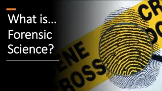
Cranio-Facial Superimposition Techniques in Forensic Anthropology
Cranio-facial superimposition is a technique used in forensic anthropology to identify a skull by overlaying an image of the skull onto a photograph or video of a missing person's face. This method involves various techniques such as photographic, video, and roentgenographic superimposition. The process includes advantages like non-invasiveness and ease of use, but has limitations requiring high-quality photographs and potential errors due to variations in positioning. Techniques range from computer-assisted to reverse superimposition, aiding in cases where clear features are visible or where images need enhancement for comparison.
- Forensic Anthropology
- Superimposition Techniques
- Skull Identification
- Image Comparison
- Facial Reconstruction

Download Presentation

Please find below an Image/Link to download the presentation.
The content on the website is provided AS IS for your information and personal use only. It may not be sold, licensed, or shared on other websites without obtaining consent from the author. Download presentation by click this link. If you encounter any issues during the download, it is possible that the publisher has removed the file from their server.
E N D
Presentation Transcript
Cranio Facial Superimposition Techniques
Introduction Craniofacial superimposition is a technique used in forensic anthropology to identify a skull by overlaying an image of the skull onto a photograph or video of the missing person's face. This technique is used when there are no other means of identification available, and is based on the assumption that the skull and the face are related in shape and size.
Photographic Superimposition, Methods of craniofacial superimposition Video Superimposition, Roentgenographic Superimposition.
Photographic Superimposition Involves overlaying an image of the skull onto a photograph of the missing person's face using a computer program or by hand. The process involves taking an image of the skull and overlaying it on a photograph at the same scale. The images are then aligned, and the features of the skull are matched with those of the photograph.
Photographic Superimposition Cont. Advantages Non-invasive nature Relative ease of use. limitations Need for high-quality photographs Potential for error due to variations in the angle and positioning of the skull and the photograph.
Some of the most common techniques include: Computer-Assisted Superimposition Direct Superimposition Indirect Superimposition Reverse Superimposition Photograph of a suspect or victim is simply placed over a photograph of a skull or face to compare the features. Used in cases where the features are clearly visible and there is a high degree of similarity between the two images. This technique involves using a photograph of a suspect or victim to create a plaster or clay model of their face. This model is then overlaid on a photograph of a skull or face to compare the features. Used in cases where the original photograph is of low quality or where the features are not clearly visible. This technique involves overlaying a photograph of a skull or face onto a photograph of a suspect or victim. Useful in cases where the photograph of the suspect or victim is of higher quality or where there are more visible features to compare. This is the most advanced form of photographic superimposition, which involves using computer software to overlay photographs and create a composite image. Used to compare multiple images and can be helpful in cases where there are multiple suspects or victims to compare.
Process Collecting photographic evidence Preparing the photographs Scaling the photographs Aligning the photographs Overlaying the photographs Comparing the features Making a determination The first step in the process is to gather photographic evidence, including photographs of the individual in question, as well as any other relevant photographs such as a skull or a composite drawing. The photographs are then prepared for superimposition by being scanned or digitized and converted into a digital format that can be easily manipulated. The next step is to scale the photographs so that they are roughly the same size. This can be done by identifying a common reference point, such as the distance between the eyes, and scaling one photograph to match the other. The two photographs are then aligned as closely as possible, using specialized software that allows for precise adjustment of position and orientation. Once the photographs are aligned, they are overlaid on top of each other so that the facial features are superimposed. The forensic expert will then carefully compare the features of the two photographs, looking for similarities and differences in the size, shape, and position of the facial features, as well as any distinctive marks or scars. Based on the degree of correspondence between the two photographs, the forensic expert will make a determination as to whether or not they are a match. This determination may be used to help identify the individual in question, or to rule out their involvement in a particular crime or other incident.
Video superimposition Involves creating a 3D digital model of the skull and overlaying it onto a 3D digital model of a potential match. The process involves capturing video footage of the skull and the photograph, which is then used to create a 3D digital model of each. The two models are then aligned, and the features of the skull are matched with those of the digital model.
Types Dynamic Superimposition Forensic Image Comparison Static Superimposition 3D Computer Modelling Overlaying a still image or photograph of a suspect or victim onto a video of a crime scene. This can be useful in cases where the video footage is of low quality or where the features of the individual are not clearly visible. Overlaying a video of a suspect or victim onto a video of a crime scene or other relevant footage. This can provide investigators with a more complete view of the crime scene and can be helpful in recreating the sequence of events leading up to the crime. Comparing images and videos from different sources to identify similarities or differences. This can be used to compare images or videos of suspects or victims to images or videos from surveillance footage or other sources. This advanced technique involves using computer software to create a 3D model of a crime scene or other relevant footage. This can be helpful in recreating the crime scene and providing investigators with a more complete view of the events leading up to the crime.
Method Collection of Evidence Enhancement of Video Selection of Images Superimposition Analysis Collect all relevant evidence, including surveillance footage, crime scene footage, and any other relevant videos. The video footage is then enhanced using specialized software to improve the quality of the video, clarify the image, and bring out important details. This step is crucial to ensure that the images are clear and accurate. The forensic investigator then selects the images or videos to be superimposed onto the crime scene footage. These images could be of suspects or victims, or any other relevant images that could provide valuable evidence. The selected images or videos are then superimposed onto the crime scene footage using specialized software. The superimposition should be precise, ensuring that the features of the individual in the image or video match up with the features of the individual in the crime scene footage. The superimposed images or videos are then analyzed by forensic investigators to identify any similarities or differences between the two. This analysis can provide valuable evidence in identifying suspects or victims, recreating crime scenes, and providing a more complete view of the events leading up to the crime.
Video Superimposition Cont. Advantages Accuracy and the ability to manipulate the digital models to match the features more precisely. Limitations Need for expensive equipment and specialized software.
Roentgenographic Superimposition Radiographic Technique involves overlaying X-ray images of the skull onto X-ray images of a potential match. The process involves taking X-rays of the skull and the photograph and overlaying them at the same scale. The images are then aligned, and the features of the skull are matched with those of the X-ray images. This technique is commonly used in dentistry and orthopedics.
Types Two-dimensional (2D) superimposition Three-dimensional (3D) superimposition This technique involves overlaying a skull X- ray or photograph of a deceased person's skull with a 2D antemortem photograph. The antemortem photograph is adjusted to match the size and orientation of the skull image, and then the two images are superimposed to determine if they match. Generally faster and less expensive than 3D superimposition, but it may not capture all of the individual features of the face. This technique involves overlaying a 3D skull model, created from a CT scan or MRI of the skull, with a 3D model of the face, created from an antemortem photograph or a 3D facial scan. The two models are adjusted to match each other in size and orientation, and then the face model is superimposed onto the skull model to determine if they match. 3D superimposition provides a more detailed and accurate comparison of the skull and face models, but it requires more specialized equipment and expertise.
Roentgenographic Superimposition Cont. Advantages Ability to reveal internal structures and features, such as dental records, that may not be visible on the surface. Limitations Need for specialized equipment and the potential for radiation exposure.
Facial Reconstruction Facial reconstruction is a technique used in forensic investigations to create a visual representation of a person's face based on their skeletal remains or other physical features. In order to create an accurate reconstruction, forensic experts often use tissue depth markers. Tissue depth markers are small pegs or markers that are placed on a skull to indicate the thickness of soft tissue in various areas of the face. These markers are based on extensive research and analysis of the average thickness of soft tissue in different parts of the face, as well as factors such as age, sex, and ethnicity.
Types In 2D facial reconstruction, forensic artists use photographs of the skull and information about the individual's age, sex, and ethnicity to create a hand-drawn or computer-generated image of what the person may have looked like in life. This method is less accurate than 3D reconstruction but can still provide useful information in cases where 3D reconstruction is not possible. Two-Dimensional Facial Reconstruction In 3D facial reconstruction, forensic experts use the skull and other skeletal remains to create a three-dimensional model of the person's face. Allows for a more realistic representation of the individual's facial features, including the size and shape of the nose, eyes, and mouth. 3D facial reconstruction is usually created using clay or computer software. Three-Dimensional Facial Reconstruction
Both methods require a high degree of skill and expertise. Forensic experts must have a deep understanding of anatomy and facial structure, as well as knowledge of the factors that can affect the appearance of an individual's face, such as age, sex, and ethnicity. They also need to take into account any injuries or trauma that may have affected the individual's appearance. Forensic facial reconstruction is often used in conjunction with other forensic methods, such as DNA analysis and dental records, to help identify individuals who have been missing or whose remains have been discovered.
Process Analysis of skeletal remains Preparation of skull Placement of markers Building the facial muscles Adding soft tissue Finishing touches The first step in facial reconstruction is to analyze the skeletal remains of the individual in question. This may involve studying the size, shape, and position of the bones, as well as any distinctive features or abnormalities. The skull is then cleaned and prepared for reconstruction, which may involve removing any soft tissue or muscle that is still attached. Replica of Skull is created to build the reconstruction without damaging the original skull. The replica is created using a variety of materials, including resin, plaster, or 3D printing technology. The next step is to place markers on the skull to indicate the position of key facial features, such as the eyes, nose, and mouth. These markers may be placed using special adhesive or by drilling small holes in the skull. A forensic artist or other expert will then use information from the analysis of the skeletal remains to build the facial muscles, which will help to create a more accurate representation of the individual's appearance. Once the facial muscles are in place, the artist will add layers of clay or other materials to simulate the thickness and texture of the individual's skin and other soft tissue. Finally, the artist will add finishing touches such as hair, eyebrows, and eyelashes to create a more realistic representation of the individual's face.
Somatoscopic and Craniometric methods Somatoscopy is a method that involves the study of external physical characteristics of an individual, such as their height, weight, and body proportions. This method is often used in cases where there is no access to skeletal remains, such as in cases of missing persons. Craniometry, on the other hand, is the measurement of the skull and its features to determine an individual's age, sex, and ethnic origin. This method has been used for centuries and involves taking precise measurements of the skull, such as its length, width, and height, as well as the shape and size of specific features such as the eye sockets, nasal aperture, and dental arches. The advantages of these methods include their ability to provide a quick estimation of the facial features without the need for specialized equipment. However, the limitations include their reliance on average measurements and the potential for error due to individual variations.
Importance of Tissue Depth in Facial Reconstruction Tissue depth is an important factor in facial reconstruction as it affects the appearance of the face. Tissue depth is the distance between the skin surface and the underlying bone, muscle, and fat. It varies depending on age, sex, and ethnicity. Inaccurate tissue depth estimation can lead to inaccurate facial reconstructions. Therefore, it's important to use accurate measurements of tissue depth when estimating the facial features.