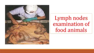
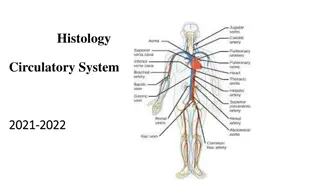
Understanding the Lymphatic System in Cattle: Nodes, Circulation, and Function
The lymphatic system in cattle plays a crucial role in maintaining overall health. It consists of lymph nodes located in various regions of the body such as the head, neck, abdomen, and hind limbs. These lymph nodes produce lymphocytes and help in filtering foreign bodies. Lymph fluid, similar to blood plasma but thinner, transports oxygen and nutrients while removing waste products from tissues. The inspection of lymph nodes is essential for assessing disease processes, considering factors like size, color, and consistency. Understanding the structure and function of the lymphatic system in cattle is vital for proper animal health management.

Download Presentation

Please find below an Image/Link to download the presentation.
The content on the website is provided AS IS for your information and personal use only. It may not be sold, licensed, or shared on other websites without obtaining consent from the author. Download presentation by click this link. If you encounter any issues during the download, it is possible that the publisher has removed the file from their server.
E N D
Presentation Transcript
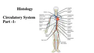
Lymphatic system Function of the lymph: Function of lymph nodes: Lymph circulation: Lymph nodes of cattle Head, neck, thorax, abdmen, pelvis& hind limbs Carcass lymph nodes Haemo lymph nodes
The lymph fluid: It is similar to the blood plasma, but thinner, more watery& poor in protein. The lymph is the medium by which oxygen and nutritive matter are transported from the blood to the body tissues, and the means by which the waste products from these tissues are removed.
Function of lymph node: Production of lymphocytes Arrest the foreign bodies that come to them through the afferent lymphatic vessels.
The inspection of lymph nodes for the presence of anatomical and pathological lesions lies mainly due to the rapid response of the lymph node to an irritant, with enlargement and congestion of its substance and possible break down of its tissue, thus the size, color and consistency of lymph nodes form a valuable guide in the estimation of disease processes in the animal body. Size: varies from that of a pinhead up to walnut. However, the posterior mediastinal L.n of cattle may reach a length of 25 cm. The size is relatively greater in young growing animals than adult, due to a relatively greater growth intensity of the lymphatic tissue.
Shape: Generally, round or oval and somewhat compressed. In ruminants, they are large and few in number, In pigs; the lymph nodes of are lobulated. In camels; the prepectoral & superficial inguinal L.n. are superficially located. Color: variable, may be white, grayish blue or almost black. The mesenteric lymph nodes of cattle are invariably black in color, while in pigs are almost white, except those of the head & neck are reddish in color.
Changes in color: In general Reddish color is due to varying degree of hyperaemia. Yellow color is due to fatty infiltration & is often seen in the mesenteric L.n. Green color may be due to eosinophilia caused by parasitic infestation in the drainage area. Consistency: varies in different parts of the body. In general, L.n of the abdomen are soft than those of the thorax. A physiological oedema of the supramammary & iliac L.n will be encountered in the lactating animals.
Lymph nodes of cattle P:position D: drainage area E: efferent
Lymph nodes of the head: 1. Submaxillary/ mandibular L.n. 2. Parotid L.n. 3. Retropharyngeal L.n. a. Medial (suprapharyngeal). b. Lateral (atlantal)
1Submaxillary L.n One on each side just inside the angel of the jaw & embedded P in the fat, 2-4.5 cm in length. skin & muscles of the head, hard and soft gum, nasal cavity, D tongue. E lateral retropharygeal L.n. Parotid L.n. One on each side of the masseter& covered by parotid salivary gland which must be incised to expose L.n. masseter muscle, head bones, anterior part of the nasal cavity& parotid gland E lateral retropharygeal L.n. 2 P D
Medial. R.Ph. L.n.( internal or suprapharyngeal L.n) 3 P 2-4 in No. & situated between the hyoid bone. 3-6cm Skin of posterior part of the head, part of soft & hard gum, and D posterior part of the nasal cavity. pharynx, m.m of the oral cavity, tonsils E lateral retropharygeal L.n. Lateral retropharyngeal L.n (atlantal L.n) Situated beneath each wing of the atlas &therefore it is usually located 4 P at the neck end of the slaughter & dressed carcass. Tongue, salivary gland, hyoid,cervical & masseter muscles. D Receive afferent vessels from the submaxillar, parotid & medial retropharyngeal. L.n. E Tracheal lymph duct.
Lymph nodes of the Clod (neck): Superficial cervical L.n. Anterior superficial cervical L.n. Posterior superficial cervical L.n. Deep cervical L.n.: Anterior deep cervical L.n. Middle deep cervical L.n. Posterior deep cervical L.n. Cost- cervical L.n.
Posterior superficial cervical or prescapular L.n. an enlarged lymph node 7-9 cm in length. P situated at the anterior border of the supraspinatus muscle or dorsocranial of the shoulder joint. It exposed by cutting an incision parallel to the shoulder in the superficial muscles just above the shoulder joint skin of the neck, shoulder and part of the thorax, scapular muscles , D tendons and muscles of the fore arm & joints of shoulder, carpus and digits (takes lymph from the distal part) on the right side it open into the end of the tracheal duct, E while those of the left side open into the terminal part of the thoracic duct or into vena cava.
Deep cervical L.n; These nodes are situated along the trachea, and can be divided into 3 groups; A- The anterior group: B- The middle group: C- The posterior group: D- cost-cervical group:
Posterior superficial cervical or prescapular L.n. an enlarged lymph node 7-9 cm in length & P situated at the anterior border of the supraspinatus muscle or dorsocranial of the shoulder joint. It exposed by cutting an incision parallel to the shoulder in the superficial muscles just above the shoulder joint skin of the neck, shoulder and part of the thorax, scapular D muscles , tendons and muscles of the fore arm & joints of shoulder, carpus and digits (takes lymph from the distal part) on the right side it open into the end of the tracheal duct, while E those of the left side open into the terminal part of the thoracic duct or into vena cava.
Deep cervical A.The anterior group: P are situated along the anterior part of the trachea, 4-6 small L.n , found in the region of the thyroid gland. ventral part of the skin of the neck, pharyngeal muscles, D larynx, salivary gland, oesophagus, trachea, thyroid gland. tracheal duct E
Deep cervical B. The middle group: Pare often absent and when present they are situated in the middle 3rd of the neck on each side of the trachea. Dventral muscles of the neck, oesophagus, trachea, cervical part of the thymus, prepectoral L.n, cost cervical L.n, axillary L.n, & prescapular L.n Etracheal duct
Deep cervical Prepectoral L.n: 2-4 on each side, and are embedded in fat along the anterior border of the 1strib. It is noted that most of the lymph from the proximal part of the forelimb enter or pass through these lymph nodes. tracheal duct, thoracic duct or common jugular vein. P D E
Costocervical L.n: P 1.5 -3 cm in length & situated on the inner side or just anterior to the 1strib and close to its junction with the 1stdorsal vertebra. This L.n is regarded as aconnecting link between the cervical and mediastinal L.n, as it receives lymph from both cervical region & pleura. D the muscle of the neck and shoulder, the costal part of the pleura, trachea and intercostals & anterior mediastinal L.n. forelimb enter or pass through these lymph nodes. E On the right side usually discharge into the right tracheal duct, while on the left side discharges into the end of thoracic duct.
1.Intercostals L.n. P 0.4-2 cm in diameter & situated in the intercostals space at the junction of the ribs with their vertebrae, and are deep seated or embedded in fat being covered by the intercostals muscles. D thoracic wall, pleura and peritoneum. E mediastinal L.n.
2.Sterna L.n. 2.a.Anterior sternal L.n.: P 1.5- 2.5 cm in length embedded in fat found anterior to the transverse thoracic muscle, in 1st intercostals space & near the sternum. D ventral part of the thoracic muscle, abdomen muscle, diaphragm, ribs, costal part of the pleura, precadium, thymus, liver. E thoracic duct. 2.b. Posterior sterna L.n.: P it is found in the loose fat at the junction of the sternum & diaphragm, at the level of the 6th rib & related antamically to the apex of the heart, the node is absent in 50% of cases. D thoracic wall, sternum, pericardium, pleura, and peritoneum. E thoracic duct.
3.Mediastinal L.n: 3.a.Anterior mediastinal L.n: P a group of L.n. about 10 in number & situated in the anterior mediastinal space in front of the aortic arch. It reached about 2.5 cm in length. It is situated also along the 2 side sof oesophagus & trachea. D the thoracic part of the oesophagus, trachea, and thymus, lungs, pericardium, heart costal & mediastinal pleura, efferents from the intercostals, apical, left bronchial & anterior sterna L.n. E thoracic duct, right tracheal duct and cost-cervical L.n.
Posterior mediastinal L.n: P are situated along the oesophagus from the aortic arch backward. The largest of the these L.n may be 20 cm or more in length, and lies near to the diaphragm, several other L.n. of smaller size lie infront of the large one. D oesophagus, lungs, pericardium, mediastinum, diaphragm, surface of the liver and spleen. E Thoracic duct. Mediastinal L.n P 2-5 L.n, 4 cm in length situated on the right side of the aortic arch. D Thoracic part of the oesophagus and trachea , lungs. E Thoracic duct
Bronchial L.n: Left bronchial L.n 2.5- 3.5 in length, often irregular in shape and found close to the left bronchus , being embedded deeply in fat and partly covered by the aorta. Right bronchial L.n Related to the right bronchus, is usually smaller than the left bronchial and partly hidden by the right lung, it is absent in 25% of cases. Middle bronchial L.n Situated in the middle line above the bifurcation of the trachea , it is absent in 50% of cases. Apical L.n Found close to the accessory bronchus where it enter the apical lobe of the right lung Inspectors lymph nodes (Reissman L.n.): Is present in 75% of the cases and situated at the junction of the 2 cardiac lobes of the right lung.
Bronchial L.n: The lungs & other thoracic tissues. D Thoracic duct & mediastinal L.n E
Lymph nodes of the abdomen, pelvis and hind limbs: Lumbar L.n Hepatic L.n Renal L.n Iliac L.n Precrural L.n Deep inguinal L.n Super ficial inguinal L.n (M) / Supramammary L.n(F) Ischiatic L.n Popliteal L.n Mesenteric L.n
1. Lumbar L.n P Situated along lumbar muscles. Some of these nodes are superficial, others are embedded in the loin suet Lumbar region and peritoneum, efferent vessels from the external & internal iliac, sacral & popliteal L.n Receptaculum chyli the abdominal aorta in the fat covering the D E
2. Hepatic or portal L.n P a group around the hepatic vein, heopatic artery and bile duct, & are covered by the pancreatas. Another group , which includes the edge of the pancreas & the caudate lobe of the liver. They are 10-15 in number. DLiver, pancreas, duodenum. EReceptaculum chyli 3. Renal L.n P Situated in the fat at the entrance of the kidney, they can be exposed by making an incision lengthwise through the blood vessels & continuing the incision 2.5 cm deep into the lumbar fat. Kidney & adrenal gland. D Receptaculum chyli E
4. Iliac L.n a. Internal iliac L.n: P A group of lymph nodes situated in relation to the terminal branches of the aorta. This group may be exposed by incision level with the junction of the sacrum & last lumbar vertebra. Muscles of the sublumber region, pelvis & thigh, femur, tibia, patella, tarsus, metatarsus, urogenital organs& receive efferent from external iliac L.n & sacral , deep inguinal & precrural L.n. Lumber trunk D E
4.b.External iliac L.n P A single or double lymph nodes which are situated lateral to the internal iliac L.n, in thefat at the bifurcation of the iliac artery, and is exposed at the level beneath the external angle of the ilium. Sometimes absent on one or both sides. Abdominal muscles, sublumber region, posterior part of the pertonium & receive some efferent from the precrural L.n. Internal iliac & Lumber trunk D E
5. Precrural / prefemoral L.n P A node which is situated in front of the tensor fascialata muscle embedded in fat 12-15 cm exposed by cutting straight in front of the cranial contour of the leg. The incision may be either oblique at the border of the abdominal muscle and fascia or vertical in the fascia. Skin, prepuce& superficial muscles. above the patella. It is D Chiefly to the deep inguinal L.n. and some to the iliac L.n. E
6. Deep inguinal L.n P situated on the middle of the shaft of the iliac bone. DAbdominal muscles, muscles of the pelvic limbs, knee and tarsl joints, urinary and genital organs. EInternal iliac L.n. and lumber trunk.
7.Superficial inguinal L.n (Male) Supramammary L.n.(Female) P Situated in the mass of fat about the neck of scrotum & behind the spermatic cord. Abdominal muscles, muscles of the pelvic limbs, knee and tarsl joints, urinary and genital organs. Deep inguinal L.n. 2 above the posterior border of the base of the mammary gland. Udder, external genital organs & part of the adjacent regions. nodes present on each side D E
8. Ischiatic L.n P Situated posteriorly on the outer surface of the sacrosciatic ligament under the biceps femoris M. Exposed from the internal surface by cutting through the sacrosciatic lig. About 4 cm in front of the posterior of the lig. Through the coccygeal m. vertical to the long axis of the muscel fibers. Skin of the hip, tail, pelvic M. . coxal joint, rectum, D posterior part of the genital organs, receive efferents from the poplitial L.n. Internal iliac L.n. E
9. Poplitial L.n Deeply seated in the midway between biceps femoris & semitendious , over the gastroenemiius M. found embedded in the intermuscular fat mass. Lower part of the leg & foot. Lumbar , iliac L.n. and ischiatic L.n.
10. Mesenteric L.n P Between the peritoneal folds of the mesentery. Divided into SmallDuodenal G.: 10-50 in no.& 0.5- 120 cm in length. The long are parallel to the intestine & small nodes are scattered throughout the mesentery between small intestine & colon. DThe duodenum E Portal L.n From the intestinal trunk to discharge later into the receptaculum chyli. jejuno-ilial group: Small intestine( jejunum& ilium)


")
:")
:")


 Supramammary L.n.(Female)")