Exploring Polydactyly and Syndactyly: Genetic Anomalies in Limb Development
Polydactyly is a condition characterized by the presence of more than five fingers or toes on a hand or foot, often associated with genetic disorders. This study delves into the types, patterns, associated anomalies, treatment plans, and outcomes related to polydactyly. Syndactyly, on the other hand, involves fused fingers or toes. The research sheds light on the genetic basis, prevalence across different demographics, and implications for hand surgeons and prenatal care.

Download Presentation

Please find below an Image/Link to download the presentation.
The content on the website is provided AS IS for your information and personal use only. It may not be sold, licensed, or shared on other websites without obtaining consent from the author. Download presentation by click this link. If you encounter any issues during the download, it is possible that the publisher has removed the file from their server.
E N D
Presentation Transcript
GROUP 6 POLYDACTYL AND SYNDACTYL
GROUP MEMBERS 1. 2. 3. 4. 5. 6. 7. 8. 9. 10. BAMBE CHIOMA 15/MHS02/018 11. ADEOLU DAMILOLA 16/MHS02/053 12. ADEDIGBE OLUWAPELUMI 15/MHS02/002 OGUNNIYI OLUWATOYIN 17/MHS02/111 OLALERE FATIMAH 17/MHS02/113 SANI RIHANAT 17/MHS02/114 SULEIMAN BINTA 17/MHS02/115 SHAIBU FAWZIYAT 15/MHS02/050 OJO LOVETH 14/MHS01/102 AKPOSIVWODOR ESEROGHENE 15/MHS02/012 OLUWASOLA BOLANLE 15/MHS02/043 MOSHESHE PRUDENCE 15/MHS02/034
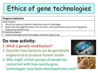
INTRODUCTION Polydactyl is characterized by more than five fingers/digits in hand/foot. It can present alone or as part of many syndrome due to genetic disorder. The defects may be due to autosomal recessive or autosomal dominant disease. The present work was conducted to analyse the type, pattern of involvement, associated anomalies, plan of treatment and outcome of this malformation.
From Raphael's paintings it was found that the father (St. Joseph) and son had postaxial polydactyly which is an autosomal dominant trait. St. Joseph found shoes uncomfortable because of his polydactyl. Pre and postaxial polydactyl are also found in Acrocallosal Syndrome, an autosomal recessive condition characterized by agenesis of corpus callosum
Molecular aspect of limb development (4) has an identifiable molecular basis. Hand surgeons should beware of the basic molecular pathways controlling limb development because they are in a unique position to be able to identify patients with such deformities. If Ultrasonographic examination can be done during 14-16 weeks of gestation, fetuses with polydactyl may be observed. USG is a valuable tool for identification and early management since there is no biochemical or histopathological markers. In utero auto-amputation (5) of extra digits can be done. Pregnancies, having polydactyl associated with other anomalies, should be terminated. Whereas isolated fetal postaxial polydactyl type II is associated with a favourable outcome.
EPIDEMIOLOGY The condition has an incidence of 1 in every 500 live births. Postaxial hand polydactyl is a common isolated disorder in African black children, and autosomal dominant transmission is suspected. Postaxial polydactyl is more frequent in native Africans living in the Eastern and Central than the Caucasians and Mongoloids and is more frequent in male children.In contrast, postaxial polydactyl seen in white children is usually syndromic and associated with an autosomal recessive transmission. This study showed incidence of all types of polydactyl to be 2.3 per 1000 in Caucasian males, 0.6 per 1000 in Caucasian females, 13.5 per 1000 in African males, and 11.1 per 1000 in African females.
Syndactyly and polydactylrespectively characterized by fused and supernumerary digits are among the most common congenital limb malformations, with syndactyly presenting at an estimated incidence of 1 in 2,000 3,000 live births and polydactyl at a frequency of 1 in approximately 700 1,000 live births. Despite their relatively regular manifestation in the clinic, the etiologies of syndactyl and polydactyl remain poorly understood because of their phenotypic and genetic diversity
TYPES OF POLYDACTYL Five types of polydactyl were encountered, Type I- cutaneous nubbin, Type II-pedunculated digit, Type III- articulating digit with fifth metacarpal, Type IV-fully developed digit with sixth metacarpal Type V- polysyndactyl. Type I and II ulnar polydactyl are more prevalent.
TYPES OF POLYDACTYL There are 3 types of polydactyl based on where the digit is: 1. Ulnar or postaxial polydactyl or small finger duplication: This is the most common form of the condition, where the extra finger is on the outside of the little finger. This side of the hand is known as the ulnar side. When this form of the condition affects the toes, it is called fibular polydactyl. 2. Radial or preaxial polydactyl or thumb duplication: This is less common, occurring in 1 in every 1,000 to 10,000 live births. The extra finger is on the outside of the thumb. This side of the hand is known as the radial side. When this form of the condition affects the toes, it is called tibial polydactyl. 3. Central polydactyl: This is a rare type of polydactyl. The extra finger is attached to the ring, middle, or most often index finger. This form of the condition has the same name when it affects the toes.
CAUSES AND SYMPTOMS Polydactyl and syndactyl are conditions that occur in the developing fetus. Most often these conditions are caused by genetic factors. Both polydactyl and syndactyl can be caused by the presence of an autosomal dominant trait. An autosomal dominant trait is a gene that is not related to the chromosome that determines gender; therefore, it affects boys and girls equally. Because the gene is dominant, when one parent has the gene, each of his or her children has a 50 percent chance of having polydactyl or syndactyl.
The primary symptom of polydactyl is the presence of extra digits on the hands or feet. Polydactyl rarely causes any difficulties for the child. The extra fingers and/or toes are usually removed for cosmetic reasons. In children with syndactyl of the hand, finger function may be impaired and, in cases where fingers of different lengths are connected by tissue, finger growth may be limited unless surgery to separate the fingers is performed
During normal embryonic development (while the baby is still in the womb), the hand initially forms in the shape of a paddle; then at about the sixth or seventh week of gestation splits into separate fingers.
SIGNS AND SYMPTOMS The way polydactyly presents can vary. - It may appear as a small, raised lump of soft tissue, containing no bones (called a nubbin) partially formed finger or toe containing some bones but no joints. -The extra fingers are usually smaller and abnormally developed than normal and can be made up of:skin and soft tissue the simplest to remove -Skin, soft tissue and bone but no joint -Skin, soft tissue and bone with joint (closer to a fully- formed finger most challenging to remove/remodel)
SIGNS AND SYMPTOMS CONTD Each person with syndactyly tends to experience different symptoms, as webbed toes may be: -Unilateral or bilateral: Impacting only one side of the body or both. -Severe, moderate, or mild: With digits almost entirely fused, with digits partially fused, or with only minor webbing between digits. - Symmetric or asymmetric: Appearing alike and in the same region on both sides of the body, or appearing dissimilar or in different places on each side of the body. - Simple or complex: Involving only two digits or a few bones, or including multiple digits or bones. - Painful or asymptomatic.
Minor cases may not interfere much with toe or foot movement and function. However, if the toes are severely webbed or fused, the condition can be disabling.
DIAGNOSIS OF POLYDACTYL Polydactyl can be seen by ultrasound prenatally and by eye (i.e. physical examination) at birth. Your doctor will use x-rays to assess the underlying structure of your baby s finger and determine a course of treatment.
Syndactyly may be seen by ultrasound prenatally and is apparent at birth. Your doctor will use x- rays to assess the underlying structure of your baby s fingers and determine a course of treatment. If the syndactyly is associated with a genetic syndrome, doctors will evaluate the baby s entire upper extremity, chest feet and head/face to detect other abnormalities.
TREATMENT OF POLYDACTYL Treatment for polydactyl depends on how and where the extra digit is connected to the hand or foot. In most cases, the extra digit is removed in the child s first two years. This gives the child typical use of their hand and allows their feet to fit into shoes. Sometimes adults will have the surgery to improve the appearance or function of their hand or foot.
Surgery is usually outpatient, with local or topical anesthesia. Different surgical techniques are a subject of ongoing research. Removal of an extra little finger or toe is usually a simple procedure. In the past, nubbins were usually just tied off, but this often left a bump. Now surgery is preferred. The child will receive stitches to close the wound. The stitches dissolve within two to four weeks.
Fifth digit: Removal of an extra little finger or toe is usually a simple procedure. In the past, nubbins were usually just tied off, but this often left a bump. Now surgery is preferred. The child will receive stitches to close the wound. The stitches dissolve within two to four weeks. Thumb or big toe: Removal of an extra thumb may be complex. The remaining thumb must have an optimal angle and shape to be functional. This may require some remodeling of the thumb, involving the soft tissues, tendons, joints, and ligaments.
Central fingers or toes: This surgery is usually more complex and requires remodeling of the hand to ensure it s fully functional. It may require more than one surgery, and the child may need to wear a cast for a few weeks after surgery. Sometimes a pin will be inserted to hold the bones together while they heal. The doctor may prescribe physical therapy to reduce scarring and help improve function. This procedure is typically performed when the child is between 1 and 2 years old. At this age, the child is old enough to tolerate anesthesia and surgery but is not at risk for missing developmental milestones such as grasping (prehension).
In general, the skin is split evenly between the two fingers with zig-zag incisions (z-plasty). Only one side of a finger is separated at a time in order to avoid complications related to the skin coverage and blood supply of the affected finger


 has an identifiable")