Clinical Microbiology Techniques: Complement Fixation Test (CFT) in Diagnostic Practice
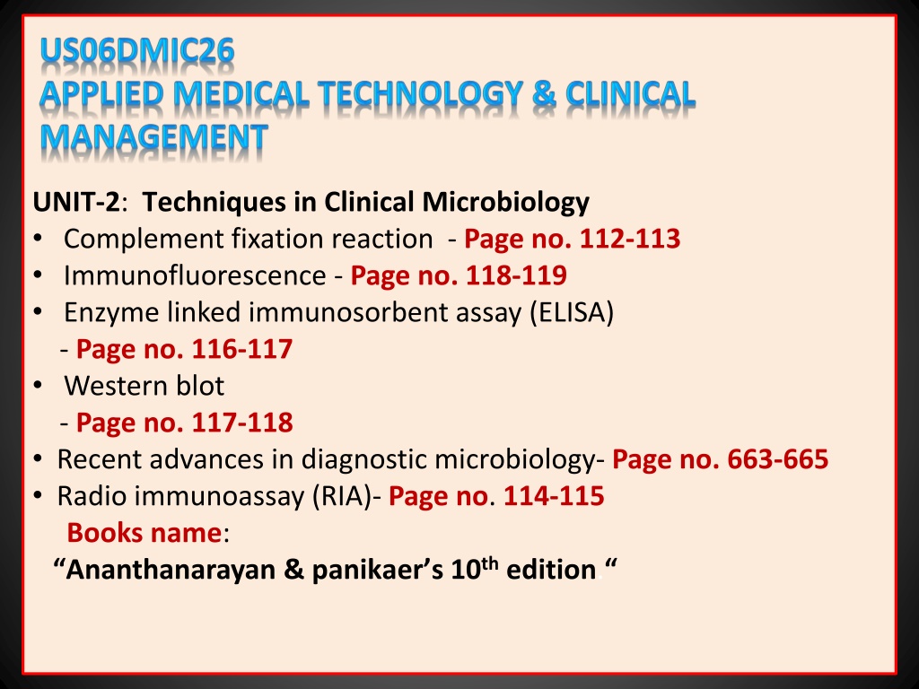
UNIT-2
:
Techniques in Clinical Microbiology
•
Complement fixation reaction
-
Page no. 112-113
•
Immunofluorescence
-
Page no. 118-119
•
Enzyme linked immunosorbent assay (ELISA)
-
Page no. 116-117
•
Western blot
-
Page no. 117-118
•
Recent advances in diagnostic microbiology
-
Page no. 663-665
•
Radio immunoassay (RIA)
-
Page
no
.
114-115
Books name
:
“
Ananthanarayan & panikaer’s 10
th
edition
.
“
COMPLEMENT FIXATION TEST (CFT)
•
Complement takes part in many immunological reactions and is
absorbed during the combination of antigens with their
antibodies.
•
In the presence of the appropriate antibodies, complement lyses
erythrocytes, kills and, in some cases, lyses bacteria, immobilises
motile organisms, promotes phagocytosis and immune
adherence and contributes to tissue damage in certain types of
hypersensitivity.
Principle
:
•
The ability of antigen-antibody complexes to 'fix' complement is
made use of in the CFT.
•
This is a very versatile and sensitive test, applicable with various
types of antigens and antibodies and capable of detecting as
little as 0.04 mg of antibody nitrogen and 0.1 mg of antigen.
•
CFT is a complex procedure consisting of two steps and five
reagents-antigen, antibody, complement, sheep erythrocytes
and amboceptor (rabbit antibody to sheep red cells). Each of
these reagents has to be separately standardized.
Procedure
:
1.
The antigen may be soluble or particulate. The antiserum
should be heated at 56°C (inactivated) for half an hour before
the test to destroy any complement activity, the serum may
have and also to remove some non-specific inhibitors of
complement present in some sera (anti-complementary
activity).
2. The source of the complement is guinea pig serum. As
complement activity is heat labile, the serum should be freshly
drawn, or preserved either in the lyophilized or frozen state or
with special preservatives, as in Richardson's method.
Standardization
:
•
The guinea pig serum should be titrated for complement activity.
•
One unit or minimum hemolytic dose (MHD) of complement is
defined as the highest dilution of the guinea pig serum that lyses
one unit volume of washed sheep erythrocytes in the presence
of excess hemolysin (amboceptor) within a fixed time (usually 30
or 60 minutes) at a fixed temperature (57°C).
•
The amboceptor should be titrated for hemolytic activity.
•
One MHD of amboceptor is defined as the least amount (or
highest dilution) of the inactivated amboceptor that lyses one
unit volume of washed sheep erythrocytes in the presence of
excess complement within a fixed time (usually 30 or 60 minutes)
at a fixed temperature (37°C).
•
The diluents used for the titrations and for CFT is physiological
saline with added calcium and magnesium ions.
Wasserman reaction:
•
The classical example of CFT is the Wassermann reaction,
formerly the routine method for the sero diagnosis of
syphilis.
Procedure
:
1.
The inactivated serum of the patient is incubated at 37 ̊C for
one hour with the Wassermann antigen and a fixed amount
(two units) of guinea pig complement. If the serum contains
syphilitic antibody, the complement will be utilized during
antigen-anti body interaction. If the serum does not contain the
antibody, no antigen-antibody reaction occurs and the
complement will therefore be left intact.
2.
Testing for complement in the post-incubation mixture will
thus indicate whether the serum had antibodies or not. This
consists of adding sensitized cells (sheep erythrocytes coated
with + MHD hemolysin), and incubating at 37°C for 30 minutes.
Interpretation of results
:
1. Lysis of the erythrocytes indicates that the complement was not
fixed in the first step and therefore, the serum did not have the
antibody (Negative CFT).
2.
Absence of erythrocyte lysis indicates that the complement was
used up in the first step and, therefore, the serum contained
the antibody (Positive CFT).
Appropriate controls should be used, including the following:
1. Antigen and serum controls to ensure that they are not anti-
complementary.
2.
Complement control to ensure that the desired amount of
complement is added
3.
Cell control to observe that sensitized erythrocytes do not
undergo lysis in the absence of complement.
Indirect complement fixation test
:
•
Certain avian (for example, duck, turkey, parrot) and
mammalian (for example, horse, cat) sera do not fix guinea pig
complement. When such sera are to be tested, the indirect
complement fixation test may be employed.
•
Here, the test is set up in duplicate and after the first step, the
standard antiserum known to fix the complement is added to
one set.
•
If the test serum contained antibody, the antigen would have
been used up in the first step and, therefore, the standard
antiserum added subsequently would not be able to fix the
complement. Therefore, in the indirect test, hemolysis indicates
a positive result.
Conglutinating complement absorption test
:
•
For systems which do not fix guinea pig complement, an
alternative method is the conglutinating complement
absorption test.
•
This uses horse complement which is non-hemolytic. The
indicator system is sensitized sheep erythrocytes mixed with
bovine serum.
•
Bovine serum contains a beta globulin component called
conglutinin, which acts as antibody to the complement.
•
Therefore, conglutinin causes agglutination of sensitized sheep
erythrocytes (conglutination) if they have combined with the
complement.
•
If the horse complement had been used up by the antigen-
antibody interaction in the first step, agglutination of sensitised
cells will not occur.
Other complement-dependent serological tests
:
•
When some bacteria (for example,
Vibrio cholerae, Treponema
pallidum
) react with the specific antibody in the presence of
complement and particulate materials such as erythrocytes or
platelets, the bacteria are aggregated and adhere to the cells.
This is known as immune adherence.
•
The immobilization test is another complement-dependent
reaction. In the
Treponema pallidum
immobilization test, a highly
specific test formerly considered the "gold standard for the
serodiagnosis of syphilis, the test serum is mixed with a live
motile suspension of
T.pallidum
in the presence of the
complement. On incubation, the specific antibody inhibits the
motility of treponemes.
•
Cytolytic or cytocidal tests are also complement-dependent.
When a suitable live bacterium, such as the
cholera vibrio,
is
mixed with its antibody in the presence of the complement, the
bacterium is killed and lysed. This forms the basis of the
vibriocidal antibody test for the measurement of anti-cholera
antibodies.
IMMUNOFLUORESCENCE
•
Fluorescence is the property of absorbing light rays of one
particular wavelength and emitting rays with a different
wavelength.
•
Fluorescent dyes show up brightly under ultraviolet light as
they convert ultraviolet into visible light.
•
Coons and his colleagues (1942) showed that fluorescent dyes
can be conjugated to antibodies and that such labeled
antibodies can be used to locate and identify antigens in
tissues.
•
This 'fluorescent antibody' or immunofluorescence technique
has several diagnostic and research applications ..
Direct immunofluorescence test
:
•
This can be used for the identification of bacteria, viruses or
other antigens, using the specific antiserum labeled with a
fluorescent dye. For example, direct immunofluorescence is
routinely used as a sensitive method of diagnosing rabies, by
detection of the rabies virus antigens in brain smears.
•
A disadvantage of this method is that separate fluorescent
conjugates have to be prepared against each antigen to be
tested.
Indirect immunofluorescence test
:
•
This test over comes the difficulty mentioned above by using an
antiglobulin fluorescent conjugate. An example is the
fluorescent treponemal antibody test for the diagnosis of
syphilis. Here, a drop of the test serum is placed on a smear of
T.pallidum
on a slide and after incubation, the slide is washed
well to remove all free serum, leaving behind only antibody
globulin, if present, coated on the surface of the treponemes.
•
The smear is then treated with a fluorescent-labelled antiserum
to human gamma globulin.
•
The fluorescent conjugate reacts with the antibody globulin
bound to the treponemes. After washing away all the unbound
fluorescent conjugate, when the slide is examined under
ultraviolet illumination, if the test is positive, the treponemes
will be seen as bright objects a dark background.
•
If the serum does not have anti-treponemal antibody, there will
be no globulin coating on the treponemes and therefore they
will not take on the fluorescent conjugates.
•
A single anti-human globulin fluorescent conjugate can be
employed for detecting human antibodies to any antigen.
•
Fluorescent dyes may also be conjugated with the complement.
•
Labeled complement is a versatile tool and can be employed for
the detection of antigen or antibody. Antigens also take
fluorescent labeling but not as well as antibodies do.
•
For detection of antibodies by immunofluorescence, the
sandwich technique can be used.
•
The antibody is first allowed to react with unlabelled antigen,
which is then treated with fluorescent-labelled antibody. A
sandwich is thus formed, the antigen being in the middle and
the labeled and unlabelled antibodies on either side.
Immuno histochemical technique
:
•
By combining the specificity of serology with the localizing
capacity of histology, immunofluorescence helps in the
visualization of antigen antibody reactions in situ and is thus
called an immuno histochemical technique. The main
disadvantage of the technique is the frequent occurrence of
non-specific fluorescence in tissues and other materials.
•
The fluorescent dyes commonly used are fluorescein
isothiocynate and rhodamine, exhibiting blue-green and
orange-red fluorescence, respectively.
Flow cytometry
:
•
This is the fluorescence technique used to identify and
enumerate cells bearing a particular antigen(s) or the surface
markers by suspending them in a stream of fluid and passing
them through an electronic detection apparatus.
•
It allows simultaneous multiparametric analysis of the physical
and/or chemical characteristics of up to thousands of particles
per second.
•
Different populations of molecules, cells or particles can be
differentiated by size and shape using forward and right-angle
light scatter.
•
These cells, particles or molecules, can be labeled with different
fluorescent labels or with dye-labelled monoclonal antibodies.
•
In this way, we can measure the amounts or isolated individual
cells or populations of particular cells from a mixed population.
•
Here, cells are made to flow in a single cell stream in a flow cell
by hydrodynamic focusing.
•
In this, the sample stream containing the cells are focused by a
surrounding layer of sheath fluid.
•
An Argon laser (at 488 nm) is focused on the cells. Various
sensors pick up the laser light reflected by the cells.
•
The parameters measured by the flow cytometer are forward
scatter (FS), a measure of cellular size, side scatter (SS), a
measure of the granularity of the cell and fluorescence sensors
(FL1-FL8), which measure light emitted by various dyes bound
to the cells. The emitted light is directed into various sensors by
a combination of filters.
Applications
:
•
Multiple parameters—the size, granularity. DNA or RNA content,
cellular antigens, receptor levels, etc.,—can be measured using
flow cytometry.
•
It is widely used in research and diagnostics, for example, to
count blood cells including differential leukocyte count (DLC), to
isolate T cell subsets (CD4 and CD8 counts in HIV patients), for
diagnosis, treatment and prognosis in cancer (especially
leukemias), to study the cell cycle and apoptosis, etc.
Enzyme linked immunosorbent assay (ELISA)
•
ELISA is so named because the technique involves the use of an
immunosorbent, an absorbing material specific for one of the
components of the reaction: the antigen or antibody.
•
This may be a particulate, for example, cellulose or agarose, or a
solid phase such as polystyrene, polyvinyl or polycarbonate
tubes or microwells, or membranes or discs of polyacrylamide,
paper or plastic.
•
ELISA is usually done using 96-well microtitre plates suitable for
automation. The principle of the test can be illustrated by
outlining its application for the detection of rotavirus antigen in
feces.
Procedure:
Sandwich ELISA:
1.
The wells of a microtitre plate are coated with goat
antirotavirus antibody. After thorough washing, the fecal
samples to be tested are added and incubated overnight at 4°C
or for two hours at 37 C. Suitable positive and negative controls
are also set up.
2.
The wells are washed and guinea pig antirotavirus antiserum,
labeled with alkaline phosphatase, is added and incubated at
37°C for one hour.
3.
After washing a suitable substrate (para-nitrophenyl
phosphate) is added and held at room temperature till the
positive controls show the development of a yellow color. The
phosphatase enzyme splits the substrate to yield a yellow
compound.
4.
If the test sample contains rotavirus, it is fixed to the antibody
coating the wells. When the enzyme labeled antibody is added
subsequently, it is in turn fixed. The presence of residual
enzyme activity, indicated by the development of yellow colour,
therefore denotes a positive test.
5.
If the sample is negative. there is no significant color change.
An ELISA reader provides quantitative color recordings which
are directly proportional to the quantity of analyte present in
the test sample.
Types
Indirect ELISA
:
The detection of antibody by ELISA can be illustrated by the anti-
HIV antibody test. Purified inactivated HIV antigen is adsorbed onto
micro assay plate wells. Test serum diluted in buffer is added to the
well and incubated at 37°C for 30 minutes. The well is then
thoroughly washed. the serum contains anti-HIV antibody, it will
form a stable complex with the HIV antigen on the plate.
A goat anti-human immunoglobulin antibody conjugated with
horseradish peroxidase enzyme is added and incubated for 30
minutes. After thorough washing, the substrate O-phenylene
diamine dihydrochloride is added and after 30 minutes, the color
that develops is read using a micro assay plate reader. Positive and
negative controls should invariably be used with test sera.
Capture ELISA
and immunometric tests are even more specific.
Several variations of the ELISA technique have been developed to
provide simple diagnostic tests, including the card and dipstick
methods suitable for clinical laboratory and bedside applications.
Sandwich ELISA
: It is used for antigen detection in patient sample.
The antigen is sandwiched between two layers of antibodies (i.e.,
capture and detection antibodies).
Competitive ELISA
:
Similar to RIA, both the unknown antigen (sample) and the known
antigen (standard) compete with each other for a fixed amount of
antibody. Competitive ELISA yields an inverse curve, where higher
values of antigen in the samples/standards yield a lower amount
of color change. It is normally used for hapten detection.
Cylinder or cassette ELISA
: A simple modification of ELISA which
has found wide application for testing one or a few samples of sera
at a time is the cylinder or cassette ELISA. Here, each specimen is
tested in a separate disposable cassette. The test is rapid (10-15
minutes). There is no need for microplate washers or readers. The
result is read visually. In-built positive and negative controls are
usually provided for validation of the test procedure. An example
of cassette ELISA is the
Dot Blot Assay
used for the detection of
HIV type I and 2 antibodies. Specific type 1 und 2 antigens are
immo bilised at separate fixed sites on the nitrocellulose
membrane in the cassette.
Procedure:
Test serum is added on the membrane and allowed to filter into
absorbent material placed below it in the cassette base. Antibody,
if present in the serum, will bind to the appropriate antigen. After
washing to remove the unbound antibody, enzyme labeled anti-
human immunoglobulin antibody is added. After additional
washing to remove the unbound conjugate, a substrate yielding a
colored product is added.
Result
:
A positive result is indicated by a colored spot developing at the
site of the antigen against which the antibody is present in the
serum. Human immunoglobulin immobilized at a spot on the
membrane acts as a control for the test procedure, as shown by
the development of color at the site.
Uses of ELISA
:
ELISA plays a major role in the diagnosis of innumerable diseases.
Some examples are given below:
•
HIV detection.
•
Infectious diseases like hepatitis, EBV, cytomegalovirus IgM/IgG,
dengue IgG. influenza, TORCH panel, etc..
•
Rotavirus detection in fecal specimens and enterotoxin of
E.coli
in feces.
•
Syphilis IgG/IgM,
H.pylori
IgG and antigen detection.
•
Food toxins like chloramphenicol, streptomycin, penicillin,
aflatoxins, etc.
•
Food adulterants including
E.coli
.
Campylobacter
and
Salmonella
antigens
•
Mycobacterial
antibody detection in tuberculosis.
•
Human allergen-specific IgE and IgA ELISA.
RADIO IMMUNOASSAY (RIA)
•
Besides fluorescent dyes, many other distinctive "labels" can also
be conjugated to antigens and antibodies.
•
The most commonly used labels are radioisotopes and enzymes.
•
A variety of tests have been devised for the measurement of
antigens and antibodies using such labeled reactants.
•
The term binder-ligand assay has been used for these reactions.
•
The substance (antigen) whose concentration is to be determined
is termed the analyte or ligand.
•
The binding protein (ordinarily, the antibody) which binds to the
ligand is called the binder. The first reaction of this type was
radioimmunoassay (RIA) described by Berson and Yallow in 1959.
•
RIA permits the measurement of analytes up to picogram (10
-12
)
quantities. The importance of RIA was acknowledged when the
Nobel Prize was awarded to Yallow for his discovery in 1977.
•
RIA is a competitive binding assay in which fixed amounts of
antibody and radio labeled antigen react in the presence of
unlabelled antigen.
•
The labeled and unlabelled antigens compete for the limited
binding sites on the antibody. This competition is determined by
the level of the unlabelled (test) antigen present in the patient's
serum samples.
•
After the reaction, the antigen is separated into free and bound'
fractions and their radioactive counts measured.
•
The concentration of the test antigen can be calculated from the
ratio of the bound and total antigen labels, using a standard
dose-response curve.
•
For any reacting system, the standard dose-response or
calibrating curve has to be prepared first. This is done by running
the reaction with fixed amounts of antibody and labeled antigen
and varying known amounts of unlabelled antigen.
•
The ratio of bound antigen to total antigen (B:T ratio plotted
against the analyte concentrations give the standard calibration
curve.
•
The concentration of antigen in the test sample is computed from
the B-T ratio of the test by interpolation from the calibration
curve.
•
RIA and its modifications have versatile applications in various
areas of biology and medicine, including the quantitation of
hormones, drugs, tumor markers, IgE and viral antigens.
IMMUNOELECTROBLOT/WESTERN BLOT TECHNIQUES
•
Immunoelectroblot or western blot techniques combine the
sensitivity of enzyme immunoassay with much greater
specificity.
•
The technique is a combination of three separate procedures:
1.
Separation of ligand-antigen components by poly- acryl amide
gel electrophoresis
2.
Blotting of the electrophoresed ligand fraction on nitrocellulose
membrane strips
3.
Enzyme immunoassay (or radioimmunoassay) to:
• detect antibody in test sera against the various ligand fraction
bands
• probe with known antisera against specific antigen bands
The western blot test, considered to be the definitive /
confirmatory test for the serodiagnosis of HIV infection, is an
example of the immunoelectroblot technique.
Recent advances in diagnostic microbiology
INTRODUCTION
Conventional laboratory techniques for diagnosis of infectious
diseases, such as culture and related methods, are widely used as
they are sensitive and inexpensive, but tend to be labor- and
resource-intensive, requiring considerable expertise. They often
require further characterization by molecular techniques to
confirm identification.
Molecular methods are useful in situations where conventional
methods are slow, insensitive or unavailable. Advances have been
made in decreasing the turnaround time (from specimen collection
to the final report reaching the patient) in culture, identification
and antibiotic susceptibility testing.
Automated and semi-automated systems
:
They fall into two main groups:
•
Blood culture systems Identification and
•
Susceptibility testing instruments
Whereas some identification and susceptibility testing instruments
take as long as traditional methods, others provide results within a
single working day. One of the newer methods to enter the chain is
the MALDI-TOFMS (matrix-assisted laser desorption/ ionisation-
time of flight mass spectrometry). It is a technology for
identification of any bacteria or fungi based on the unique protein
composition of the bacterial cell. Its advantage is rapid (in minutes)
and reliable results.
The most advancement in diagnostic microbiology has been made
in the application of molecular techniques.
MOLECULAR METHODS
They can be broadly classified into one of three categories:
• Hybridization
• Amplification
• Sequencing and enzymatic digestion of nucleic acid
Hybridization
This is based on the ability of two nucleic acid strands that have
complementary base sequences to bind specifically with each
other and form a double-stranded molecule or hybrid. The assay
requires one nucleic acid strand (probe) from an organism of
known identity and another (target) from an unknown organism.
Hybridization is detected by the use of a reporter molecule that
forms a complex with the single-stranded probe DNA. Probes may
be labeled with a variety of molecules-radioactive, biotin-avidin
and chemiluminescent labels.
One of the major advancements of this technique has been in the
application of line probe assay for diagnosis of tuberculosis and
detection of drug resistance.
Amplification
Polymerase chain reaction (PCR):
This is the most widely used target nucleic acid amplification
method. It combines the principles of complementary nucleic acid
hybridization and replication.
Conventional PCR involves the following steps:
1.
Extraction and denaturation
: The nucleic acid is first extracted
from the organism or clinical sample by heat, chemical or
enzymatic method, and the target nucleic acid is then added
and placed in a thermal cycler to undergo amplification.
2.
Annealing
: Short strands of DNA sequences (primers) are
selected to specifically hybridize (anneal) to a particular nucleic
acid target.
3.
Extension of the primer
: Taq polymerase is the enzyme
commonly used for primer extension at a temperature of 72°C.
Annealing provides a template that allows DNA polymerase to
add nucleotides and produce by extension a sequence that is
complementary to the target template.
4.
Detection of PCR products
: The amplification product of PCR
(amplicon) contains the target nucleic acid of interest. A
labeled probe is used for detecting specific amplicons.
Types of PCR used in diagnostic microbiology
Multiplex PCR:
Two or more unique target sequences can be amplified
simultaneously. One primer pair is directed at sequences present
in
all clinically relevant bacteria (control primer) and the second is
directed at a specific sequence (test primer). This is used to detect
infectious agents where the exact cause is not known.
Nested PCR
: This involves the sequential use of two primer sets.
The first set is used for amplification of a target sequence. The
second set of primers are used to amplify a particular region within
the first target sequence. This gives a more specific test result and
is used for specific disease detection. Gene Expert for detecting
M.tuberculosis
and resistance to Rifampin is widely used and has
become a mandated test by the Revised National Tuberculosis
Program (RNTCP) as a diagnostic tool and for monitoring of drug
resistance.
Quantitative PCR
: In addition to detection of the agent, the actual
number of targets in the clinical sample is detected, for example,
detecting viral load in HIV.
Reverse transcriptase PCR (RT-PCR):
Conventional PCR amplifies
dsDNA, but not RNA. The enzyme reverse transcriptase directs the
synthesis of DNA from the viral RNA template. The DNA thus
obtained is subjected to routine PCR method. This is used to detect
H1N1 virus from nasopharyngeal swab.
Real-time PCR:
This combines nucleic acid ampli fication with
qualitative or quantitative measurement of the amplified product.
Most molecular detection methods used in clinical microbiology
use this technique.
Transcription-mediated amplification (TMA)
This is an isothermal RNA amplification and uses three enzymes:
RNase, DNA-dependent RNA polymerase and reverse transcriptase
enzymes. The RNA target is converted to c DNA using reverse
transcriptase and then RNA copies are synthesized with the help of
RNA polymerase.
Nucleic acid sequence-based amplification (NASBA)
This is similar to TMA and is also an isothermal RNA amplification
method. The RNA target is converted to cDNA using reverse
transcriptase and then RNA cop ies are synthesised with the help of
RNA polymerase.
Ligase chain reaction (LCR)
This reaction is based on the ligation of two adjacent synthetic
oligonucleotide primers, which uniquely hybridize to one of the
strands of the target DNA. A second pair of oligonucleotides is
designed to hybridize to the complementary DNA, in the same
region. When the nucleotides are present, the DNA polymerase
and the ligase create a gap between the adjacent primers, which
will then be filled with the appropriate nucleotides, leading to
ligation of the primers. LCR allows the discrimination of DNA
sequences differing in only a single base pair. Hence, it is used to
differentiate products.
SEQUENCING AND ENZYMATIC DIGESTIONOF NUCLEIC ACID
Nucleic acid sequencing
: This involves methods that determine the
exact nucleotide sequence of a gene or gene fragment of an
organism.
High-density DNA probes
: This uses hybridization of a fluorescent-
labelled nucleic acid target to a set of oligonucleotides synthesized
on a miniature glass substrate or 'chip'.
Methods of typing isolates
Ribotyping
: This involves the enzymatic digestion of chromosomal
DNA followed by hybridization using probes that encode ribosomal
RNA.
Restriction fragment length polymorphism(RFLP):
Restriction enzyme analysis is the process by which enzyme
digestion patterns are analyzed. Restriction patterns are obtained
after gel electrophoresis, the differences in restriction patterns of
microorganism is known as restriction fragment length
polymorphism.
Pulsed-field gel electrophoresis (PFGE):
Specialized electrophoresis devices are used to separate chromo
somal fragments obtained by enzymatic digestion of intact
bacterial chromosomal DNA. The DNA is then digested using
restriction end nuclease enzymes.
Use of a pulsed electrical field across the agrose gel subjects the
DNA fragment to different voltages from varying angles at different
time intervals.
Applications
There is increasing application of molecular techniques in
diagnostic microbiology.
Diagnosis
•
Detection of infectious agents from clinical specimens, CSF and
body fluids.
•
Identification of bacteria, antimicrobial resistance (tuberculosis).
•
Diagnosis of infections due to non-cultivable organisms.
Epidemiology and disease detection
•
Typing of isolates for epidemiological purposes and tracing path
of transmission Research.
Research
•
Research
into understanding genetic makeup of infectious
agents.
•
Drug discovery and newer diagnostics
Complement Fixation Test (CFT) is a key technique in clinical microbiology, utilizing antigen-antibody complexes to fix complement for various immunological reactions. The test involves multiple steps and reagents, with guinea pig serum for complement activation. Standardization is crucial for accurate results, with applications like the Wasserman reaction for syphilis diagnosis showcasing its significance in diagnostic microbiology.

Download Presentation

Please find below an Image/Link to download the presentation.
The content on the website is provided AS IS for your information and personal use only. It may not be sold, licensed, or shared on other websites without obtaining consent from the author. Download presentation by click this link. If you encounter any issues during the download, it is possible that the publisher has removed the file from their server.
E N D
Presentation Transcript
UNIT-2: Techniques in Clinical Microbiology Complement fixation reaction - Page no. 112-113 Immunofluorescence - Page no. 118-119 Enzyme linked immunosorbent assay (ELISA) - Page no. 116-117 Western blot - Page no. 117-118 Recent advances in diagnostic microbiology- Page no. 663-665 Radio immunoassay (RIA)- Page no. 114-115 Books name: Ananthanarayan & panikaer s 10th edition.
COMPLEMENT FIXATION TEST (CFT) Complement takes part in many immunological reactions and is absorbed during the combination of antigens with their antibodies. In the presence of the appropriate antibodies, complement lyses erythrocytes, kills and, in some cases, lyses bacteria, immobilises motile organisms, promotes phagocytosis and immune adherence and contributes to tissue damage in certain types of hypersensitivity. Principle: The ability of antigen-antibody complexes to 'fix' complement is made use of in the CFT. This is a very versatile and sensitive test, applicable with various types of antigens and antibodies and capable of detecting as little as 0.04 mg of antibody nitrogen and 0.1 mg of antigen.
CFT is a complex procedure consisting of two steps and five reagents-antigen, antibody, complement, sheep erythrocytes and amboceptor (rabbit antibody to sheep red cells). Each of these reagents has to be separately standardized. Procedure: 1. The antigen may be soluble or particulate. The antiserum should be heated at 56 C (inactivated) for half an hour before the test to destroy any complement activity, the serum may have and also to remove some non-specific inhibitors of complement present in some sera (anti-complementary activity). 2. The source of the complement is guinea pig serum. As complement activity is heat labile, the serum should be freshly drawn, or preserved either in the lyophilized or frozen state or with special preservatives, as in Richardson's method.
Standardization: The guinea pig serum should be titrated for complement activity. One unit or minimum hemolytic dose (MHD) of complement is defined as the highest dilution of the guinea pig serum that lyses one unit volume of washed sheep erythrocytes in the presence of excess hemolysin (amboceptor) within a fixed time (usually 30 or 60 minutes) at a fixed temperature (57 C). The amboceptor should be titrated for hemolytic activity. One MHD of amboceptor is defined as the least amount (or highest dilution) of the inactivated amboceptor that lyses one unit volume of washed sheep erythrocytes in the presence of excess complement within a fixed time (usually 30 or 60 minutes) at a fixed temperature (37 C). The diluents used for the titrations and for CFT is physiological saline with added calcium and magnesium ions.
Wasserman reaction: The classical example of CFT is the Wassermann reaction, formerly the routine method for the sero diagnosis of syphilis. Procedure: 1. The inactivated serum of the patient is incubated at 37 C for one hour with the Wassermann antigen and a fixed amount (two units) of guinea pig complement. If the serum contains syphilitic antibody, the complement will be utilized during antigen-anti body interaction. If the serum does not contain the antibody, no antigen-antibody reaction occurs and the complement will therefore be left intact. 2. Testing for complement in the post-incubation mixture will thus indicate whether the serum had antibodies or not. This consists of adding sensitized cells (sheep erythrocytes coated with + MHD hemolysin), and incubating at 37 C for 30 minutes.
Interpretation of results : 1. Lysis of the erythrocytes indicates that the complement was not fixed in the first step and therefore, the serum did not have the antibody (Negative CFT). 2. Absence of erythrocyte lysis indicates that the complement was used up in the first step and, therefore, the serum contained the antibody (Positive CFT). Appropriate controls should be used, including the following: 1. Antigen and serum controls to ensure that they are not anti- complementary. 2. Complement control to ensure that the desired amount of complement is added 3. Cell control to observe that sensitized erythrocytes do not undergo lysis in the absence of complement.
Indirect complement fixation test: Certain avian (for example, duck, turkey, parrot) and mammalian (for example, horse, cat) sera do not fix guinea pig complement. When such sera are to be tested, the indirect complement fixation test may be employed. Here, the test is set up in duplicate and after the first step, the standard antiserum known to fix the complement is added to one set. If the test serum contained antibody, the antigen would have been used up in the first step and, therefore, the standard antiserum added subsequently would not be able to fix the complement. Therefore, in the indirect test, hemolysis a positive result.
Conglutinating complement absorption test: For systems which do not fix guinea pig complement, an alternative method is the conglutinating complement absorption test. This uses horse complement which is non-hemolytic. The indicator system is sensitized sheep erythrocytes mixed with bovine serum. Bovine serum contains a beta globulin component called conglutinin, which acts as antibody to the complement. Therefore, conglutinin causes agglutination of sensitized sheep erythrocytes (conglutination) if they have combined with the complement. If the horse complement had been used up by the antigen- antibody interaction in the first step, agglutination of sensitised cells will not occur.
Other complement-dependent serological tests: When some bacteria (for example, Vibrio cholerae, Treponema pallidum) react with the specific antibody in the presence of complement and particulate materials such as erythrocytes or platelets, the bacteria are aggregated and adhere to the cells. This is known as immune adherence. The immobilization test is another complement-dependent reaction. In the Treponema pallidum immobilization test, a highly specific test formerly considered the "gold standard for the serodiagnosis of syphilis, the test serum is mixed with a live motile suspension of T.pallidum in the presence of the complement. On incubation, the specific antibody inhibits the motility of treponemes.
Cytolytic or cytocidal tests are also complement-dependent. When a suitable live bacterium, such as the cholera vibrio, is mixed with its antibody in the presence of the complement, the bacterium is killed and lysed. This forms the basis of the vibriocidal antibody test for the measurement of anti-cholera antibodies.
IMMUNOFLUORESCENCE Fluorescence is the property of absorbing light rays of one particular wavelength and emitting rays with a different wavelength. Fluorescent dyes show up brightly under ultraviolet light as they convert ultraviolet into visible light. Coons and his colleagues (1942) showed that fluorescent dyes can be conjugated to antibodies and that such labeled antibodies can be used to locate and identify antigens in tissues. This 'fluorescent antibody' or immunofluorescence technique has several diagnostic and research applications ..
Direct immunofluorescence test: This can be used for the identification of bacteria, viruses or other antigens, using the specific antiserum labeled with a fluorescent dye. For example, direct immunofluorescence is routinely used as a sensitive method of diagnosing rabies, by detection of the rabies virus antigens in brain smears. A disadvantage of this method is that separate fluorescent conjugates have to be prepared against each antigen to be tested. Indirect immunofluorescence test: This test over comes the difficulty mentioned above by using an antiglobulin fluorescent conjugate. An example is the fluorescent treponemal antibody test for the diagnosis of syphilis. Here, a drop of the test serum is placed on a smear of T.pallidum on a slide and after incubation, the slide is washed well to remove all free serum, leaving behind only antibody globulin, if present, coated on the surface of the treponemes.
The smear is then treated with a fluorescent-labelled antiserum to human gamma globulin. The fluorescent conjugate reacts with the antibody globulin bound to the treponemes. After washing away all the unbound fluorescent conjugate, when the slide is examined under ultraviolet illumination, if the test is positive, the treponemes will be seen as bright objects a dark background. If the serum does not have anti-treponemal antibody, there will be no globulin coating on the treponemes and therefore they will not take on the fluorescent conjugates. A single anti-human globulin fluorescent conjugate can be employed for detecting human antibodies to any antigen. Fluorescent dyes may also be conjugated with the complement. Labeled complement is a versatile tool and can be employed for the detection of antigen or antibody. Antigens also take fluorescent labeling but not as well as antibodies do.
For detection of antibodies by immunofluorescence, the sandwich technique can be used. The antibody is first allowed to react with unlabelled antigen, which is then treated with fluorescent-labelled antibody. A sandwich is thus formed, the antigen being in the middle and the labeled and unlabelled antibodies on either side. Immuno histochemical technique: By combining the specificity of serology with the localizing capacity of histology, immunofluorescence helps in the visualization of antigen antibody reactions in situ and is thus called an immuno histochemical technique. The main disadvantage of the technique is the frequent occurrence of non-specific fluorescence in tissues and other materials. The fluorescent dyes commonly used are fluorescein isothiocynate and rhodamine, exhibiting blue-green and orange-red fluorescence, respectively.
Flow cytometry: This is the fluorescence technique used to identify and enumerate cells bearing a particular antigen(s) or the surface markers by suspending them in a stream of fluid and passing them through an electronic detection apparatus. It allows simultaneous multiparametric analysis of the physical and/or chemical characteristics of up to thousands of particles per second. Different populations of molecules, cells or particles can be differentiated by size and shape using forward and right-angle light scatter. These cells, particles or molecules, can be labeled with different fluorescent labels or with dye-labelled monoclonal antibodies. In this way, we can measure the amounts or isolated individual cells or populations of particular cells from a mixed population. Here, cells are made to flow in a single cell stream in a flow cell by hydrodynamic focusing.
In this, the sample stream containing the cells are focused by a surrounding layer of sheath fluid. An Argon laser (at 488 nm) is focused on the cells. Various sensors pick up the laser light reflected by the cells. The parameters measured by the flow cytometer are forward scatter (FS), a measure of cellular size, side scatter (SS), a measure of the granularity of the cell and fluorescence sensors (FL1-FL8), which measure light emitted by various dyes bound to the cells. The emitted light is directed into various sensors by a combination of filters.
Applications: Multiple parameters the size, granularity. DNA or RNA content, cellular antigens, receptor levels, etc., can be measured using flow cytometry. It is widely used in research and diagnostics, for example, to count blood cells including differential leukocyte count (DLC), to isolate T cell subsets (CD4 and CD8 counts in HIV patients), for diagnosis, treatment and prognosis in cancer (especially leukemias), to study the cell cycle and apoptosis, etc.
Enzyme linked immunosorbent assay (ELISA) ELISA is so named because the technique involves the use of an immunosorbent, an absorbing material specific for one of the components of the reaction: the antigen or antibody. This may be a particulate, for example, cellulose or agarose, or a solid phase such as polystyrene, polyvinyl or polycarbonate tubes or microwells, or membranes or discs of polyacrylamide, paper or plastic. ELISA is usually done using 96-well microtitre plates suitable for automation. The principle of the test can be illustrated by outlining its application for the detection of rotavirus antigen in feces.
Procedure: Sandwich ELISA: 1. The wells of a microtitre plate are coated with goat antirotavirus antibody. After thorough washing, the fecal samples to be tested are added and incubated overnight at 4 C or for two hours at 37 C. Suitable positive and negative controls are also set up. 2. The wells are washed and guinea pig antirotavirus antiserum, labeled with alkaline phosphatase, is added and incubated at 37 C for one hour. 3. After washing a suitable substrate (para-nitrophenyl phosphate) is added and held at room temperature till the positive controls show the development of a yellow color. The phosphatase enzyme splits the substrate to yield a yellow compound.
4. If the test sample contains rotavirus, it is fixed to the antibody coating the wells. When the enzyme labeled antibody is added subsequently, it is in turn fixed. The presence of residual enzyme activity, indicated by the development of yellow colour, therefore denotes a positive test. 5. If the sample is negative. there is no significant color change. An ELISA reader provides quantitative color recordings which are directly proportional to the quantity of analyte present in the test sample.
Types Indirect ELISA: The detection of antibody by ELISA can be illustrated by the anti- HIV antibody test. Purified inactivated HIV antigen is adsorbed onto micro assay plate wells. Test serum diluted in buffer is added to the well and incubated at 37 C for 30 minutes. The well is then thoroughly washed. the serum contains anti-HIV antibody, it will form a stable complex with the HIV antigen on the plate.
A goat anti-human immunoglobulin antibody conjugated with horseradish peroxidase enzyme is added and incubated for 30 minutes. After thorough washing, the substrate O-phenylene diamine dihydrochloride is added and after 30 minutes, the color that develops is read using a micro assay plate reader. Positive and negative controls should invariably be used with test sera. Capture ELISA and immunometric tests are even more specific. Several variations of the ELISA technique have been developed to provide simple diagnostic tests, including the card and dipstick methods suitable for clinical laboratory and bedside applications. Sandwich ELISA: It is used for antigen detection in patient sample. The antigen is sandwiched between two layers of antibodies (i.e., capture and detection antibodies).
Competitive ELISA: Similar to RIA, both the unknown antigen (sample) and the known antigen (standard) compete with each other for a fixed amount of antibody. Competitive ELISA yields an inverse curve, where higher values of antigen in the samples/standards yield a lower amount of color change. It is normally used for hapten detection.
Cylinder or cassette ELISA: A simple modification of ELISA which has found wide application for testing one or a few samples of sera at a time is the cylinder or cassette ELISA. Here, each specimen is tested in a separate disposable cassette. The test is rapid (10-15 minutes). There is no need for microplate washers or readers. The result is read visually. In-built positive and negative controls are usually provided for validation of the test procedure. An example cassette ELISA is the Dot Blot Assay used for the detection of HIV type I and 2 antibodies. Specific type 1 und 2 antigens are immo bilised at separate fixed sites on the nitrocellulose membrane in the cassette.
Procedure: Test serum is added on the membrane and allowed to filter into absorbent material placed below it in the cassette base. Antibody, if present in the serum, will bind to the appropriate antigen. After washing to remove the unbound antibody, enzyme labeled anti- human immunoglobulin antibody is added. After additional washing to remove the unbound conjugate, a substrate yielding a colored product is added. Result: A positive result is indicated by a colored spot developing at the site of the antigen against which the antibody is present in the serum. Human immunoglobulin immobilized at a spot on the membrane acts as a control for the test procedure, as shown by development of color at the site.
Uses of ELISA: ELISA plays a major role in the diagnosis of innumerable diseases. Some examples are given below: HIV detection. Infectious diseases like hepatitis, EBV, cytomegalovirus IgM/IgG, dengue IgG. influenza, TORCH panel, etc.. Rotavirus detection in fecal specimens and enterotoxin of E.coli in feces. Syphilis IgG/IgM, H.pylori IgG and antigen detection. Food toxins like chloramphenicol, streptomycin, penicillin, aflatoxins, etc. Food adulterants including E.coli. Campylobacter and Salmonella antigens Mycobacterial antibody detection in tuberculosis. Human allergen-specific IgE and IgA ELISA.
RADIO IMMUNOASSAY (RIA) Besides fluorescent dyes, many other distinctive "labels" can also be conjugated to antigens and antibodies. The most commonly used labels are radioisotopes and enzymes. A variety of tests have been devised for the measurement of antigens and antibodies using such labeled reactants. The term binder-ligand assay has been used for these reactions. The substance (antigen) whose concentration is to be determined is termed the analyte or ligand. The binding protein (ordinarily, the antibody) which binds to the ligand is called the binder. The first reaction of this type was radioimmunoassay (RIA) described by Berson and Yallow in 1959. RIA permits the measurement of analytes up to picogram (10-12) quantities. The importance of RIA was acknowledged when the Nobel Prize was awarded to Yallow for his discovery in 1977.
RIA is a competitive binding assay in which fixed amounts of antibody and radio labeled antigen react in the presence of unlabelled antigen. The labeled and unlabelled antigens compete for the limited binding sites on the antibody. This competition is determined by the level of the unlabelled (test) antigen present in the patient's serum samples. After the reaction, the antigen is separated into free and bound' fractions and their radioactive counts measured. The concentration of the test antigen can be calculated from the ratio of the bound and total antigen labels, using a standard dose-response curve. For any reacting system, the standard dose-response or calibrating curve has to be prepared first. This is done by running the reaction with fixed amounts of antibody and labeled antigen and varying known amounts of unlabelled antigen.
The ratio of bound antigen to total antigen (B:T ratio plotted against the analyte concentrations give the standard calibration curve. The concentration of antigen in the test sample is computed from the B-T ratio of the test by interpolation from the calibration curve. RIA and its modifications have versatile applications in various areas of biology and medicine, including the quantitation of hormones, drugs, tumor markers, IgE and viral antigens.
IMMUNOELECTROBLOT/WESTERN BLOT TECHNIQUES Immunoelectroblot or western blot techniques combine the sensitivity of enzyme immunoassay with much greater specificity. The technique is a combination of three separate procedures: 1. Separation of ligand-antigen components by poly- acryl amide gel electrophoresis 2. Blotting of the electrophoresed ligand fraction on nitrocellulose membrane strips 3. Enzyme immunoassay (or radioimmunoassay) to: detect antibody in test sera against the various ligand fraction bands probe with known antisera against specific antigen bands The western blot test, considered to be the definitive / confirmatory test for the serodiagnosis of HIV infection, is an example of the immunoelectroblot technique.
Recent advances in diagnostic microbiology INTRODUCTION Conventional laboratory techniques for diagnosis of infectious diseases, such as culture and related methods, are widely used as they are sensitive and inexpensive, but tend to be labor- and resource-intensive, requiring considerable expertise. They often require further characterization by molecular techniques to confirm identification. Molecular methods are useful in situations where conventional methods are slow, insensitive or unavailable. Advances have been made in decreasing the turnaround time (from specimen collection to the final report reaching the patient) in culture, identification and antibiotic susceptibility testing. Automated and semi-automated systems: They fall into two main groups: Blood culture systems Identification and Susceptibility testing instruments
Whereas some identification and susceptibility testing instruments take as long as traditional methods, others provide results within a single working day. One of the newer methods to enter the chain is the MALDI-TOFMS (matrix-assisted laser desorption/ ionisation- time of flight mass spectrometry). It is a technology for identification of any bacteria or fungi based on the unique protein composition of the bacterial cell. Its advantage is rapid (in minutes) and reliable results. The most advancement in diagnostic microbiology has been made in the application of molecular techniques. MOLECULAR METHODS They can be broadly classified into one of three categories: Hybridization Amplification Sequencing and enzymatic digestion of nucleic acid
Hybridization This is based on the ability of two nucleic acid strands that have complementary base sequences to bind specifically with each other and form a double-stranded molecule or hybrid. The assay requires one nucleic acid strand (probe) from an organism of known identity and another (target) from an unknown organism. Hybridization is detected by the use of a reporter molecule that forms a complex with the single-stranded probe DNA. Probes may be labeled with a variety of molecules-radioactive, biotin-avidin and chemiluminescent labels. One of the major advancements of this technique has been in the application of line probe assay for diagnosis of tuberculosis and detection of drug resistance.
Amplification Polymerase chain reaction (PCR): This is the most widely used target nucleic acid amplification method. It combines the principles of complementary nucleic acid hybridization and replication. Conventional PCR involves the following steps: 1. Extraction and denaturation: The nucleic acid is first extracted from the organism or clinical sample by heat, chemical or enzymatic method, and the target nucleic acid is then added and placed in a thermal cycler to undergo amplification. 2. Annealing: Short strands of DNA sequences (primers) are selected to specifically hybridize (anneal) to a particular nucleic acid target. 3. Extension of the primer: Taq polymerase is the enzyme commonly used for primer extension at a temperature of 72 C.
Annealing provides a template that allows DNA polymerase to add nucleotides and produce by extension a sequence that is complementary to the target template. 4. Detection of PCR products: The amplification product of PCR (amplicon) contains the target nucleic acid of interest. A labeled probe is used for detecting specific amplicons. Types of PCR used in diagnostic microbiology Multiplex PCR: Two or more unique target sequences can be amplified simultaneously. One primer pair is directed at sequences present in all clinically relevant bacteria (control primer) and the second is directed at a specific sequence (test primer). This is used to detect infectious agents where the exact cause is not known.
Nested PCR: This involves the sequential use of two primer sets. The first set is used for amplification of a target sequence. The second set of primers are used to amplify a particular region within the first target sequence. This gives a more specific test result and is used for specific disease detection. Gene Expert for detecting M.tuberculosis and resistance to Rifampin is widely used and has become a mandated test by the Revised National Tuberculosis Program (RNTCP) as a diagnostic tool and for monitoring of drug resistance. Quantitative PCR: In addition to detection of the agent, the actual number of targets in the clinical sample is detected, for example, detecting viral load in HIV. Reverse transcriptase PCR (RT-PCR): Conventional PCR amplifies dsDNA, but not RNA. The enzyme reverse transcriptase directs the synthesis of DNA from the viral RNA template. The DNA thus obtained is subjected to routine PCR method. This is used to detect H1N1 virus from nasopharyngeal swab.
Real-time PCR: This combines nucleic acid ampli fication with qualitative or quantitative measurement of the amplified product. Most molecular detection methods used in clinical microbiology use this technique. Transcription-mediated amplification (TMA) This is an isothermal RNA amplification and uses three enzymes: RNase, DNA-dependent RNA polymerase and reverse transcriptase enzymes. The RNA target is converted to c DNA using reverse transcriptase and then RNA copies are synthesized with the help of RNA polymerase. Nucleic acid sequence-based amplification (NASBA) This is similar to TMA and is also an isothermal RNA amplification method. The RNA target is converted to cDNA using reverse transcriptase and then RNA cop ies are synthesised with the help of RNA polymerase.
Ligase chain reaction (LCR) This reaction is based on the ligation of two adjacent synthetic oligonucleotide primers, which uniquely hybridize to one of the strands of the target DNA. A second pair of oligonucleotides is designed to hybridize to the complementary DNA, in the same region. When the nucleotides are present, the DNA polymerase and the ligase create a gap between the adjacent primers, which will then be filled with the appropriate nucleotides, leading to ligation of the primers. LCR allows the discrimination of DNA sequences differing in only a single base pair. Hence, it is used to differentiate products.
SEQUENCING AND ENZYMATIC DIGESTIONOF NUCLEIC ACID Nucleic acid sequencing: This involves methods that determine the exact nucleotide sequence of a gene or gene fragment of an organism. High-density DNA probes: This uses hybridization of a fluorescent- labelled nucleic acid target to a set of oligonucleotides synthesized on a miniature glass substrate or 'chip'. Methods of typing isolates Ribotyping: This involves the enzymatic digestion of chromosomal DNA followed by hybridization using probes that encode ribosomal RNA. Restriction fragment length polymorphism(RFLP): Restriction enzyme analysis is the process by which enzyme digestion patterns are analyzed. Restriction patterns are obtained after gel electrophoresis, the differences in restriction patterns of microorganism is known as restriction fragment length polymorphism.
Pulsed-field gel electrophoresis (PFGE): Specialized electrophoresis devices are used to separate chromo somal fragments obtained by enzymatic digestion of intact bacterial chromosomal DNA. The DNA is then digested using restriction end nuclease enzymes. Use of a pulsed electrical field across the agrose gel subjects the DNA fragment to different voltages from varying angles at different time intervals.
Applications There is increasing application of molecular techniques in diagnostic microbiology. Diagnosis Detection of infectious agents from clinical specimens, CSF and body fluids. Identification of bacteria, antimicrobial resistance (tuberculosis). Diagnosis of infections due to non-cultivable organisms. Epidemiology and disease detection Typing of isolates for epidemiological purposes and tracing path of transmission Research. Research Research into understanding genetic makeup of infectious agents. Drug discovery and newer diagnostics

")


")


")


")

:")