Tension Pneumocephalus Following Sinus Surgery: Clinical Case Study

A 75-year-old male post-sinus surgery developed tension pneumocephalus, causing intracranial air with mass effect and midline shift. Learn about its presentation, diagnosis, and management, including surgical decompression and conservative measures. Despite not needing surgery, the patient improved and was discharged with precautions.

Download Presentation

Please find below an Image/Link to download the presentation.
The content on the website is provided AS IS for your information and personal use only. It may not be sold, licensed, or shared on other websites without obtaining consent from the author. Download presentation by click this link. If you encounter any issues during the download, it is possible that the publisher has removed the file from their server.
E N D
Presentation Transcript
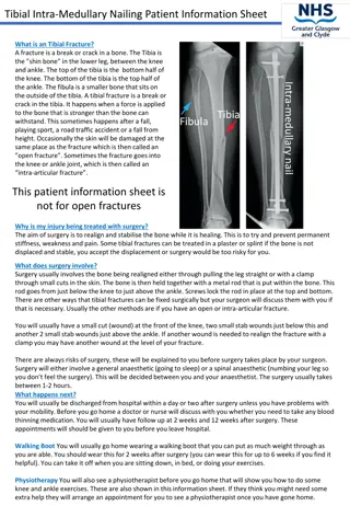
75 yo male s/p sinus surgery Luis Goity
Clinical Scenario 75 yom POD1 from b/l maxillary antrostomy, ethmoidectomy, sphenoidotomy, frontal sinusotomy with balloon dilation at OSH for chronic sinusitis. He developed HA, motor and sensory deficits in L foot and suffered two ground level falls after attempting to stand from sitting. Denies dizziness/ lightheadedness preceding the falls.
Findings Intracranial air representing pneumocephalus, with mass effect on brain parenchyma Leftward mild midline shift Discontinuous area of bone at right ethmoid roof, likely representing disruption after surgical exploration
Tension Pneumocephalus Arises from communication between extracranial and intracranial compartments Ball-valve mechanism leads to trapped intracranial air and increased ICP, leading to mass effect on parenchyma Must have neurologic symptoms from increased ICP Most common cause is trauma to frontal and ethmoid sinuses with associated dural defect, sinus infection, and ENT procedures CT gold standard requires only .55 mL air
Mt. Fuji Sign but should probably be called the Millennium Falcon sign
Next Steps Often requires surgical decompression, especially if there is significant widening of the interhemispheric space, which indicates more severe pneumocephalus than simple peaking of the frontal lobe tips Conservative treatment includes Fowler position, avoiding Valsalva, and osmotic diuretics to encourage absorption
Patient Hospital Course Did not require surgical decompression due to improvement in symptoms and small size of defect (<1 mm) F/u CT 5 days later demonstrated interval decrease in intracranial air and improvement of midline shift Discharged with precautions
Sources Loevner, Laurie. Brain Imaging Case Review Series. Mosby, Philadelphia; 2009. pp 271, 272. http://surgicalneurologyint.com/surgicalint- articles/review-of-the-management-of- pneumocephalus/ https://www.jtbgenesis.com/pic/tour/141231Mt. fuji.Mitsutouge.jpg http://i1119.photobucket.com/albums/k637/jait eastu/5%20Foot%20Millennium%20Falcon/Unde rside/ANH%20Underside/5ft%20ANH%20Bottom %20View.jpg