Understanding Lasers in Periodontal Therapy

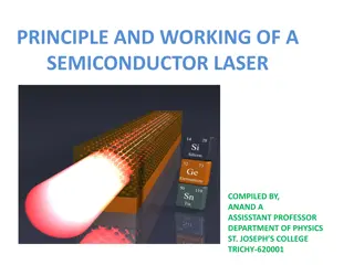
Laser technology in periodontal therapy harnesses the energy of photons to provide precise and effective treatment. Originally developed as MASER, the laser produces monochromatic, coherent light with unique properties such as directionality. Key components include the active medium, power supply, and optical resonator. Different laser systems are classified based on their operation modes. Laser delivery systems ensure accurate treatment delivery for improved oral health outcomes.

Download Presentation

Please find below an Image/Link to download the presentation.
The content on the website is provided AS IS for your information and personal use only. It may not be sold, licensed, or shared on other websites without obtaining consent from the author. Download presentation by click this link. If you encounter any issues during the download, it is possible that the publisher has removed the file from their server.
E N D
Presentation Transcript
LASERS IN PERIODONTAL THERAPY
INTRODUCTION LASER is an acronym for light amplification by stimulated emission of radiation. Light is a form of energy that travels in a wave and exists as a particle. This particle is called a photon. Photons are the smallest units of energy and are generally regarded as having zero mass or charge. When subjected to higher energy levels they undergo amplification, stimulation which consequently emits a monochromatic coherent energy photons called LASER.
HISTORICAL BACKGROUND Laser initially was called Maser . MASER is an acronym for Microwave Amplification by Stimulated Emission of Radiation. This principle was invented in 1958 by Schawlow and Townes of the Massachusetts Institute of Technology. Nobel Prize for development of laser was given to Towne, Basov, Prokhorov in 1964
MonochroMatic Light Mono chromatic light has a very narrow range of frequency and single wavelength (i.e. it is only made of light of one colour)
COHERENCE All the emitted photons are in phase with eachother and have identical peaks and valleys.
PARTS OF A LASER Active medium/Gain: Gas , solid, liquid suspended in an optical cavity. Power supply: external energy source- flash lamp/ electrical energy. Optical resonator: mirrors for amplification. Cooling system, Control system, Delivery system.
MAIN COMPONENTS OF LASER
CLASSIFICATON OF LASER SYSTEM
MODES OF OPERATION OF LASER Continuous wave Gated pulsed mode (Physical gating of beam) Free running pulsed mode (Property of the active medium)
THEORETICAL ZONES OF TISSUE CHANGE ASSOCIATED WITH SOFT TISSUE EXPOSURE TO LASER LIGHT
THEORETICAL ZONE OF TISSUE CHANGE ASSOCIATED WITH HARD DENTAL TISSUE EXPOSURE TO LASER LIGHT
CHARACTERISTICS OF LASER THERAPY Penetration depth of lasers Thermal side effects and hemostasis Tissue ablation Disinfection and detoxification effects Biostimulation (photobiomodulation)
TISSUE ABLATION Soft tissue ablation- CO2, Nd:YAG, Diode. Photo-thermal effect Hard tissue ablation- Er Group Thermo-mechanical effect
THERMAL SIDE EFFECTS AND HEMOSTASIS LASER COAGULATION DEPTH Nd:YAG 0.3-0.8mm (Perry et al and white et al) Diode 1mm (White et al) CO2 100-300 m (Arashiro et al) Er:YAG 10-50 m (Walsh et al) Er: YAG Hard tissue 5-30 m on cementum, dentin and bone
DISINFECTION AND DETOXIfiCATION EFFECTS Killing bacteria by photo-thermal effects . Bacteria are evaporated, destroyed or denatured by laser irradiation, resulting in their devitalization or inactivation. Nd:YAG laser exhibits selective absorption in pigments. Ablate or inactivate toxic substances, such as bacterial endotoxins (lipopolysaccharide).
BIOSTIMULATION (PHOTOBIOMODULATION) Biostimulatory effects may allow faster or more favorable wound healing, relative to conventional mechanical therapy. Photobiomodulation effects, such as promotion of cell proliferation and differentiation, anti-inflammatory effects positively modulate wound healing. Rate of wound healing is strongly influenced by the degree of remaining collateral thermal injuries. Low level co2 laser has a good tissue remodelling property.
WHAT DOES THE OPERATOR CONTROL
LASERS Advantages Disadvantages Hard tissuedamage (bone) High cost. Risk of pulpaldamage. No single wavelength can treat all diseases Hemostasis. Ablation. Detoxification. Bactericidal activity. Osseous tissueremoval and contouring easy with Er family
WHY LASERS IN PERIODONTICS Conventional methods LASER Effectivehemostasis. No sutures. (conceptof tissue welding). Topical anesthetic-some procedures. Fasterhealing. Minimal/no postoperative complications. Laser sterilizationof wound site. Laserbandage. Bleeding- surgicalfield. Suturing. Localanesthesia. Post-operativediscomfort. Healingtime. Post-operative complications. Infection. Periodontaldressings.
PERIODONTAL APPLICATIONS Calculus removal Soft tissue excision Incision Ablation Decontamination of root and implant surfaces Biostimulation Bacteria reduction Bone removal (osseous surgery). Removal of gingival pigmentation Reduction of periodontopathogenic black-pigmented bacteria. Management of dentinal hypersensitivity
GINGIVAL SOFT TISSUE PROCEDURES Advantages of lasers over conventional: Hemostasis. Ablation. Little wound contraction/ minimal scarring. Faster healing. Less post-operative discomfort. Less risk of damage to underlying structures as compared to cautery.
GINGIVAL SOFT TISSUE PROCEDURES Indications: Gingivectomy. Gingivoplasty. Frenectomy/ frenotomy. Vestibuloplasty. Operculectomy. Depigmentation. Lasers used; Diode. Nd YAG. Er YAG. Co2. Diode and Nd YAG:deep penetration . Er YAG, Co2:superficial action.
GINGIVAL SOFT TISSUE PROCEDURES Diode and Nd YAG: Co2 laser: Effective for cuttingand reshaping of soft tissue. Good hemostasis Greater thermal effects. Thicker coagulated layer. Rapid ablation of soft tissue. Good hemostasis. Effective even forthick tissue. Risk of charring- thermal damage.
GINGIVAL SOFT TISSUE PROCEDURES. Er YAG : Fine cutting can be done. Less hemostasis as compared to other lasers. Very less thermal damage: use with irrigation. Width of thermally affected layer: 5-20 microns Er YAG: Safer even in thin tissues. Useful to remove melanin and metal tattoos.
NON SURGICALTHERAPY Introduction: Primarily aimed at efficient removal of plaque and calculus and reduction of bacterial load, inflammation. Conventional therapy limitations: Incomplete removal of calculus. Incomplete elimination of inflamed pocket lining. Lasers used: Diode, Nd YAG, Er YAG, Co2 lasers.
SUBGINGIVAL CALCULUS DETECTION- UNIQUE APPLICATION FOR LASER Conventional method- tactile feel. Latest: Er YAG laser with fluorescent feedback system for calculus detection. Rationale: Difference in the fluorescence emission properties of calculus and dental hard tissue when subjected to irradiation with 655 nm diode laser.
ROOT SURFACEALTERATIONS Nd YAG laser surface pitting, craters, melting, carbonization of root surface. decrease in protein/mineral ratio, production of proteinby- products. NdYAG treated root surface not favorable for fibroblast attachment. Laser followed by SRP- restores the biocompatibility of root surface Diode laser Dry or saline moistened root surfaces- no detectable alterations. Blood coatedspecimens- charring
CO2 Laser Erbium family Carbonized layer on root surface. Cyanamide , cyanate ions- detected on the carbonized layer- FTIS method. Char layer periodontal soft attachment. Co2 laser contraindicated for root surface treatment in focused mode. No thermal effects such as cracking, fissuring. No major chemical or compositional change- on root cementum or dentin. Biocompatability of root surface: micro-irregularity offers better attachment to fibroblasts inhibits tissue
BACTERIAL REDUCTION The only two soft tissue wavelengths that currently meet the criterion of having a delivery system able to deliver laser energy efficiently and effectively to the periodontal pockets for nonsurgical periodontal therapy are Nd:YAG and diode. Well absorbed by melanin and hemoglobin and other chormophores present in periodontally diseased tissues. The laser energy is transmitted through water and poorly absorbed in hydroxyapitite.
Both of these wavelengths have been shown to be extremely effective against periodontal pathogens in vivo and in vitro diode laser revealed inflammation, and pockets through the elimination of bacteria. a bactericidal effect, helped reduce supported healing of the periodontal
SURGICAL POCKET THERAPY- LASERS Lasers used: Co2 and Erbium family Involves use of lasers for calculus removal, osseous surgery, de-toxification of the root surface and bone, granulation tissue removal Advantage of Laser: Better access in furcation areas, hemostasis, less post- operative discomfort, faster healing.
LASER-ASSISTED GUIDED TISSUE REGENERATION. a b c (a) Preoperative clinical condition; (b) clinical condition after laser-assisted scaling and root planing in conjunction with a de-epithelialization of the oral and sulcular epithelium for pocket reduction using an Nd: YAG laser; (c) stable long-term clinical condition (5 years postoperative)
IMPLANT THERAPY- MANAGEMENT OF PERI- IMPLANTITIS Peri-implantitis Management options- Conventional- plastic curettes and antibiotics. New option-Laser Rationale: Disinfection and de-contamination of implant surface. Granulation tissue removal. Lasers used: Diode, Co2, Erbium family. Lasers contra-indicated: Nd YAG (Implant damage).
LOW LEVEL LASERS Helium neonlaser Gallium aluminum arsenide diodelaser Gallium arsenide diodelaser Argon ionlaser Defocused Co2laser Defocused Nd:YAG laser
BIOSTIMULATION EFFECTS OF LOW LEVEL LASER Reduction of discomfort / pain Promotion of wound healing Bone regeneration Suppression of inflammatory process. Activation of gingival and periodontal ligament fibroblast, growth factor release Alteration of gene expression of inflammatory cytokines Photobiostimulaation
PHOTODYNAMIC THERAPY IN LASER Main objective of periodontal therapy: eliminate the deposits of bacteria. Conventional mechanical therapy: due to Anatomical complexity of root. Deep periodontal pockets. incomplete elimination
PRINCIPLES BEHIND PDT PS light 1O1O 2 2 TARGET CELL 1O 1O 2 2 1O1O 2 2 1O1O 22 Celldeath
DESTRUCTION OF PERIODONTOPATHOGENIC BACTERIA Polysaccharides oxygen. in biofilm are highly sensitive to singlet During inflammation reduced O2consumption change in pH growth of anaerobes
tissue blood flow and venous congestion PDT oxygenation of gingival tissues 21 47 % The activity of PDT ..been reported in vitro and in vivo . Greater bacterial reduction of S.sanguis numbers compared with A.actinomycetamcomitans.
HEALING AFTER LASERTHERAPY o Laser created wounds heal more quickly and produce less scar tissue than conventional scalpel surgery. o more initial tissue damage may result, and that wounds have less tensile strength during the early phase of healing.
LASER SAFETY Regulatory organizations: CDRH center for devices and radiologic health ANSI American National Standards Institute OSHA occupational safety and health administration Laser safety officer. Environment: warning signs, restricted access, reflective surface minimized. Laser use documentation. Training. Eye and tissue protection.
CLASSIFICATION OF LASER- BASED ON SAFETY Based on the potential of the primary laser beam or the reflected beam to cause biologic damage to the eye or skin. Four basic classes: Class I. Class II: a,b Class III: a, b Class IV.