Color Tests for Proteins and Amino Acids

This experiment involves employing color tests to detect specific amino acids like tyrosine and tryptophan in proteins. Various reagents are used to observe distinctive color changes, indicating the presence of these amino acids. Detailed procedures, materials, and results are documented for each test.

Download Presentation

Please find below an Image/Link to download the presentation.
The content on the website is provided AS IS for your information and personal use only. It may not be sold, licensed, or shared on other websites without obtaining consent from the author. Download presentation by click this link. If you encounter any issues during the download, it is possible that the publisher has removed the file from their server.
E N D
Presentation Transcript
CLS281 Exp.:2 Color Tests for Proteins and Amino Acids (Test for Specific Amino Acids) Done by: Daheeya Alenazi Haifa Altwaijry
1/Millon's Test: Used for detecting the presence of monohydroxybenzene derivatives (e.g: Tyrosine, Tyrosine derivatives, Phenol). *Millon's reagent is a solution of mercuric and mercurous ions in nitric and nitrous acids. *Phenolic group of tyrosine reacts with Millon's reagent and give a red color, which is due to a mercury salt of nitrated tyrosine. File:Phenol chemical structure.png
Materials: Water Egg albumin: protein contains tyrosine Salicylic acid:is a monohydroxybenzoic acid, a type of phenolic acid Tyrosine Gelatin: is a translucent, colorless, flavorless solid substance, derived from collagen. It contains tyrosine Phenylalanine : a.a Phenol
Procedure: Tube 1 Tube 2 Tube 3 Tube 4 Tube 5 Tube 6 Tube7 0.02% phenol 0.02% phenylalanin e 1% gelatin 0.02% tyrosine 0.02% salicylic acid 1% egg albumin H2O 2 ml 2 ml 2 ml 2 ml 2 ml 2 ml 2 ml Millon's Reagent 3-5 drops 3-5 drops 3-5 drops 3-5 drops 3-5 drops 3-5 drops 3-5 drops Incubate in boiling water bath for 2 min. Note the color formed
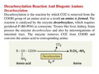
2/Hopkins-Cole Test: The hopkins-cole test is used to determine the presence of the amino acid tryptophan. Tryptophan has an indole nucleus which is responsible for the violet ring found at the junction between the two layers Tryptophan + Hopkins-Cole reagent purple ring *Hopkins-Cole reagent contain magnesium glyoxylate (Magnesium powder and Oxalic acid). Skeletal formula with numbering scheme Indole
sulfuric acid glyoxylic acid + indole ring purple ring Purple ring is formed by the reaction of glyoxylic acid ( CHO- COOH ) with the indole ring of tryptophan in presence of sulfuric acid (H2SO4)
Materials: Tyrosine Tryptophan Casein: protein contains tryptophan Gelatin: incomplete protein, because it lacks the essential amino acid tryptophan Egg albumin: protein contains tryptophan Water : as control
Procedure: 1 2 3 4 5 6 0.02% tyrosine 0.02% tryptophan 1% casein 1% gelatin 1%egg albumin H2O 2 ml 2 ml 2 ml 2 ml 2 ml 2 ml Hopkins-Cole Reagent 3 ml 3 ml 3 ml 3 ml 3 ml 3 ml *Concentrated H2SO4 5 ml 5 ml 5 ml 5 ml 5 ml 5 ml *Carefully add 5 ml of con. H2SO4 from buret by touching the tip of the stopcock to the inside wall of the test tube and allowing the acid to slowly drain down the tube so that the two liquids form separate layers. Add 5 ml of con. H2SO4 to each tube. Observe the color at the zone of contact of the two fluids. If no color appears, swirl the tube gently, but do not mix.
Results: Observe the color formed at the junction of the two liquids. If necessary, gently rotate the tube to develop the colored ring.
3/Lead Acetate Test: *Used for detecting the presence of Sulfur in Cystine , free cysteine and methionine *Proteins or peptides containing these amino acid heated with NaOH to split the sulfide group from amino acids. *Then, lead acetate reacts with free sulfide ions resulting in the formation of lead sulfide ( pbs ) a brown to black color.
Procedure: 1 2 3 4 0.02% methionine 0.02% cystine 1% egg albumin H2O 2 ml 2 ml 2 ml 2 ml 5% NaOH 5 ml 5 ml 5 ml 5 ml Lead Acetate Few crystals Few crystals Few crystals Few crystals Incubate in boiling water bath for 10 minutes with occasional mixing. Describe the color changes in each test tubes.
4/Sakaguchi Test: *This test is specific for the guanidine group of arginine (H2N-C-NH), but a non-amino acid like creatine which contains this group will also answer this test. http://upload.wikimedia.org/wikipedia/commons/thumb/f/f4/Arginin_-_Arginine.svg/200px-Arginin_-_Arginine.svg.png http://upload.wikimedia.org/wikipedia/commons/thumb/e/e5/Guanidine-2D-skeletal.png/100px-Guanidine-2D-skeletal.png guanidine group arginine *This test positive for all proteins containing arginine and for arginine itself. *When arginine reacts with -Naphthol and Sodium hypobromite (NaOBr) a red color result.
Red color is due to a reaction between the hypobromite and the NH2 group of the guanidino part of arginine
Materials: Urea: is an organic compound Creatine: contains guanidine group Arginine Gelatin: contains arginine Water: as control
Procedure: 0.02% urea 0.02% creatine 0.02% arginine 0.1% gelatin H2O 5 ml 5 ml 5 ml 5 ml 5 ml 10% NaOH 1 ml 1 ml 1 ml 1 ml 1 ml 0.02% -Naphthol 1 ml 1 ml 1 ml 1 ml 1 ml Mix, incubate at room temperature for 3 minutes NaOBr 4 drops 4 drops 4 drops 4 drops 4 drops Note the color formed
5/Folin's Test: *This test for free cystine, therefore protein or peptide must be first hydrolyzed by sodium carbonate and sodium sulfite. *Then, Folin's uric acid reagent (phosphotungstic acid) is added which is reduced to tungsten blue. *This can be used for the quantitative determination of cystine.
Procedure: 1 2 3 4 0.1% 1% egg albumin 0.1% cystine H2O methionine 1 ml 1 ml 1 ml 1 ml Sodium Carbonate (saturated) 7 ml 7 ml 7 ml 7 ml 20% Sodium Sulfite 3 ml 3 ml 3 ml 3 ml Incubate at R.T for 5 min. Uric acid reagent 1 ml 1 ml 1 ml 1 ml Note the color formed