Amino Acids: Disposal of Nitrogen
Overview of nitrogen metabolism with a focus on the disposal of nitrogen through amino acid catabolism. It explains the process of removing the α-amino groups from amino acids, the synthesis of urea, and the conversion of carbon skeletons to energy-producing pathways.
- nitrogen metabolism
- amino acid catabolism
- disposal of nitrogen
- α-amino groups
- urea synthesis
- carbon skeletons
- protein turnover

Download Presentation

Please find below an Image/Link to download the presentation.
The content on the website is provided AS IS for your information and personal use only. It may not be sold, licensed, or shared on other websites without obtaining consent from the author. Download presentation by click this link. If you encounter any issues during the download, it is possible that the publisher has removed the file from their server.
E N D
Presentation Transcript
NITROGEN METABOLISM Amino Acids: Disposal of Nitrogen
I. Overview Unlike fats and carbohydrates, amino acids are not stored by the body, i.e., no protein exists whose sole function is to maintain a supply of amino acids for future use. Therefore, amino acids must be obtained from the diet, synthesized de novo, or produced from normal protein degradation. Any amino acids in excess of the biosynthetic needs of the cell are rapidly degraded. The first phase of catabolism involves the removal of the -amino groups (usually by transamination and subsequent oxidative deamination), forming ammonia and the corresponding skeletons of amino acids. -keto acid the carbon
A portion of the free ammonia is excreted in the urine, but most is used in the synthesis of urea, which is quantitatively important route for disposing of nitrogen from the body. In the second phase of amino acid catabolism, the carbon skeletons of the -keto acids are converted to common intermediates of energy producing, metabolic pathways. These compounds can be metabolized to CO2 and water, glucose, fatty acids, or ketone bodies by the central pathways of metabolism. the most
II. Overall Nitrogen Metabolism Amino acid catabolism is part of the larger process of the metabolism of nitrogen- containing molecules. Nitrogen enters the body in a variety of compounds present important being amino acids contained in dietary protein. Nitrogen leaves the body as urea, ammonia, and other products derived from amino acid metabolism. The role of body transformations involves concepts: the amino acid pool and protein turnover. in food, the most proteins in important these two
A. Amino acid pool Free amino acids are present throughout the body, for example, in cells, blood, and the extracellular fluids. For the purpose of this discussion, envision all these amino acids as if they belonged to a single entity, called the amino acid pool. This pool is supplied by three sources: 1) amino acids provided by the degradation of body proteins, 2) amino acids derived from dietary protein, and 3) synthesis of nonessential amino acids from simple intermediates of metabolism (Figure 19.2). : In healthy, well fed individuals, the input to the amino acid pool is balanced by the output, that is, the amount of amino acids contained in the pool is constant. The amino acid pool is said to be in a steady state.
Conversely, the amino pool is depleted by three routes: 1) synthesis of body protein, 2) amino acids consumed as precursors of essential nitrogen-containing small molecules, and 3) conversion of amino acids to glucose, glycogen, fatty acids or CO2 . Although the amino acid pool is small (comprised of about 90 100 g of amino acids) in comparison with the amount of protein in the body (about 12 kg in a 70-kg man), it is conceptually at the center of whole- body nitrogen metabolism.
B. Protein turnover Most proteins in the body are constantly being synthesized permitting the removal of abnormal or unneeded proteins. For many proteins, regulation of synthesis determines the concentration of protein in the cell, with protein degradation assuming a minor role. For other proteins, the rate of synthesis is constitutive, that is, relatively constant, and cellular levels of the protein are controlled by selective degradation. and then degraded,
2. Protein degradation: There are two major enzyme systems responsible for degrading damaged or unneeded proteins: the energy-dependent mechanism, and the degradative enzymes (acid lysosomes. Proteasomes mainly degrade endogenous proteins, that is, proteins that were synthesized within the cell. Lysosomal enzymes degrade primarily extracellular proteins, such as plasma proteins that are taken into the cell by endocytosis, and cell-surface membrane proteins that are used endocytosis. ubiquitin-proteasome non-energy-dependent hydrolases) of the in receptor-mediated
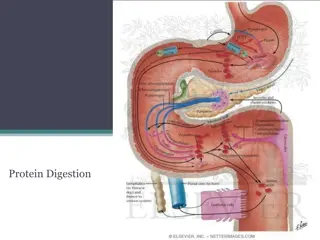
Digestion of Dietary Proteins : Most of the nitrogen in the diet is consumed in the form of protein, typically amounting to 70 100 g/day in the American diet (see Figure 19.2). Proteins are generally too large to be absorbed by the intestine. [Note: An example of an exception to this rule is that newborns can take up maternal antibodies in breast milk.] They must, therefore, be hydrolyzed to yield their constituent amino acids, which can be absorbed. Proteolytic enzymes responsible for degrading proteins are produced by three different organs: the stomach, the pancreas, and the small intestine (Figure 19.4).
Figure 19.4 Digestion of dietary proteins by the proteolytic enzymes of the gastrointestinal
A.Digestion of proteins by gastric secretion The digestion of proteins begins in the stomach, which secretes gastric juice a unique solution containing hydrochloric acid and the proenzyme, pepsinogen. 1. Hydrochloric acid: Stomach acid is too dilute (pH 2 3) to hydrolyze proteins. The acid functions instead to kill some bacteria and to denature proteins, thus making them more susceptible hydrolysis by proteases. 2. Pepsin: This acid-stable endopeptidase is secreted by the serous cells of the stomach as an inactive zymogen (or proenzyme), pepsinogen. In general, zymogens contain extra amino acids in their sequences, which prevent catalytically active. : to subsequent them from being
[Note: Removal of these amino acids permits the proper folding required for an active enzyme.]. Pepsinogen is activated to pepsin, either by HCl, or autocatalytically by other pepsin molecules that have already been activated. Pepsin releases peptides and a few free amino acids from dietary proteins. B. Digestion of proteins by pancreatic enzymes On entering the small intestine, large polypeptides produced in the stomach by the action of pepsin are further cleaved to oligopeptides and amino acids by a group of pancreatic proteases. :
1. Specificity: - Each of these enzymes has a different specificity for the amino acid R-groups adjacent to the susceptible peptide bond (Figure 19.5). - For example, trypsin cleaves only when the carbonyl group of the peptide bond is contributed by arginine or lysine. - These enzymes, like pepsin described above, are synthesized and secreted as inactive zymogens. 2. Release of zymogens: The release and activation of the pancreatic zymogens is mediated by the secretion of cholecystokinin and secretin, two polypeptide hormones of the digestive tract .
3. Activation of zymogens: Enteropeptidase (formerly called enterokinase) an enzyme synthesized by and present on the luminal surface of intestinal mucosal cells of the brush border membrane converts the trypsinogen to trypsin by removal of a hexapeptide from the NH2-terminus of trypsinogen. Trypsin subsequently converts molecules to trypsin by cleaving a limited number of specific peptide bonds in the zymogen. Enteropeptidase thus unleashes a cascade of proteolytic activity, because trypsin is the common activator of all the pancreatic zymogens (see Figure 19.5). pancreatic zymogen other trypsinogen
4. Abnormalities in protein digestion: In individuals with a deficiency in pancreatic secretion (for example, due to chronic pancreatitis, cystic fibrosis, or surgical removal of the pancreas), the digestion and absorption of fat and protein is incomplete. This results in the abnormal appearance of lipids (called steatorrhea) and undigested protein in the feces.
Figure 19.5 Cleavage of dietary protein by proteases from the pancreas. The peptide bonds susceptible to hydrolysis are shown for each of the five major pancreatic proteases. [Note: Enteropeptidaseis synthesized in the intestine.]
C. Digestion of oligopeptides by enzymes of the : small intestine The aminopeptidase an cleaves the N-terminal residue from oligopeptides to produce free amino acids and smaller peptides. luminal surface of the intestine that contains repeatedly exopeptidase
D. dipeptides: Free amino acids are taken into the enterocytes up by a Na+-linked secondary transport system. Di- and tripeptides, however, are taken up by a H+- linked transport system. hydrolyzed in the cytosol to amino acids before being released into the portal system. Thus, only free amino acids are found in the portal vein after a meal containing protein. These amino acids are either metabolized by the liver or released into the general circulation. [Note:Branched-chain amino examples of amino acids that are not metabolized by the liver, but instead are sent from the liver into the blood.] Absorption of amino acids and There, the peptides are acids are important
: Transport of Amino Acids into Cells The extracellular fluids is significantly lower than that within the cells of the body. This concentration gradient is maintained because active transport systems, driven by the hydrolysis of ATP, are required for movement of amino acids from the extracellular space into cells. At least seven different transport systems are known that have overlapping specificities for different amino acids. concentration of free amino acids in the
The small intestine and the proximal tubule of the kidney have common transport systems for amino acid uptake; therefore, a defect in any one of these systems results in an inability to absorb particular amino acids into the gut and into the kidney tubules. For example, one system is responsible for the uptake of cystine and the dibasic amino acids, ornithine, arginine, and lysine (represented as COAL ). In the inherited disorder cystinuria, this carrier system is defective, and all four amino acids appear in the urine . Cystinuria occurs at a frequency individuals, making it one of the most common inherited diseases, and the most common genetic error of amino acid transport. of 1 in 7,000
The disease expresses itself clinically by the precipitation of cystine to form kidney stones (calculi), which can block the urinary tract. Oral hydration is an important part of treatment for this disorder. [Note: Defects in the transport of tryptophan (and other neutral amino acids) can result in Hartnup disorder and pellagra-like (see p. 380) dermatologic and neurologic symptoms.
Essential and non essential amino acid essential Non essential *Arginine Alanine Histidine Asparagine Isoleucine Aspartate Leucine Cysteine Lysine Glutamate Methionine* Glutamine Phenylalanine* Glycine Threonine Proline Tryptophan Serine valine Tyrosine
Central role of glutamate Four of the amino acids : glutamate, aspartate, alanine and glutamine are present in cells at much higher concentration than the others 16 . All four have major metabolic function in addition to their roles in proteins but glutamate occupies the prime position .
Alanin Glutamic e acid Glutamine Aspartic acid
Function of four amino acids Glutamate and aspartate function as excitatory neurotransmitters in the CNS Glutamate is partly responsible for the flavor of food . Glutamine also occupies especial position in amino acids breakdown ,and most of the nitrogen from dietary protein is ultimately excreted from the body via glutamate pool.
Glutamate is a special because it is chemically related to 2- oxoglutarate which is a key intermediate in the citric acid cycle . Glutamate can be reversibly into oxoglutarate by transaminases or by glutamate dehydrogenase. Glutamate can be converted into glutamine , an important nitrogen carrier ,and the most common free amino acid in human blood plasma.
Transamination: the funneling of amino groups to glutamate The first step in the catabolism of most amino acids is the transfer of their -amino group to -ketoglutarate. The products are an -keto acid (derived from the original amino acid) and glutamate. -Ketoglutarate metabolism by accepting the amino groups from other amino acids, thus becoming glutamate. plays a pivotal role in amino acid Glutamate produced by transamination can be oxidatively deaminated (see below), or used as an amino group donor in the synthesis of nonessential amino acids.
R2 R2 R1 R1 CH- NH3 C=O CH- NH3 C=O + = COO- COO- COO- COO- Amino acid1 Keto acid2 Keto acid1 Amino acid2
This transfer of amino groups from one carbon skeleton to another is catalyzed by a family of enzymes called aminotransferases (formerly called transaminases). These enzymes are found in the cytosol and mitochondria of cells throughout the body especially those of the liver, kidney, intestine, and muscle. All amino acids, with the exception of lysine and threonine, participate in transamination at some point in their catabolism. These two amino acids lose their -amino groups by deamination.
Aspartate aminotransferase (AST): AST formerly called glutamate-oxaloacetate transaminase, AST is an exception to the rule that aminotransferases funnel amino groups to form glutamate. During amino acid catabolism, AST transfers amino groups from glutamate to oxaloacetate, forming aspartate, which is .used as a source of nitrogen in the urea cycle .
Alanine aminotransferase (ALT): Formerly transaminase, ALT is present in many tissues. called glutamate-pyruvate The enzyme catalyzes the transfer of the amino group of alanine to -ketoglutarate, resulting in the formation of pyruvate and glutamate. The reaction is readily reversible. However, during amino acid catabolism, this enzyme (like most aminotransferases) direction of glutamate synthesis. functions in the Thus, glutamate, in effect, acts as a collector of . nitrogen from alanine
Reactions catalyzed during amino acid catabolism. A. Alanine aminotransferase (ALT). B. Aspartate aminotransferase (AST).
Diagnostic value of plasma aminotransferases: Aminotransferases are normally intracellular enzymes, with the low levels found in the plasma representing the release of cellular contents during normal cell turnover. The presence of elevated plasma levels of aminotransferases indicates damage to cells rich in these enzymes. For example, physical trauma or a disease process can cause cell lysis, resulting in release of intracellular enzymes into the blood. Two aminotransferases AST and ALT are of particular diagnostic value when they are found in the plasma.
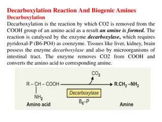
Glutamate dehydrogenase: the oxidative deamination of amino acids In transfer amino groups, oxidative deamination by glutamate dehydrogenase results in the liberation of the amino group as free ammonia . These reactions occur primarily in the liver and kidney. They provide -keto acids that can enter the central pathway of energy ammonia, which is a source of nitrogen in urea synthesis. contrast to transamination reactions that metabolism, and
1-Glutamate dehydrogenase: As described above, the amino groups of most amino acids are ultimately funneled to glutamate by means of transamination with -ketoglutarate. Glutamate is unique in that it is the only amino acid that undergoes rapid oxidative deamination a reaction catalyzed by glutamate dehydrogenase . Therefore, the sequential action of transamination (resulting in the collection of amino groups from other amino acids onto -ketoglutarate to produce glutamate) and the oxidative deamination of that glutamate (regenerating -ketoglutarate) provide a pathway whereby the amino groups of most amino acids can be released as ammonia.
2. D-Amino acid oxidase: D-Amino acids are found in plants and in the cell walls of microorganisms, but are not used in the synthesis of mammalian proteins. D-Amino acids are, however, present in the diet, and are efficiently metabolized by the kidney and liver. D-Amino acid oxidase is an FAD-dependent peroxisomal enzyme that catalyzes the oxidative deamination of these amino acid isomers. The resulting -keto acids can enter the general pathways of amino acid metabolism, and be reaminated to L-isomers, or catabolized for energy.
Transport of ammonia to the liver Two mechanisms are available in humans for the transport of ammonia from the peripheral tissues to the liver for its ultimate conversion to urea. The first, found in most tissues, uses glutamine synthetase to combine ammonia with glutamate to form glutamine a nontoxic transport form of ammonia . The glutamine is transported in the blood to the liver where it is cleaved by glutaminase to produce glutamate and free ammonia
The second transport mechanism, used primarily by muscle, involves transamination of pyruvate (the end product of aerobic glycolysis) to form alanine . Alanine is transported by the blood to the liver, where it is converted to pyruvate, again by transamination. In the liver, the pathway of gluconeogenesis can use the pyruvate to synthesize glucose, which can enter the blood and be used by muscle a pathway called the glucose-alanine cycle.
Urea Cycle Urea is the major disposal form of amino groups derived from amino acids, and accounts for about 90% of the nitrogen-containing components of urine. One nitrogen of the urea molecule is supplied by free NH3, and the other nitrogen by aspartate. [Note: Glutamate is the immediate precursor of both ammonia (through oxidative deamination by glutamate dehydrogenase) and aspartate transamination of oxaloacetate by AST).] The carbon and oxygen of urea are derived from CO2. Urea is produced by the liver, and then is transported in the blood to the kidneys for excretion in the urine. nitrogen (through
Reactions of the cycle The first two reactions leading to the synthesis of urea occur in the mitochondria, whereas the remaining cycle enzymes are located in the cytosol . Formation of carbamoyl phosphate: Formation of carbamoyl phosphate by carbamoyl phosphate synthetase I is driven by cleavage of two molecules of ATP. Ammonia incorporated into carbamoyl phosphate is provided primarily by the oxidative deamination of glutamate by mitochondrial glutamate dehydrogenase . Ultimately, the nitrogen atom derived from this ammonia becomes one of the nitrogens of urea.
Carbamoyl phosphate synthetase I requires N- acetylglutamate as a positive allosteric activator . [Note: Carbamoyl phosphate synthetase II participates in the biosynthesis of pyrimidines .It does not require N- acetylglutamate, and occurs in the cytosol.]
2. Formation of citrulline: Ornithine and citrulline are basic amino acids that participate in the urea cycle. [Note: They are not incorporated into cellular proteins, because there are no codons for these amino acids.] Ornithine is regenerated with each turn of the urea cycle, much in the same way that oxaloacetate is regenerated by the reactions of the citric acid cycle . The release of the high-energy carbamoyl phosphate as inorganic phosphate drives the reaction in the forward direction. The reaction product, citrulline, is transported to the cytosol. phosphate of
3. Synthesis of argininosuccinate: Citrulline condenses with aspartate to form argininosuccinate. The -amino group of aspartate provides the second nitrogen that is ultimately incorporated into urea. The formation of argininosuccinate is driven by the cleavage of ATP to adenosine monophosphate (AMP) and pyrophosphate. This is the third and final molecule of ATP consumed in the formation of urea.
4. Cleavage of argininosuccinate: Argininosuccinate is cleaved to yield arginine and fumarate. The arginine formed by this reaction serves as the immediate precursor of urea. Fumarate produced in the urea cycle is hydrated to malate, providing a link with several metabolic pathways. For example, the malate can be transported into the mitochondria via the malate shuttle and reenter the tricarboxylic acid cycle. Alternatively, cytosolic malate can be oxidized to oxaloacetate, which can be converted to aspartate or glucose .
5. Cleavage of arginine to ornithine and urea: Arginase cleaves arginine to ornithine and urea, and occurs almost exclusively in the liver. Thus, whereas other tissues, such as the kidney, can synthesize arginine by these reactions, only the liver can cleave arginine and, thereby, synthesize urea.
Fate of urea: Urea diffuses from the liver, and is transported in the blood to the kidneys, where it is filtered and excreted in the urine. A portion of the urea diffuses from the blood into the intestine, and is cleaved to CO2 and NH3 by bacterial urease. This ammonia is partly lost in the feces, and is partly reabsorbed into the blood. In patients with kidney failure, plasma urea levels are elevated, promoting a greater transfer of urea from blood into the gut. The intestinal action of urease on this urea becomes a clinically important source of ammonia, contributing to the hyperammonemia often seen in these patients. Oral administration of neomycin reduces the number of intestinal bacteria responsible for this NH3 production.


:")
:")