Understanding Digestion and Absorption in the Gastrointestinal Tract


DIGESTION AND ABSORPTION
PROFESSOR DR.NOORI MOHAMMED LUAIBI
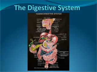
Digestion and Absorption in the Gastrointestinal Tract
The major foods on which the body lives (with the exception of small quantities of substances such as vitamins
and minerals) can be classified as carbohydrates, fats,and proteins. They generally cannot be absorbed in their
natural forms through the gastrointestinal mucosa, and for this reason, they are useless as nutrients without
preliminary digestion. This chapter discusses the processes by which carbohydrates, fats, and proteins are
digested into small enough compounds for absorption and the mechanisms by which the digestive end products,
as well as water, electrolytes, and other substances, are absorbed.
DIGESTION OF THE VARIOUS FOODS BY HYDROLYSIS
Hydrolysis of Carbohydrates.
Almost all the carbohydrates of the diet are either large polysaccharides or disaccharides, which
are combinations of monosaccharides bound to one another by condensation. This phenomenon
means that a hydrogen ion (H+ ) has been removed from one of the monosaccharides, and a
hydroxyl ion (OH− ) has been removed from the next one. The two monosaccharides then
combine with each other at these sites of removal, and the hydrogen and hydroxyl ions combine
to form water (H2O). When carbohydrates are digested, this process is reversed and the
carbohydrates are converted into monosaccharides. Specific enzymes in the digestive juices of
the gastrointestinal tract return the hydrogen and hydroxyl ions from water to the polysaccharides
and thereby separate the monosaccharides from each other. This process, called hydrolysis, is
the following (in which R″-R′ is a disaccharide):
R′′ − R′ + H2O
Digestive /Enzyme
R ′′ O H + R ′H
Hydrolysis of Fats
.
Almost the entire fat portion of the diet consists of triglycerides (neutral fats), which are combinations of three fatty acid
molecules condensed with a single glycerol molecule. During condensation, three molecules of water are removed. Hydrolysis
(digestion) of the triglycerides consists of the reverse process: the fat-digesting enzymes return three molecules of water to
the triglyceride molecule and thereby split the fatty acid molecules away from the glycerol.
Hydrolysis of Proteins
.
Proteins are formed from multiple amino acids that are bound together by peptide linkages. At each linkage, a hydroxyl ion
has been removed from one amino acid and a hydrogen ion has been removed from the succeeding one; thus, the successive
amino acids in the protein chain are also bound together by condensation, and digestion occurs by the reverse effect:
hydrolysis. That is, the proteolytic enzymes return hydrogen and hydroxyl ions from water molecules to the protein molecules
to split them into their constituent amino acids. Therefore, the chemistry of digestion is simple because, in the case of all three
major types of food, the same basic process of hydrolysis is involved. The only difference lies in the types of enzymes
required to promote the hydrolysis reactions for each type of food. All the digestive enzymes are proteins.
DIGESTION OF CARBOHYDRATES
Carbohydrate Foods of the Diet
.
Only three major sources of carbohydrates exist in the normal human diet. They are sucrose, which is the disaccharide known
popularly as cane sugar; lactose, which is a disaccharide found in milk; and starches, which are large polysaccharides
present in almost all non-animal foods, particularly in potatoes and different types of grains. Other carbohydrates ingested
to a slight extent are amylose, glycogen, alcohol, lactic acid, pyruvic acid, pectins, dextrins, and minor quantities of
carbohydrate derivatives in meats. The diet also contains a large amount of cellulose, which is a carbohydrate. However,
enzymes capable of hydrolyzing cellulose are not secreted in the human digestive tract. Consequently, cellulose cannot be
considered a food for humans.
Digestion of Carbohydrates Begins in the Mouth and Stomach
.
When food is chewed, it is mixed with saliva, which contains the digestive enzyme ptyalin (an α-amylase)
secreted mainly by the parotid glands. This enzyme hydrolyzes starch into the disaccharide maltose and other
small polymers of glucose that contain three to nine glucose molecules, as shown in Figure 66-1. However, the
food remains in the mouth only a short time, so probably not more than 5 percent of all the starches will have
become hydrolyzed by the time the food is swallowed. Starch digestion sometimes continues in the body and
fundus of the stomach for as long as 1 hour before the food becomes mixed with the stomach secretions.
Activity of the salivary amylase is then blocked by acid of the gastric secretions because the amylase is
essentially inactive as an enzyme once the pH of the medium falls below about 4.0. Nevertheless, on average,
before food and its accompanying saliva become completely mixed with the gastric secretions, as much as 30 to
40 percent of the starches will have been hydrolyzed, mainly to form maltose.
DIGESTION OF CARBOHYDRATES IN THE SMALL INTESTINE
Digestion by Pancreatic Amylase
.
Pancreatic secretion, like saliva, contains a large quantity of α-amylase that is almost identical in its function to the α-
amylase of saliva but is several times as powerful. Therefore, within 15 to 30 minutes after the chyme empties from
the stomach into the duodenum and mixes with pancreatic juice, virtually all the carbohydrates will have become
digested. In general, the carbohydrates are almost totally converted into maltose and/or other small glucose polymers
before passing beyond the duodenum or upper jejunum.
Hydrolysis of Disaccharides and Small Glucose Polymers Into Monosaccharides by
Intestinal Epithelial Enzymes.
The enterocytes lining the villi of the small intestine contain four enzymes (lactase, sucrase, maltase, and
α-
dextrinase),
which are capable of splitting the disaccharides lactose, sucrose, and maltose, plus other small glucose polymers, into their
constituent monosaccharides. These enzymes are located in the enterocytes covering the intestinal microvilli brush border,
so the disaccharides are digested as they come in contact with these enterocytes. Lactose splits into a molecule of
galactose and a molecule of glucose. Sucrose splits into a molecule of fructose and a molecule of glucose. Maltose and
other small glucose polymers all split into multiple molecules of glucose. Thus, the final products of carbohydrate digestion
are all monosaccharides. They are all water soluble and are absorbed immediately into the portal blood. In the ordinary
diet, which contains far more starches than all other carbohydrates combined, glucose represents more than 80 percent of
the final products of carbohydrate digestion, and galactose and fructose each seldom represent more than 10 percent.
The major steps in carbohydrate digestion are summarized in Figure 66-1
DIGESTION OF PROTEINS
Proteins of the Diet. Dietary proteins are chemically long chains of amino acids bound together by peptide linkages. A
typical linkage is the following:
The characteristics of each protein are determined by the types of amino acids in the protein molecule and by the sequential
arrangements of these amino acids.
Digestion of Proteins in the Stomach
.
Pepsin, an important peptic enzyme of the stomach, is most active at a pH of 2.0 to 3.0 and is inactive at a pH above about
5.0. Consequently, for this enzyme to cause digestion of protein, the stomach juices must be acidic. As explained in Chapter
65, the gastric glands secrete a large quantity of hydrochloric acid. This hydrochloric acid is secreted by the parietal (oxyntic)
cells in the glands at a pH of about 0.8, but by the time it is mixed with the stomach contents and with secretions from the non-
oxyntic glandular cells of the stomach, the pH then averages around 2.0 to 3.0, a highly favorable range of acidity for
pepsin activity. One of the important features of pepsin digestion is its ability to digest the protein collagen, an albuminoid
type of protein that is affected little by other digestive enzymes. Collagen is a major constituent of the intercellular connective
tissue of meats; therefore, for the digestive enzymes to penetrate meats and digest the other meat proteins, it is necessary
that the collagen fibers be digested. Consequently, in persons who lack pepsin in the stomach juices, the ingested meats are
less well penetrated by the other digestive enzymes and, therefore, may be poorly digested. As shown in Figure 66-2, pepsin
only initiates the process of protein digestion, usually providing only 10 to 20 percent of the total protein digestion to convert
the protein to proteoses, peptones, and a few polypeptides. This splitting of proteins occurs as a result of hydrolysis at the
peptide linkages between amino acids.
Figure 66-2. Digestion of proteins.
Most Protein Digestion Results From Actions of Pancreatic Proteolytic Enzymes
.
Most protein digestion occurs in the upper small intestine, in the duodenum and jejunum, under the influence of
proteolytic enzymes from pancreatic secretion. Immediately upon entering the small intestine from the stomach, the
partial breakdown products of the protein foods are attacked by the major proteolytic pancreatic enzymes trypsin,
chymotrypsin, carboxypolypeptidase, and elastase, as shown in Figure 66-2. Both trypsin and chymotrypsin split
protein molecules into small polypeptides; carboxypolypeptidase then cleaves individual amino acids from the
carboxyl ends of the polypeptides. Proelastase, in turn, is converted into elastase, which then digests elastin fibers that
partially hold meats together. Only a small percentage of the proteins are digested all the way to their constituent
amino acids by the pancreatic juices. Most remain as dipeptides and tripeptides.
Digestion of Peptides by Peptidases in the Enterocytes That Line the Small Intestinal Villi
.
The last digestive stage of the proteins in the intestinal lumen is achieved by the enterocytes that line the villi of the
small intestine, mainly in the duodenum and jejunum.
These cells have a brush border that consists of hundreds of microvilli projecting from the surface of each cell. In the
membrane of each of these microvilli are multiple peptidases that protrude through the membranes to the exterior, where
they come in contact with the intestinal fluids. Two types of peptidase enzymes are especially important,
aminopolypeptidase and several dipeptidases. They split the remaining larger polypeptides into tripeptides and
dipeptides and a few into amino acids. The amino acids, dipeptides, and tripeptides are easily transported through the
microvillar membrane to the interior of the enterocyte. Finally, inside the cytosol of the enterocyte are multiple other
peptidases that are specific for the remaining types of linkages between amino acids. Within minutes, virtually all the last
dipeptides and tripeptides are digested to the final stage to form single amino acids, which then pass on through to the
other side of the enterocyte and thence into the blood. More than 99 percent of the final protein digestive products that
are absorbed are individual amino acids, with only rare absorption of peptides and very rare absorption of whole
protein molecules. Even these few absorbed molecules of whole protein can sometimes cause serious allergic or
immunologic disturbances.
DIGESTION OF FATS
Fats of the Diet
.
By far the most abundant fats of the diet are the neutral fats, also known as triglycerides, each molecule of which is
composed of a glycerol nucleus and three fatty acid side chains, as shown in Figure 66-3. Neutral fat is a major
constituent in food of animal origin but much less so in food of plant origin. Small quantities of phospholipids, cholesterol,
and cholesterol esters are also present in the usual diet. The phospholipids and cholesterol esters contain fatty acid and
therefore can be considered fats. Cholesterol is a sterol compound that contains no fatty acid, but it does exhibit some of
the physical and chemical characteristics of fats; in addition, it is derived from fats and is metabolized similarly to fats.
Therefore, cholesterol is considered, from a dietary point of view, to be a fat.
Digestion of Fats Occurs Mainly in the Small Intestine.
A small amount of triglycerides is digested in the stomach by lingual lipase secreted by lingual glands in the mouth and swallowed
with the saliva. This amount of digestion is less than 10 percent and is generally unimportant. Instead, essentially all fat digestion
occurs in the small intestine as follows.
The First Step in Fat Digestion Is Emulsification by Bile Acids and Lecithin
.
The first step in fat digestion is to physically break the fat globules into small sizes so that the water-soluble digestive enzymes can
act on the globule surfaces. This process is called emulsification of the fat, and it begins by agitation in the stomach to mix the fat
with the products of stomach digestion. Most of the emulsification then occurs in the duodenum under the influence of bile, the
secretion from the liver that does not contain any digestive enzymes. However, bile does contain a large quantity of bile salts, as well
as the phospholipid lecithin. Both of these substances, but especially the lecithin, are extremely important for emulsification of the fat.
The polar parts (i.e., the points where ionization occurs in water) of the bile salts and lecithin molecules are highly soluble in water,
whereas most of the remaining portions of their molecules are highly soluble in fat. Therefore, the fat-soluble portions of these liver
secretions dissolve in the surface layer of the fat globules, with the polar portions projecting. The polar projections, in turn, are
soluble in the surrounding watery fluids, which greatly decreases the interfacial tension of the fat and makes it soluble as well.
When the interfacial tension of a globule of nonmiscible fluid is low, this non-miscible fluid, upon agitation, can be broken up into
many tiny particles far more easily than it can when the interfacial tension is great. Consequently, a major function of the bile salts
and lecithin (especially the lecithin) in the bile is to make the fat globules readily fragmentable by agitation with the water in the
small bowel. This action is the same as that of many detergents that are widely used in household cleaners for removing grease.
Each time the diameters of the fat globules are significantly decreased as a result of agitation in the small intestine, the total
surface area of the fat increases manyfold. Because the average diameter of the fat particles in the intestine after emulsification
has occurred is less than 1 micrometer, this represents an increase of as much as 1000-fold in total surface areas of the fats caused
by the emulsification process. The lipase enzymes are water-soluble compounds and can attack the fat globules only on their
surfaces. Consequently, this detergent function of bile salts and lecithin is very important for digestion of fats.
Triglycerides Are Digested by Pancreatic Lipase
.
By far the most important enzyme for digestion of the triglycerides is pancreatic lipase, present in enormous quantities in pancreatic
juice, enough to digest within 1 minute all triglycerides that it can reach. The enterocytes of the small intestine contain additional
lipase, known as enteric lipase, but it is usually not needed.
End Products of Fat Digestion Are Free Fatty Acids.
Most of the triglycerides of the diet are split by pancreatic lipase into free fatty acids and 2-monoglycerides, as shown in Figure
66-4.
Bile Salts Form Micelles That Accelerate Fat Digestion. The hydrolysis of triglycerides is a highly reversible process; therefore,
accumulation of monoglycerides and free fatty acids in the vicinity of digesting fats quickly blocks further digestion. However, the
bile salts play the additional important role of removing the monoglycerides and free fatty acids from the vicinity of the digesting
fat globules almost as rapidly as these end products of digestion are formed. This process occurs in the following way. When bile
salts are of a high enough concentration in water, they have the propensity to form micelles, which are small spherical, cylindrical
globules 3 to 6 nanometers in diameter composed of 20 to 40 molecules of bile salt. These micelles develop because each bile salt
molecule is composed of a sterol nucleus that is highly fat-soluble and a polar group that is highly water-soluble. The sterol nucleus
encompasses the fat digestate, forming a small fat globule in the middle of a resulting micelle, with polar groups of bile salts
projecting outward to cover the surface of the micelle. Because these polar groups are negatively charged, they allow the entire
micelle globule to dissolve in the water of the digestive fluids and to remain in stable solution until the fat is absorbed into the
blood. The bile salt micelles also act as a transport medium to carry the monoglycerides and free fatty acids, both of which would
otherwise be relatively insoluble, to the brush borders of the intestinal epithelial cells. There the monoglycerides and free fatty acids
are absorbed into the blood, as discussed later, but the bile salts are released back into the chyme to be used again and again for
this “ferrying” process.
Digestion of Cholesterol Esters and Phospholipids
.
Most cholesterol in the diet is in the form of cholesterol esters, which are combinations of free cholesterol and one molecule of fatty
acid. Phospholipids also contain fatty acid within their molecules. Both the cholesterol esters and the phospholipids are hydrolyzed
by two other lipases in the pancreatic secretion that free the fatty acids—the enzyme cholesterol ester hydrolase to hydrolyze the
cholesterol ester, and phospholipase A2 to hydrolyze the phospholipid. The bile salt micelles play the same role in “ferrying” free
cholesterol and phospholipid molecule digestates that they play in ferrying monoglycerides and free fatty acids. Indeed, essentially
no cholesterol is absorbed without this function of the micelles.
BASIC PRINCIPLES OF GASTROINTESTINAL ABSORPTION
The following paragraphs present specialized applications of these transport processes during gastrointestinal absorption
ANATOMICAL BASIS OF ABSORPTION
The total quantity of fluid that must be absorbed each day by the intestines is equal to the ingested fluid (about 1.5 liters) plus
that secreted in the various gastrointestinal secretions (about 7 liters), which comes to a total of 8 to 9 liters. All but about 1.5 liters
of this fluid is absorbed in the small intestine, leaving only 1.5 liters to pass through the ileocecal valve into the colon each day.
The stomach is a poor absorptive area of the gastrointestinal tract because it lacks the typical villus type of absorptive membrane,
and also because the junctions between the epithelial cells are tight junctions. Only a few highly lipid-soluble substances, such as
alcohol and some drugs like aspirin, can be absorbed in small quantities.
Folds of Kerckring, Villi, and Microvilli Increase the Mucosal Absorptive Area by Nearly 1000-Fold
Figure 66-5
demonstrates the absorptive surface of the small intestinal mucosa, showing many folds called valvulae conniventes (or
folds of Kerckring), which increase the surface area of the absorptive mucosa about threefold. These folds extend circularly most
of the way around the intestine and are especially well developed in the duodenum and jejunum, where they often protrude up to
8 millimeters into the lumen. Also located on the epithelial surface of the small intestine all the way down to the ileocecal valve are
millions of small villi. These villi project about 1 millimeter from the surface of the mucosa, as shown on the surfaces of the valvulae
conniventes in
Figure 66-5
and in individual detail in
Figure 66-6
. The villi lie so close to one another in the upper small intestine that
they touch in most areas, but their distribution is less profuse in the distal small intestine. The presence of villi on the mucosal surface
enhances the total absorptive area another 10-fold. Finally, each intestinal epithelial cell on each villus is characterized by a brush
border, consisting of as many as 1000 microvilli that are 1 micrometer in length and 0.1 micrometer in diameter and protrude into
the intestinal chyme. These microvilli are shown in the electron micrograph in
Figure
66-7.
This brush border increases the surface area
exposed to the intestinal materials at least another 20-fold.
Figure 66-6
. Functional organization of the villus.
A
, Longitudinal section.
B
, Cross section showing a
basement membrane beneath the epithelial cells and a brush border at the other ends of these cells.
Thus, the combination of the folds of Kerckring, the villi, and the microvilli increases the total absorptive area of the mucosa
perhaps 1000-fold, making a tremendous total area of 250 or more square meters for the entire small intestine—about the
surface area of a tennis court.
Figure 66-6A
shows in longitudinal section the general organization of the villus, emphasizing
(1)
the
advantageous arrangement of the vascular system for absorption of fluid and dissolved material into the portal blood and
(2)
the
arrangement of the “central lacteal” lymph vessel for absorption into the lymph.
Figure 66-6B
shows a cross section of the villus, and
Figure 66-7
shows many small pinocytic vesicles, which are pinched-off portions of infolded enterocyte membrane forming vesicles of
absorbed fluids that have been entrapped. Small amounts of substances are absorbed by this physical process of pinocytosis.
Extending from the epithelial cell body into each microvillus of the brush border are multiple actin filaments that contract
rhythmically to cause continual movement of the microvilli, keeping them constantly exposed to new quantities of intestinal fluid.
ABSORPTION IN THE SMALL INTESTINE
Absorption from the small intestine each day consists of several hundred grams of carbohydrates, 100 or moregrams of fat, 50 to
100 grams of amino acids, 50 to 100 grams of ions, and 7 to 8 liters of water. The absorptive capacity of the normal small
intestine is far greater than this; each day as much as several kilograms of carbohydrates, 500 grams of fat, 500 to 700 grams
of proteins, and 20 or more liters of water can be absorbed. The large intestine can absorb still more water and ions, although it
can absorb very few nutrients.
ISOSMOTIC ABSORPTION OF WATER
Water is transported through the intestinal membrane entirely by diffusion. Furthermore, this diffusion obeys the usual laws of
osmosis. Therefore, when the chyme is dilute enough, water is absorbed through the intestinal mucosa into the blood of the villi
almost entirely by osmosis. Conversely, water can also be transported in the opposite direction—from plasma into the chyme. This
type of transport occurs especially when hyperosmotic solutions are discharged from the stomach into the duodenum. Within
minutes, sufficient water usually will be transferred by osmosis to make the chyme isosmotic with the plasma.
ABSORPTION OF IONS
Sodium Is Actively Transported Through the Intestinal Membrane
.
Twenty to 30 grams of sodium are secreted in the intestinal secretions each day. In addition, the average person eats 5 to 8
grams of sodium each day. Therefore, to prevent net loss of sodium into the feces, the intestines must absorb 25 to 35 grams of
sodium each day, which is equal to about one seventh of all the sodium present in the body. Whenever significant amounts of
intestinal secretions are lost to the exterior, as in extreme diarrhea, the sodium reserves of the body can sometimes be depleted to
lethal levels within hours. Normally, however, less than 0.5 percent of the intestinal sodium is lost in the feces each day because it
is rapidly absorbed through the intestinal mucosa. Sodium also plays an important role in helping to absorb sugars and amino
acids, as subsequent discussions reveal. The basic mechanism of sodium absorption from the intestine is shown in
Figure 66-8
.
Sodium absorption is powered by active transport of sodium from inside
the epithelial cells through the basal and lateral walls of these cells into
paracellular spaces. This active transport obeys the usual laws of active
transport: It requires energy, and the energy process is catalyzed by
appropriate adenosine triphosphatase (ATPase) enzymes in the cell
membrane. Part of the sodium is absorbed along
with chloride ions; in fact, the negatively charged chloride ions are
mainly passively “dragged” by the positive electrical charges of the
sodium ions. Active transport of sodium through the basolateral
membranes of the cell reduces the sodium concentration inside
the cell to a low value (≈50 mEq/L). Because the sodium
concentration in the chyme is normally about 142 mEq/L
(i.e., about equal to that in plasma), sodium moves down this steep
electrochemical gradient from the chyme through the brush border
of the epithelial cell into the epithelial cell cytoplasm.
Sodium is also co-transported through the brush border membrane by several specific carrier proteins, including (1) the sodium-
glucose co-transporter, (2) sodium-amino acid co-transporters, and (3) the sodium-hydrogen exchanger. These transporters
function similarly as in the renal tubules, described in Chapter 28, and provide still more sodium ions to be transported by the
epithelial cells into the interstitial fluid and paracellular spaces. At the same time, they also provide secondary active absorption
of glucose and amino acids, powered by the active sodium-potassium (Na+ -K+ ) ATPase pump on the basolateral membrane.
Osmosis of the Water
.
The next step in the transport process is osmosis of water by transcellular and paracellular pathways. This osmosis occurs because
a large osmotic gradient has been created by the elevated concentration of ions in the paracellular space. Much of this osmosis
occurs through the tight junctions between the apical borders of the epithelial cells (the paracellular pathway), but much also
occurs through the cells themselves (the transcellular pathway). Osmotic movement of water creates flow of fluid into and through
the paracellular spaces and, finally, into the circulating blood of the villus.
Aldosterone Greatly Enhances Sodium Absorption
.
When a person becomes dehydrated, large amounts of aldosterone are secreted by the cortices of the adrenal glands. Within 1
to 3 hours this aldosterone causes increased activation of the enzyme and transport mechanisms for all aspects of sodium
absorption by the intestinal epithelium. The increased sodium absorption in turn causes secondary increases in absorption of
chloride ions, water, and some other substances. This effect of aldosterone is especially important in the colon because it allows
virtually no loss of sodium chloride in the feces and also little water loss. Thus, the function of aldosterone in the intestinal tract is
the same as that achieved by aldosterone in the renal tubules, which also serves to conserve sodium chloride and water in the
body when a person becomes depleted of sodium chloride and dehydrated.
Absorption of Chloride Ions in the Small Intestine
.
In the upper part of the small intestine, chloride ion absorption is rapid and occurs mainly by diffusion (i.e., absorption of sodium
ions through the epithelium creates electronegativity in the chyme and electropositivity in the paracellular spaces between the
epithelial cells). Chloride ions then move along this electrical gradient to “follow” the sodium ions. Chloride is also absorbed
across the brush border membrane of parts of the ileum and large intestine by a brush border membrane chloridebicarbonate
exchanger. Chloride exits the cell on the basolateral membrane through chloride channels.
Absorption of Bicarbonate Ions in the Duodenum and Jejunum
.
Often large quantities of bicarbonate ions must be reabsorbed from the upper small intestine because large amounts of
bicarbonate ions have been secreted into the duodenum in both pancreatic secretion and bile. The bicarbonate ion is absorbed in
an indirect way as follows: When sodium ions are absorbed, moderate amounts of hydrogen ions are secreted into the lumen of
the gut in exchange for some of the sodium. These hydrogen ions, in turn, combine with the bicarbonate ions to form carbonic acid
(H2CO3), which then dissociates to form water and carbon dioxide. The water remains as part of the chyme in the intestines, but
the carbon dioxide is readily absorbed into the blood and subsequently expired through the lungs. This process is the so-called
“active absorption of bicarbonate ions.” It is the same mechanism that occurs in the tubules of the kidneys.
Secretion of Bicarbonate and Absorption of Chloride Ions in the Ileum and Large Intestine
.
The epithelial cells on the surfaces of the villi in the ileum, as well as on all surfaces of the large intestine, have a special
capability of secreting bicarbonate ions in exchange for absorption of chloride ions
(see Figure 66-8).
This capability is important
because it provides alkaline bicarbonate ions that neutralize acid products formed by bacteria in the large intestine.
Active Absorption of Calcium, Iron, Potassium, Magnesium, and Phosphate
.
Calcium ions are actively absorbed into the blood, especially from the duodenum, and the amount of calcium ion absorption is
exactly controlled to supply the daily need of the body for calcium. One important factor controlling calcium absorption is
parathyroid hormone secreted by the parathyroid glands, and another is vitamin D. Parathyroid hormone activates vitamin D, and
the activated vitamin D in turn greatly enhances calcium absorption. Iron ions are also actively absorbed from the small intestine.
The principles of iron absorption and regulation of its absorption in proportion to the body’s need for iron, especially for the
formation of hemoglobin.
Potassium, magnesium, phosphate, and probably still other ions can also be actively absorbed through the intestinal mucosa. In
general, the monovalent ions are absorbed with ease and in great quantities. Bivalent ions are normally absorbed in only small
amounts; for example, maximum absorption of calcium ions is only 1/50 as great as the normal absorption of sodium ions.
Fortunately, only small quantities of the bivalent ions are normally required daily by the body.
ABSORPTION OF NUTRIENTS
Carbohydrates Are Mainly Absorbed as Monosaccharides
Essentially all the carbohydrates in food are absorbed in the form of monosaccharides; only a small fraction is absorbed as
disaccharides and almost none is absorbed as larger carbohydrate compounds. By far the most abundant of the absorbed
monosaccharides is glucose, which usually accounts for more than 80 percent of the carbohydrate calories absorbed. The reason
for this high percentage is that glucose is the final digestion product of our most abundant carbohydrate food, the starches. The
remaining 20 percent of absorbed monosaccharides is composed almost entirely of galactose and fructose—the galactose
derived from milk and the fructose as one of the monosaccharides digested from cane sugar. Virtually all the monosaccharides
are absorbed by a secondary active transport process. We will first discuss the absorption of glucose.
Glucose Is Transported by a Sodium Co-Transport Mechanism
.
In the absence of sodium transport through the intestinal membrane, virtually no glucose can be absorbed because glucose
absorption occurs in a co-transport mode with active transport of sodium (
see Figure 66-8
). The transport of sodium through the
intestinal membrane occurs in two stages. First is active transport of sodium ions through the basolateral membranes of the
intestinal epithelial cells into the interstitial fluid, thereby depleting sodium inside the epithelial cells. Second, a decrease of
sodium inside the cells causes sodium from the intestinal lumen to move through the brush border of the epithelial cells to the cell
interiors by a process of secondary active transport. That is, a sodium ion combines with a transport protein, but the transport
protein will not transport the sodium to the interior of the cell until the protein also combines with some other appropriate
substance such as glucose. Intestinal glucosealso combines simultaneously with the same transport protein and both the sodium ion
and glucose molecule are then transported together to the interior of the cell. Thus, the low concentration of sodium inside the cell
literally “drags” sodium to the interior of the cell, and glucose is dragged along with it. Once inside the epithelial cell, other
transport proteins and enzymes cause facilitated diffusion of the glucose through the cell’s basolateral membrane into the
paracellular space and from there into the blood. To summarize, it is the initial active transport of sodium through the basolateral
membranes of the intestinal epithelial cells that provides the eventual force for moving glucose through the membranes as well.
Absorption of Other Monosaccharides
.
Galactose is transported by almost exactly the same mechanism as glucose. Fructose transport does not occur by the sodium co-
transport mechanism. Instead, fructose is transported by facilitated diffusion all the way through the intestinal epithelium and is
not coupled with sodium transport. Much of the fructose, upon entering the cell, becomes phosphorylated. It is then converted to
glucose and finally transported in the form of glucose the rest of the way into the blood. Because fructose is not co-transported
with sodium, its overall rate of transport is only about one half that of glucose or galactose.
Absorption of Proteins as Dipeptides, Tripeptides, or Amino Acids
As explained earlier, most proteins, after digestion, are absorbed through the luminal membranes of the intestinal epithelial cells in
the form of dipeptides, tripeptides, and a few free amino acids. The energy for most of this transport is supplied by a sodium co-
transport mechanism in the same way that sodium co-transport of glucose occurs. That is, most peptide or amino acid molecules bind
in the cell’s microvillus membrane with a specific transport protein that requires sodium binding before transport can occur. After
binding, the sodium ion then moves down its electrochemical gradient to the interior of the cell and pulls the amino acid or peptide
along with it. This process is called co-transport (or secondary active transport) of the amino acids and peptides (
see Figure 66-8
). A
few amino acids do not require this sodium co-transport mechanism but instead are transported by special membrane transport
proteins in the same way that fructose is transported, by facilitated diffusion. At least five types of transport proteins for amino
acids and peptides have been found in the luminal membranes of intestinal epithelial cells. This multiplicity of transport proteins is
required because of the diverse binding properties of different amino acids and peptides.
Absorption of Fats
Earlier in this chapter, it was pointed out that when fats are digested to form monoglycerides and free fatty acids, both of these
digestive end products first become dissolved in the central lipid portions of bile micelles. Because the molecular dimensions of these
micelles are only 3 to 6 nanometers in diameter, and because of their highly charged exterior, they are soluble in chyme. In this
form, the monoglycerides and free fatty acids are carried to the surfaces of the microvilli of the intestinal cell brush border and
then penetrate into the recesses among the moving, agitating microvilli. Here, both the monoglycerides and fatty acids diffuse
immediately out of the micelles and into the interior of the epithelial cells, which is possible because the lipids are also soluble in the
epithelial cell membrane. This process leaves the bile micelles still in the chyme, where they function again and again to help absorb
still more monoglycerides and fatty acids. Thus, the micelles perform a “ferrying” function that is highly important for fat absorption.
In the presence of an abundance of bile micelles, about 97 percent of the fat is absorbed; in the absence of the bile micelles, only
40 to 50 percent can be absorbed. After entering the epithelial cell, the fatty acids and monoglycerides are taken up by the cell’s
smooth endoplasmic reticulum; here, they are mainly used to form new triglycerides that are subsequently released in the form of
chylomicrons through the base of the epithelial cell, to flow upward through the thoracic lymph duct and empty into the circulating blood.
Direct Absorption of Fatty Acids Into the Portal Blood
.
Small quantities of short- and medium-chain fatty acids, such as those from butterfat, are absorbed directly into the portal blood
rather than being converted into triglycerides and absorbed by way of the lymphatics. The cause of this difference between short-
and long-chain fatty acid absorption is that the short-chain fatty acids are more water soluble and mostly are not reconverted into
triglycerides by the endoplasmic reticulum. This phenomenon allows direct diffusion of these short-chain fatty acids from the
intestinal epithelial cells directly into the capillary blood of the intestinal villi.
ABSORPTION IN THE LARGE INTESTINE: FORMATION OF FECES
About 1500 milliliters of chyme normally pass through the ileocecal valve into the large intestine each day. Most of the water and
electrolytes in this chyme are absorbed in the colon, usually leaving less than 100 milliliters of fluid to be excreted in the feces. Also,
essentially all the ions are absorbed, leaving only 1 to 5 mEq each of sodium and chloride ions to be lost in the feces. Most of the
absorption in the large intestine occurs in the proximal one half of the colon, giving this portion the name absorbing colon, whereas
the distal colon functions principally for feces storage until a propitious time for feces excretion and is therefore called the storage
colon.
Absorption and Secretion of Electrolytes and Water
.
The mucosa of the large intestine, like that of the small intestine, has a high capability for active absorption of sodium, and the
electrical potential gradient created by sodium absorption causes chloride absorption as well. The tight junctions between the
epithelial cells of the large intestinal epithelium are much tighter than those of the small intestine. This characteristic prevents
significant amounts of back-diffusion of ions through these junctions, thus allowing the large intestinal mucosa to absorb sodium ions
far more completely—that is, against a much higher concentration gradient—than can occur in the small intestine. This is especially
true when large quantities of aldosterone are available because aldosterone greatly enhances sodium transport capability. In
addition, as occurs in the distal portion of the small intestine, the mucosa of the large intestine secretes bicarbonate ions while it
simultaneously absorbs an equal number of chloride ions in an exchange transport process already described. The bicarbonate
helps neutralize the acidic end products of bacterial action in the large intestine. Absorption of sodium and chloride ions creates an
osmotic gradient across the large intestinal mucosa, which in turn causes absorption of water.
Maximum Absorption Capacity of the Large Intestine
.
The large intestine can absorb a maximum of 5 to 8 liters of fluid and electrolytes each day. When the total quantity entering
the large intestine through the ileocecal valve or by way of large intestine secretion exceeds this amount, the excess appears in the
feces as diarrhea. As noted earlier, toxins from cholera or certain other bacterial infections often cause the crypts in the terminal
ileum and large intestine to secrete 10 or more liters of fluid each day, leading to severe and sometimes lethal diarrhea
Composition of the Feces.
The feces normally are about three-fourths water and one-fourth solid matter that is composed of about 30 percent dead
bacteria, 10 to 20 percent fat, 10 to 20 percent inorganic matter, 2 to 3 percent protein, and 30 percent undigested roughage
from the food and dried constituents of digestive juices, such as bile pigment and sloughed epithelial cells. The brown color of
feces is caused by stercobilin and urobilin, derivatives of bilirubin. The odor is caused principally by products of bacterial action;
these products vary from one person to another, depending on each person’s colonic bacterial flora and on the type of food
eaten. The actual odoriferous products include indole, skatole, mercaptans, and hydrogen sulfide.
Digestion and absorption in the gastrointestinal tract are crucial processes for breaking down carbohydrates, fats, and proteins into smaller compounds that can be absorbed by the body. Carbohydrates undergo hydrolysis to convert into monosaccharides, fats are broken down from triglycerides, and proteins are digested into amino acids. These processes involve specific enzymes and mechanisms for effective nutrient absorption.

Download Presentation

Please find below an Image/Link to download the presentation.
The content on the website is provided AS IS for your information and personal use only. It may not be sold, licensed, or shared on other websites without obtaining consent from the author. Download presentation by click this link. If you encounter any issues during the download, it is possible that the publisher has removed the file from their server.
E N D
Presentation Transcript
DIGESTION AND ABSORPTION PROFESSOR DR.NOORI MOHAMMED LUAIBI
Digestion and Absorption in the Gastrointestinal Tract The major foods on which the body lives (with the exception of small quantities of substances such as vitamins and minerals) can be classified as carbohydrates, fats,and proteins. They generally cannot be absorbed in their natural forms through the gastrointestinal mucosa, and for this reason, they are useless as nutrients without preliminary digestion. This chapter discusses the processes by which carbohydrates, fats, and proteins are digested into small enough compounds for absorption and the mechanisms by which the digestive end products, as well as water, electrolytes, and other substances, are absorbed. DIGESTION OF THE VARIOUS FOODS BY HYDROLYSIS Hydrolysis of Carbohydrates. Almost all the carbohydrates of the diet are either large polysaccharides or disaccharides, which are combinations of monosaccharides bound to one another by condensation. This phenomenon means that a hydrogen ion (H+ ) has been removed from one of the monosaccharides, and a hydroxyl ion (OH ) has been removed from the next one. The two monosaccharides then combine with each other at these sites of removal, and the hydrogen and hydroxyl ions combine to form water (H2O). When carbohydrates are digested, this process is reversed and the carbohydrates are converted into monosaccharides. Specific enzymes in the digestive juices of the gastrointestinal tract return the hydrogen and hydroxyl ions from water to the polysaccharides and thereby separate the monosaccharides from each other. This process, called hydrolysis, is the following (in which R -R is a disaccharide): R R + H2O Digestive /Enzyme R O H + R H
Hydrolysis of Fats. Almost the entire fat portion of the diet consists of triglycerides (neutral fats), which are combinations of three fatty acid molecules condensed with a single glycerol molecule. During condensation, three molecules of water are removed. Hydrolysis (digestion) of the triglycerides consists of the reverse process: the fat-digesting enzymes return three molecules of water to the triglyceride molecule and thereby split the fatty acid molecules away from the glycerol. Hydrolysis of Proteins. Proteins are formed from multiple amino acids that are bound together by peptide linkages. At each linkage, a hydroxyl ion has been removed from one amino acid and a hydrogen ion has been removed from the succeeding one; thus, the successive amino acids in the protein chain are also bound together by condensation, and digestion occurs by the reverse effect: hydrolysis. That is, the proteolytic enzymes return hydrogen and hydroxyl ions from water molecules to the protein molecules to split them into their constituent amino acids. Therefore, the chemistry of digestion is simple because, in the case of all three major types of food, the same basic process of hydrolysis is involved. The only difference lies in the types of enzymes required to promote the hydrolysis reactions for each type of food. All the digestive enzymes are proteins. DIGESTION OF CARBOHYDRATES Carbohydrate Foods of the Diet. Only three major sources of carbohydrates exist in the normal human diet. They are sucrose, which is the disaccharide known popularly as cane sugar; lactose, which is a disaccharide found in milk; and starches, which are large polysaccharides present in almost all non-animal foods, particularly in potatoes and different types of grains. Other carbohydrates ingested to a slight extent are amylose, glycogen, alcohol, lactic acid, pyruvic acid, pectins, dextrins, and minor quantities of carbohydrate derivatives in meats. The diet also contains a large amount of cellulose, which is a carbohydrate. However, enzymes capable of hydrolyzing cellulose are not secreted in the human digestive tract. Consequently, cellulose cannot be considered a food for humans.
Digestion of Carbohydrates Begins in the Mouth and Stomach. When food is chewed, it is mixed with saliva, which contains the digestive enzyme ptyalin (an -amylase) secreted mainly by the parotid glands. This enzyme hydrolyzes starch into the disaccharide maltose and other small polymers of glucose that contain three to nine glucose molecules, as shown in Figure 66-1. However, the food remains in the mouth only a short time, so probably not more than 5 percent of all the starches will have become hydrolyzed by the time the food is swallowed. Starch digestion sometimes continues in the body and fundus of the stomach for as long as 1 hour before the food becomes mixed with the stomach secretions. Activity of the salivary amylase is then blocked by acid of the gastric secretions because the amylase is essentially inactive as an enzyme once the pH of the medium falls below about 4.0. Nevertheless, on average, before food and its accompanying saliva become completely mixed with the gastric secretions, as much as 30 to 40 percent of the starches will have been hydrolyzed, mainly to form maltose.
DIGESTION OF CARBOHYDRATES IN THE SMALL INTESTINE Digestion by Pancreatic Amylase. Pancreatic secretion, like saliva, contains a large quantity of -amylase that is almost identical in its function to the - amylase of saliva but is several times as powerful. Therefore, within 15 to 30 minutes after the chyme empties from the stomach into the duodenum and mixes with pancreatic juice, virtually all the carbohydrates will have become digested. In general, the carbohydrates are almost totally converted into maltose and/or other small glucose polymers before passing beyond the duodenum or upper jejunum. Hydrolysis of Disaccharides and Small Glucose Polymers Into Monosaccharides by Intestinal Epithelial Enzymes. The enterocytes lining the villi of the small intestine contain four enzymes (lactase, sucrase, maltase, and -dextrinase), which are capable of splitting the disaccharides lactose, sucrose, and maltose, plus other small glucose polymers, into their constituent monosaccharides. These enzymes are located in the enterocytes covering the intestinal microvilli brush border, so the disaccharides are digested as they come in contact with these enterocytes. Lactose splits into a molecule of galactose and a molecule of glucose. Sucrose splits into a molecule of fructose and a molecule of glucose. Maltose and other small glucose polymers all split into multiple molecules of glucose. Thus, the final products of carbohydrate digestion are all monosaccharides. They are all water soluble and are absorbed immediately into the portal blood. In the ordinary diet, which contains far more starches than all other carbohydrates combined, glucose represents more than 80 percent of the final products of carbohydrate digestion, and galactose and fructose each seldom represent more than 10 percent. The major steps in carbohydrate digestion are summarized in Figure 66-1
DIGESTION OF PROTEINS Proteins of the Diet. Dietary proteins are chemically long chains of amino acids bound together by peptide linkages. A typical linkage is the following: The characteristics of each protein are determined by the types of amino acids in the protein molecule and by the sequential arrangements of these amino acids. Digestion of Proteins in the Stomach. Pepsin, an important peptic enzyme of the stomach, is most active at a pH of 2.0 to 3.0 and is inactive at a pH above about 5.0. Consequently, for this enzyme to cause digestion of protein, the stomach juices must be acidic. As explained in Chapter 65, the gastric glands secrete a large quantity of hydrochloric acid. This hydrochloric acid is secreted by the parietal (oxyntic) cells in the glands at a pH of about 0.8, but by the time it is mixed with the stomach contents and with secretions from the non- oxyntic glandular cells of the stomach, the pH then averages around 2.0 to 3.0, a highly favorable range of acidity for pepsin activity. One of the important features of pepsin digestion is its ability to digest the protein collagen, an albuminoid type of protein that is affected little by other digestive enzymes. Collagen is a major constituent of the intercellular connective tissue of meats; therefore, for the digestive enzymes to penetrate meats and digest the other meat proteins, it is necessary that the collagen fibers be digested. Consequently, in persons who lack pepsin in the stomach juices, the ingested meats are less well penetrated by the other digestive enzymes and, therefore, may be poorly digested. As shown in Figure 66-2, pepsin only initiates the process of protein digestion, usually providing only 10 to 20 percent of the total protein digestion to convert the protein to proteoses, peptones, and a few polypeptides. This splitting of proteins occurs as a result of hydrolysis at the peptide linkages between amino acids.
Figure 66-2. Digestion of proteins. Most Protein Digestion Results From Actions of Pancreatic Proteolytic Enzymes. Most protein digestion occurs in the upper small intestine, in the duodenum and jejunum, under the influence of proteolytic enzymes from pancreatic secretion. Immediately upon entering the small intestine from the stomach, the partial breakdown products of the protein foods are attacked by the major proteolytic pancreatic enzymes trypsin, chymotrypsin, carboxypolypeptidase, and elastase, as shown in Figure 66-2. Both trypsin and chymotrypsin split protein molecules into small polypeptides; carboxypolypeptidase then cleaves individual amino acids from the carboxyl ends of the polypeptides. Proelastase, in turn, is converted into elastase, which then digests elastin fibers that partially hold meats together. Only a small percentage of the proteins are digested all the way to their constituent amino acids by the pancreatic juices. Most remain as dipeptides and tripeptides. Digestion of Peptides by Peptidases in the Enterocytes That Line the Small Intestinal Villi. The last digestive stage of the proteins in the intestinal lumen is achieved by the enterocytes that line the villi of the small intestine, mainly in the duodenum and jejunum.
These cells have a brush border that consists of hundreds of microvilli projecting from the surface of each cell. In the membrane of each of these microvilli are multiple peptidases that protrude through the membranes to the exterior, where they come in contact with the intestinal fluids. Two types of peptidase enzymes are especially important, aminopolypeptidase and several dipeptidases. They split the remaining larger polypeptides into tripeptides and dipeptides and a few into amino acids. The amino acids, dipeptides, and tripeptides are easily transported through the microvillar membrane to the interior of the enterocyte. Finally, inside the cytosol of the enterocyte are multiple other peptidases that are specific for the remaining types of linkages between amino acids. Within minutes, virtually all the last dipeptides and tripeptides are digested to the final stage to form single amino acids, which then pass on through to the other side of the enterocyte and thence into the blood. More than 99 percent of the final protein digestive products that are absorbed are individual amino acids, with only rare absorption of peptides and very rare absorption of whole protein molecules. Even these few absorbed molecules of whole protein can sometimes cause serious allergic or immunologic disturbances. DIGESTION OF FATS Fats of the Diet. By far the most abundant fats of the diet are the neutral fats, also known as triglycerides, each molecule of which is composed of a glycerol nucleus and three fatty acid side chains, as shown in Figure 66-3. Neutral fat is a major constituent in food of animal origin but much less so in food of plant origin. Small quantities of phospholipids, cholesterol, and cholesterol esters are also present in the usual diet. The phospholipids and cholesterol esters contain fatty acid and therefore can be considered fats. Cholesterol is a sterol compound that contains no fatty acid, but it does exhibit some of the physical and chemical characteristics of fats; in addition, it is derived from fats and is metabolized similarly to fats. Therefore, cholesterol is considered, from a dietary point of view, to be a fat.
Digestion of Fats Occurs Mainly in the Small Intestine. A small amount of triglycerides is digested in the stomach by lingual lipase secreted by lingual glands in the mouth and swallowed with the saliva. This amount of digestion is less than 10 percent and is generally unimportant. Instead, essentially all fat digestion occurs in the small intestine as follows. The First Step in Fat Digestion Is Emulsification by Bile Acids and Lecithin. The first step in fat digestion is to physically break the fat globules into small sizes so that the water-soluble digestive enzymes can act on the globule surfaces. This process is called emulsification of the fat, and it begins by agitation in the stomach to mix the fat with the products of stomach digestion. Most of the emulsification then occurs in the duodenum under the influence of bile, the secretion from the liver that does not contain any digestive enzymes. However, bile does contain a large quantity of bile salts, as well as the phospholipid lecithin. Both of these substances, but especially the lecithin, are extremely important for emulsification of the fat. The polar parts (i.e., the points where ionization occurs in water) of the bile salts and lecithin molecules are highly soluble in water, whereas most of the remaining portions of their molecules are highly soluble in fat. Therefore, the fat-soluble portions of these liver secretions dissolve in the surface layer of the fat globules, with the polar portions projecting. The polar projections, in turn, are soluble in the surrounding watery fluids, which greatly decreases the interfacial tension of the fat and makes it soluble as well.
When the interfacial tension of a globule of nonmiscible fluid is low, this non-miscible fluid, upon agitation, can be broken up into many tiny particles far more easily than it can when the interfacial tension is great. Consequently, a major function of the bile salts and lecithin (especially the lecithin) in the bile is to make the fat globules readily fragmentable by agitation with the water in the small bowel. This action is the same as that of many detergents that are widely used in household cleaners for removing grease. Each time the diameters of the fat globules are significantly decreased as a result of agitation in the small intestine, the total surface area of the fat increases manyfold. Because the average diameter of the fat particles in the intestine after emulsification has occurred is less than 1 micrometer, this represents an increase of as much as 1000-fold in total surface areas of the fats caused by the emulsification process. The lipase enzymes are water-soluble compounds and can attack the fat globules only on their surfaces. Consequently, this detergent function of bile salts and lecithin is very important for digestion of fats. Triglycerides Are Digested by Pancreatic Lipase. By far the most important enzyme for digestion of the triglycerides is pancreatic lipase, present in enormous quantities in pancreatic juice, enough to digest within 1 minute all triglycerides that it can reach. The enterocytes of the small intestine contain additional lipase, known as enteric lipase, but it is usually not needed. End Products of Fat Digestion Are Free Fatty Acids. Most of the triglycerides of the diet are split by pancreatic lipase into free fatty acids and 2-monoglycerides, as shown in Figure 66-4.
Bile Salts Form Micelles That Accelerate Fat Digestion. The hydrolysis of triglycerides is a highly reversible process; therefore, accumulation of monoglycerides and free fatty acids in the vicinity of digesting fats quickly blocks further digestion. However, the bile salts play the additional important role of removing the monoglycerides and free fatty acids from the vicinity of the digesting fat globules almost as rapidly as these end products of digestion are formed. This process occurs in the following way. When bile salts are of a high enough concentration in water, they have the propensity to form micelles, which are small spherical, cylindrical globules 3 to 6 nanometers in diameter composed of 20 to 40 molecules of bile salt. These micelles develop because each bile salt molecule is composed of a sterol nucleus that is highly fat-soluble and a polar group that is highly water-soluble. The sterol nucleus encompasses the fat digestate, forming a small fat globule in the middle of a resulting micelle, with polar groups of bile salts projecting outward to cover the surface of the micelle. Because these polar groups are negatively charged, they allow the entire micelle globule to dissolve in the water of the digestive fluids and to remain in stable solution until the fat is absorbed into the blood. The bile salt micelles also act as a transport medium to carry the monoglycerides and free fatty acids, both of which would otherwise be relatively insoluble, to the brush borders of the intestinal epithelial cells. There the monoglycerides and free fatty acids are absorbed into the blood, as discussed later, but the bile salts are released back into the chyme to be used again and again for this ferrying process. Digestion of Cholesterol Esters and Phospholipids. Most cholesterol in the diet is in the form of cholesterol esters, which are combinations of free cholesterol and one molecule of fatty acid. Phospholipids also contain fatty acid within their molecules. Both the cholesterol esters and the phospholipids are hydrolyzed by two other lipases in the pancreatic secretion that free the fatty acids the enzyme cholesterol ester hydrolase to hydrolyze the cholesterol ester, and phospholipase A2 to hydrolyze the phospholipid. The bile salt micelles play the same role in ferrying free cholesterol and phospholipid molecule digestates that they play in ferrying monoglycerides and free fatty acids. Indeed, essentially no cholesterol is absorbed without this function of the micelles.
BASIC PRINCIPLES OF GASTROINTESTINAL ABSORPTION The following paragraphs present specialized applications of these transport processes during gastrointestinal absorption ANATOMICAL BASIS OF ABSORPTION The total quantity of fluid that must be absorbed each day by the intestines is equal to the ingested fluid (about 1.5 liters) plus that secreted in the various gastrointestinal secretions (about 7 liters), which comes to a total of 8 to 9 liters. All but about 1.5 liters of this fluid is absorbed in the small intestine, leaving only 1.5 liters to pass through the ileocecal valve into the colon each day. The stomach is a poor absorptive area of the gastrointestinal tract because it lacks the typical villus type of absorptive membrane, and also because the junctions between the epithelial cells are tight junctions. Only a few highly lipid-soluble substances, such as alcohol and some drugs like aspirin, can be absorbed in small quantities. Folds of Kerckring, Villi, and Microvilli Increase the Mucosal Absorptive Area by Nearly 1000-Fold Figure 66-5 demonstrates the absorptive surface of the small intestinal mucosa, showing many folds called valvulae conniventes (or folds of Kerckring), which increase the surface area of the absorptive mucosa about threefold. These folds extend circularly most of the way around the intestine and are especially well developed in the duodenum and jejunum, where they often protrude up to 8 millimeters into the lumen. Also located on the epithelial surface of the small intestine all the way down to the ileocecal valve are millions of small villi. These villi project about 1 millimeter from the surface of the mucosa, as shown on the surfaces of the valvulae conniventes in Figure 66-5 and in individual detail in Figure 66-6. The villi lie so close to one another in the upper small intestine that they touch in most areas, but their distribution is less profuse in the distal small intestine. The presence of villi on the mucosal surface enhances the total absorptive area another 10-fold. Finally, each intestinal epithelial cell on each villus is characterized by a brush border, consisting of as many as 1000 microvilli that are 1 micrometer in length and 0.1 micrometer in diameter and protrude into the intestinal chyme. These microvilli are shown in the electron micrograph in Figure66-7. This brush border increases the surface area exposed to the intestinal materials at least another 20-fold.
Figure 66-6. Functional organization of the villus. A, Longitudinal section. B, Cross section showing a basement membrane beneath the epithelial cells and a brush border at the other ends of these cells.
Thus, the combination of the folds of Kerckring, the villi, and the microvilli increases the total absorptive area of the mucosa perhaps 1000-fold, making a tremendous total area of 250 or more square meters for the entire small intestine about the surface area of a tennis court. Figure 66-6A shows in longitudinal section the general organization of the villus, emphasizing (1) the advantageous arrangement of the vascular system for absorption of fluid and dissolved material into the portal blood and (2) the arrangement of the central lacteal lymph vessel for absorption into the lymph. Figure 66-6B shows a cross section of the villus, and Figure 66-7 shows many small pinocytic vesicles, which are pinched-off portions of infolded enterocyte membrane forming vesicles of absorbed fluids that have been entrapped. Small amounts of substances are absorbed by this physical process of pinocytosis. Extending from the epithelial cell body into each microvillus of the brush border are multiple actin filaments that contract rhythmically to cause continual movement of the microvilli, keeping them constantly exposed to new quantities of intestinal fluid. ABSORPTION IN THE SMALL INTESTINE Absorption from the small intestine each day consists of several hundred grams of carbohydrates, 100 or moregrams of fat, 50 to 100 grams of amino acids, 50 to 100 grams of ions, and 7 to 8 liters of water. The absorptive capacity of the normal small intestine is far greater than this; each day as much as several kilograms of carbohydrates, 500 grams of fat, 500 to 700 grams of proteins, and 20 or more liters of water can be absorbed. The large intestine can absorb still more water and ions, although it can absorb very few nutrients. ISOSMOTIC ABSORPTION OF WATER Water is transported through the intestinal membrane entirely by diffusion. Furthermore, this diffusion obeys the usual laws of osmosis. Therefore, when the chyme is dilute enough, water is absorbed through the intestinal mucosa into the blood of the villi almost entirely by osmosis. Conversely, water can also be transported in the opposite direction from plasma into the chyme. This type of transport occurs especially when hyperosmotic solutions are discharged from the stomach into the duodenum. Within minutes, sufficient water usually will be transferred by osmosis to make the chyme isosmotic with the plasma.
ABSORPTION OF IONS Sodium Is Actively Transported Through the Intestinal Membrane. Twenty to 30 grams of sodium are secreted in the intestinal secretions each day. In addition, the average person eats 5 to 8 grams of sodium each day. Therefore, to prevent net loss of sodium into the feces, the intestines must absorb 25 to 35 grams of sodium each day, which is equal to about one seventh of all the sodium present in the body. Whenever significant amounts of intestinal secretions are lost to the exterior, as in extreme diarrhea, the sodium reserves of the body can sometimes be depleted to lethal levels within hours. Normally, however, less than 0.5 percent of the intestinal sodium is lost in the feces each day because it is rapidly absorbed through the intestinal mucosa. Sodium also plays an important role in helping to absorb sugars and amino acids, as subsequent discussions reveal. The basic mechanism of sodium absorption from the intestine is shown in Figure 66-8. Sodium absorption is powered by active transport of sodium from inside the epithelial cells through the basal and lateral walls of these cells into paracellular spaces. This active transport obeys the usual laws of active transport: It requires energy, and the energy process is catalyzed by appropriate adenosine triphosphatase (ATPase) enzymes in the cell membrane. Part of the sodium is absorbed along with chloride ions; in fact, the negatively charged chloride ions are mainly passively dragged by the positive electrical charges of the sodium ions. Active transport of sodium through the basolateral membranes of the cell reduces the sodium concentration inside the cell to a low value ( 50 mEq/L). Because the sodium concentration in the chyme is normally about 142 mEq/L (i.e., about equal to that in plasma), sodium moves down this steep electrochemical gradient from the chyme through the brush border of the epithelial cell into the epithelial cell cytoplasm.
Sodium is also co-transported through the brush border membrane by several specific carrier proteins, including (1) the sodium- glucose co-transporter, (2) sodium-amino acid co-transporters, and (3) the sodium-hydrogen exchanger. These transporters function similarly as in the renal tubules, described in Chapter 28, and provide still more sodium ions to be transported by the epithelial cells into the interstitial fluid and paracellular spaces. At the same time, they also provide secondary active absorption of glucose and amino acids, powered by the active sodium-potassium (Na+ -K+ ) ATPase pump on the basolateral membrane. Osmosis of the Water. The next step in the transport process is osmosis of water by transcellular and paracellular pathways. This osmosis occurs because a large osmotic gradient has been created by the elevated concentration of ions in the paracellular space. Much of this osmosis occurs through the tight junctions between the apical borders of the epithelial cells (the paracellular pathway), but much also occurs through the cells themselves (the transcellular pathway). Osmotic movement of water creates flow of fluid into and through the paracellular spaces and, finally, into the circulating blood of the villus. Aldosterone Greatly Enhances Sodium Absorption. When a person becomes dehydrated, large amounts of aldosterone are secreted by the cortices of the adrenal glands. Within 1 to 3 hours this aldosterone causes increased activation of the enzyme and transport mechanisms for all aspects of sodium absorption by the intestinal epithelium. The increased sodium absorption in turn causes secondary increases in absorption of chloride ions, water, and some other substances. This effect of aldosterone is especially important in the colon because it allows virtually no loss of sodium chloride in the feces and also little water loss. Thus, the function of aldosterone in the intestinal tract is the same as that achieved by aldosterone in the renal tubules, which also serves to conserve sodium chloride and water in the body when a person becomes depleted of sodium chloride and dehydrated.
Absorption of Chloride Ions in the Small Intestine. In the upper part of the small intestine, chloride ion absorption is rapid and occurs mainly by diffusion (i.e., absorption of sodium ions through the epithelium creates electronegativity in the chyme and electropositivity in the paracellular spaces between the epithelial cells). Chloride ions then move along this electrical gradient to follow the sodium ions. Chloride is also absorbed across the brush border membrane of parts of the ileum and large intestine by a brush border membrane chloridebicarbonate exchanger. Chloride exits the cell on the basolateral membrane through chloride channels. Absorption of Bicarbonate Ions in the Duodenum and Jejunum. Often large quantities of bicarbonate ions must be reabsorbed from the upper small intestine because large amounts of bicarbonate ions have been secreted into the duodenum in both pancreatic secretion and bile. The bicarbonate ion is absorbed in an indirect way as follows: When sodium ions are absorbed, moderate amounts of hydrogen ions are secreted into the lumen of the gut in exchange for some of the sodium. These hydrogen ions, in turn, combine with the bicarbonate ions to form carbonic acid (H2CO3), which then dissociates to form water and carbon dioxide. The water remains as part of the chyme in the intestines, but the carbon dioxide is readily absorbed into the blood and subsequently expired through the lungs. This process is the so-called active absorption of bicarbonate ions. It is the same mechanism that occurs in the tubules of the kidneys. Secretion of Bicarbonate and Absorption of Chloride Ions in the Ileum and Large Intestine. The epithelial cells on the surfaces of the villi in the ileum, as well as on all surfaces of the large intestine, have a special capability of secreting bicarbonate ions in exchange for absorption of chloride ions (see Figure 66-8). This capability is important because it provides alkaline bicarbonate ions that neutralize acid products formed by bacteria in the large intestine.
Active Absorption of Calcium, Iron, Potassium, Magnesium, and Phosphate. Calcium ions are actively absorbed into the blood, especially from the duodenum, and the amount of calcium ion absorption is exactly controlled to supply the daily need of the body for calcium. One important factor controlling calcium absorption is parathyroid hormone secreted by the parathyroid glands, and another is vitamin D. Parathyroid hormone activates vitamin D, and the activated vitamin D in turn greatly enhances calcium absorption. Iron ions are also actively absorbed from the small intestine. The principles of iron absorption and regulation of its absorption in proportion to the body s need for iron, especially for the formation of hemoglobin. Potassium, magnesium, phosphate, and probably still other ions can also be actively absorbed through the intestinal mucosa. In general, the monovalent ions are absorbed with ease and in great quantities. Bivalent ions are normally absorbed in only small amounts; for example, maximum absorption of calcium ions is only 1/50 as great as the normal absorption of sodium ions. Fortunately, only small quantities of the bivalent ions are normally required daily by the body. ABSORPTION OF NUTRIENTS Carbohydrates Are Mainly Absorbed as Monosaccharides Essentially all the carbohydrates in food are absorbed in the form of monosaccharides; only a small fraction is absorbed as disaccharides and almost none is absorbed as larger carbohydrate compounds. By far the most abundant of the absorbed monosaccharides is glucose, which usually accounts for more than 80 percent of the carbohydrate calories absorbed. The reason for this high percentage is that glucose is the final digestion product of our most abundant carbohydrate food, the starches. The remaining 20 percent of absorbed monosaccharides is composed almost entirely of galactose and fructose the galactose derived from milk and the fructose as one of the monosaccharides digested from cane sugar. Virtually all the monosaccharides are absorbed by a secondary active transport process. We will first discuss the absorption of glucose.
Glucose Is Transported by a Sodium Co-Transport Mechanism. In the absence of sodium transport through the intestinal membrane, virtually no glucose can be absorbed because glucose absorption occurs in a co-transport mode with active transport of sodium (see Figure 66-8). The transport of sodium through the intestinal membrane occurs in two stages. First is active transport of sodium ions through the basolateral membranes of the intestinal epithelial cells into the interstitial fluid, thereby depleting sodium inside the epithelial cells. Second, a decrease of sodium inside the cells causes sodium from the intestinal lumen to move through the brush border of the epithelial cells to the cell interiors by a process of secondary active transport. That is, a sodium ion combines with a transport protein, but the transport protein will not transport the sodium to the interior of the cell until the protein also combines with some other appropriate substance such as glucose. Intestinal glucosealso combines simultaneously with the same transport protein and both the sodium ion and glucose molecule are then transported together to the interior of the cell. Thus, the low concentration of sodium inside the cell literally drags sodium to the interior of the cell, and glucose is dragged along with it. Once inside the epithelial cell, other transport proteins and enzymes cause facilitated diffusion of the glucose through the cell s basolateral membrane into the paracellular space and from there into the blood. To summarize, it is the initial active transport of sodium through the basolateral membranes of the intestinal epithelial cells that provides the eventual force for moving glucose through the membranes as well. Absorption of Other Monosaccharides. Galactose is transported by almost exactly the same mechanism as glucose. Fructose transport does not occur by the sodium co- transport mechanism. Instead, fructose is transported by facilitated diffusion all the way through the intestinal epithelium and is not coupled with sodium transport. Much of the fructose, upon entering the cell, becomes phosphorylated. It is then converted to glucose and finally transported in the form of glucose the rest of the way into the blood. Because fructose is not co-transported with sodium, its overall rate of transport is only about one half that of glucose or galactose.
Absorption of Proteins as Dipeptides, Tripeptides, or Amino Acids As explained earlier, most proteins, after digestion, are absorbed through the luminal membranes of the intestinal epithelial cells in the form of dipeptides, tripeptides, and a few free amino acids. The energy for most of this transport is supplied by a sodium co- transport mechanism in the same way that sodium co-transport of glucose occurs. That is, most peptide or amino acid molecules bind in the cell s microvillus membrane with a specific transport protein that requires sodium binding before transport can occur. After binding, the sodium ion then moves down its electrochemical gradient to the interior of the cell and pulls the amino acid or peptide along with it. This process is called co-transport (or secondary active transport) of the amino acids and peptides (see Figure 66-8). A few amino acids do not require this sodium co-transport mechanism but instead are transported by special membrane transport proteins in the same way that fructose is transported, by facilitated diffusion. At least five types of transport proteins for amino acids and peptides have been found in the luminal membranes of intestinal epithelial cells. This multiplicity of transport proteins is required because of the diverse binding properties of different amino acids and peptides. Absorption of Fats Earlier in this chapter, it was pointed out that when fats are digested to form monoglycerides and free fatty acids, both of these digestive end products first become dissolved in the central lipid portions of bile micelles. Because the molecular dimensions of these micelles are only 3 to 6 nanometers in diameter, and because of their highly charged exterior, they are soluble in chyme. In this form, the monoglycerides and free fatty acids are carried to the surfaces of the microvilli of the intestinal cell brush border and then penetrate into the recesses among the moving, agitating microvilli. Here, both the monoglycerides and fatty acids diffuse immediately out of the micelles and into the interior of the epithelial cells, which is possible because the lipids are also soluble in the epithelial cell membrane. This process leaves the bile micelles still in the chyme, where they function again and again to help absorb still more monoglycerides and fatty acids. Thus, the micelles perform a ferrying function that is highly important for fat absorption. In the presence of an abundance of bile micelles, about 97 percent of the fat is absorbed; in the absence of the bile micelles, only 40 to 50 percent can be absorbed. After entering the epithelial cell, the fatty acids and monoglycerides are taken up by the cell s smooth endoplasmic reticulum; here, they are mainly used to form new triglycerides that are subsequently released in the form of chylomicrons through the base of the epithelial cell, to flow upward through the thoracic lymph duct and empty into the circulating blood.
Direct Absorption of Fatty Acids Into the Portal Blood. Small quantities of short- and medium-chain fatty acids, such as those from butterfat, are absorbed directly into the portal blood rather than being converted into triglycerides and absorbed by way of the lymphatics. The cause of this difference between short- and long-chain fatty acid absorption is that the short-chain fatty acids are more water soluble and mostly are not reconverted into triglycerides by the endoplasmic reticulum. This phenomenon allows direct diffusion of these short-chain fatty acids from the intestinal epithelial cells directly into the capillary blood of the intestinal villi. ABSORPTION IN THE LARGE INTESTINE: FORMATION OF FECES About 1500 milliliters of chyme normally pass through the ileocecal valve into the large intestine each day. Most of the water and electrolytes in this chyme are absorbed in the colon, usually leaving less than 100 milliliters of fluid to be excreted in the feces. Also, essentially all the ions are absorbed, leaving only 1 to 5 mEq each of sodium and chloride ions to be lost in the feces. Most of the absorption in the large intestine occurs in the proximal one half of the colon, giving this portion the name absorbing colon, whereas the distal colon functions principally for feces storage until a propitious time for feces excretion and is therefore called the storage colon. Absorption and Secretion of Electrolytes and Water. The mucosa of the large intestine, like that of the small intestine, has a high capability for active absorption of sodium, and the electrical potential gradient created by sodium absorption causes chloride absorption as well. The tight junctions between the epithelial cells of the large intestinal epithelium are much tighter than those of the small intestine. This characteristic prevents significant amounts of back-diffusion of ions through these junctions, thus allowing the large intestinal mucosa to absorb sodium ions far more completely that is, against a much higher concentration gradient than can occur in the small intestine. This is especially true when large quantities of aldosterone are available because aldosterone greatly enhances sodium transport capability. In addition, as occurs in the distal portion of the small intestine, the mucosa of the large intestine secretes bicarbonate ions while it simultaneously absorbs an equal number of chloride ions in an exchange transport process already described. The bicarbonate helps neutralize the acidic end products of bacterial action in the large intestine. Absorption of sodium and chloride ions creates an osmotic gradient across the large intestinal mucosa, which in turn causes absorption of water.
Maximum Absorption Capacity of the Large Intestine. The large intestine can absorb a maximum of 5 to 8 liters of fluid and electrolytes each day. When the total quantity entering the large intestine through the ileocecal valve or by way of large intestine secretion exceeds this amount, the excess appears in the feces as diarrhea. As noted earlier, toxins from cholera or certain other bacterial infections often cause the crypts in the terminal ileum and large intestine to secrete 10 or more liters of fluid each day, leading to severe and sometimes lethal diarrhea Composition of the Feces. The feces normally are about three-fourths water and one-fourth solid matter that is composed of about 30 percent dead bacteria, 10 to 20 percent fat, 10 to 20 percent inorganic matter, 2 to 3 percent protein, and 30 percent undigested roughage from the food and dried constituents of digestive juices, such as bile pigment and sloughed epithelial cells. The brown color of feces is caused by stercobilin and urobilin, derivatives of bilirubin. The odor is caused principally by products of bacterial action; these products vary from one person to another, depending on each person s colonic bacterial flora and on the type of food eaten. The actual odoriferous products include indole, skatole, mercaptans, and hydrogen sulfide.