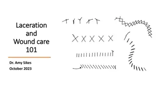
Understanding Surgical Suture Materials and Techniques
Surgical sutures play a vital role in wound repair by providing support and promoting healing of different tissues. An ideal suture should be easy to handle, minimize tissue reaction, inhibit bacterial growth, and be nonreactive after healing. Suture size, flexibility, and material are key factors to consider when choosing the right suture for a procedure. Surgeons must carefully select the suture and needle combination that best suits the tissue type and healing duration.

Download Presentation

Please find below an Image/Link to download the presentation.
The content on the website is provided AS IS for your information and personal use only. It may not be sold, licensed, or shared on other websites without obtaining consent from the author. Download presentation by click this link. If you encounter any issues during the download, it is possible that the publisher has removed the file from their server.
E N D
Presentation Transcript
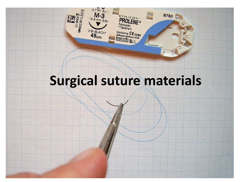
Surgical suture materials Sutures and needles
Sutures Suture plays an important role in wound repair by providing hemostasis and support for healing tissue. Tissues have different requirements for suture support, depending on the type of tissue and anticipated duration of healing. Some tissues need support for only a few days (e.g., muscle, subcutaneous tissue, require weeks (fascia) or months (tendon) to heal. skin), whereas others
an ideal suture is one that will lose its tensile strength at a rate similar to that with which the tissue gains strength, and it will be absorbed by the tissue so that no foreign material remains in the wound.
The ideal suture is Easy to handle, Reacts minimally in tissue, Inhibits bacterial growth, Holds securely when knotted, Resists shrinking in tissue, Absorbs with minimal reaction after the tissue has healed, Is noncapillary, nonallergenic, noncarcinogenic, and nonferromagnetic but such a material does not exist. Therefore, surgeons must choose a suture that most closely approximates the ideal for a given procedure and tissue to be sutured. A wide variety of suture and needle combinations are available.
Suture size. The smallest diameter suture that will adequately secure wounded tissue should be used in order to : 1. minimize trauma as the suture is passed through the tissue and 2. to reduce the amount of foreign material left in the wound. There is no advantage to using a suture that is stronger than the tissue to be sutured The most commonly used standard for suture size is the United States Pharmacopeia (USP), which denotes dimensions from fine to coarse (with diameters in inches) according to a numeric scale, with 12-0 being the smallest and 7 the largest. The smaller the suture size, the less tensile strength it has.
Flexibility. It is determined by its torsional stiffness and diameter which influence its handling and use. Flexible sutures are indicated for ligating vessels or performing continuous suture patterns. Less flexible sutures (e.g., wire) cannot be used to ligate small bleeders. Nylon and surgical gut are relatively stiff compared with silk suture; braided polyester sutures have intermediate stiffness.
Surface characteristics and coating. The surface characteristics of a suture influence the ease with which it is pulled through tissue (i.e., the amount of friction or drag ) and the amount of trauma caused. Rough sutures cause more injury than smooth sutures. Smooth surfaces are particularly important in delicate tissues, such as the eye. However, sutures with smooth surfaces also require greater tension to ensure good apposition of tissues and have less knot security Braided materials have more drag than monofilament sutures. Braided materials often are coated to reduce capillarity, but this also provides a smooth surface. Teflon, silicone, wax, paraffin wax, and calcium stearate are used for coating sutures
Capillarity. Capillarity is the process by which fluid and bacteria are carried into the interstices of multifilament fibers. Because neutrophils and macrophages are too large to enter the interstices of the fiber, infection may persist, particularly in nonabsorbable sutures. All braided materials (e.g., polyglycolic acid, silk) have degrees of capillarity, whereas monofilament sutures are considered noncapillary. Coating reduces the capillarity of some sutures, but regardless, capillary suture materials should not be used in contaminated or infected sites.
Knot tensile strength. Knot tensile strength is measured by the force in pounds that the suture strand can withstand before it breaks when knotted. Sutures should be as strong as the normal tissue through which they are being placed; however, the tensile strength of the suture should not greatly exceed the tensile strength of the tissue.
Relative knot security. Relative knot security is the holding capacity of a suture expressed as a percentage of its tensile strength. The knot-holding capacity of a suture material is the strength required to untie or break a defined knot by loading the part of the suture that forms the loop, whereas the suture material s tensile strength is the strength required to break an untied fiber with a force applied in the direction of its length
Specific Suturing Materials Suture materials may be classified according to 1. their behavior in tissue (absorbable or nonabsorbable), 2. their structure (monofilament or multifilament), or 3. their origin (synthetic, organic, or metallic)
Two major mechanisms of absorption result in the degradation of absorbable sutures. 1. Sutures of organic origin, such as surgical gut, are gradually digested by tissue enzymes and phagocytized, 2. sutures manufactured from synthetic polymers are principally broken down by hydrolysis. Nonabsorbable sutures are ultimately encapsulated or walled off by fibrous tissue.
Monofilament sutures are made of a single strand of material and therefore have less tissue drag than multifilament sutures and do not have interstices that may harbor bacteria or fluid. Care should be used in handling monofilament suture because damaging the material with forceps or needle holders may weaken the suture and predispose it to breakage. Multifilament sutures consist of several strands of suture that are twisted or braided together. Multifilament sutures generally are more pliable and flexible than monofilament sutures. They may be coated to reduce tissue drag and enhance handling characteristics.
Absorbable Suture Materials Absorbable suture materials (e.g., surgical gut, polyglycolic acid [Dexon, Covidien, Mansfield, Mass.], polyglactin 910 [Vicryl, Ethicon, Somerville, N.J.], polydioxanone [PDS II, Ethicon, Somerville, N.J.], polyglyconate [Maxon, Covidien, Mansfield Mass.], poliglecaprone 25 [Monocryl, Ethicon, Somerville, N.J.], glycomer 631 [Biosyn, Covidien, Mansfield, Mass.]) lose most of their tensile strength within 60 days and eventually disappear from the tissue implantation site because they have been phagocytized or hydrolyzed. The time to loss of strength and for complete absorption varies among suture materials.
Organic absorbable materials. Catgut (surgical gut) is the most common nonsynthetic absorbable suture material. Although once very popular, its use has decreased substantially in veterinary medicine with the advent of strong, monofilament synthetic absorbable suture materials. The word catgut is derived from the term kitgut or kitstring (the string used on a kit, or fiddle). Misinterpretation of the word kit as referring to a young cat led to the use of the term catgut. Surgical gut is in fact made from the submucosa of sheep intestine or the serosa of bovine intestine and is approximately 90% collagen. It is broken down by phagocytosis and, in contrast with other suture materials, elicits a notable inflammatory reaction. Surgical gut is available as plain, medium chromic, or chromic; increased tanning generally implies prolonged strength and reduced tissue reaction. Surgical gut is rapidly removed from infected sites or areas where it is exposed to digestive enzymes and is quickly degraded in catabolic patients. The knots may loosen when wet.
Synthetic absorbable materials. Synthetic absorbable materials (see Table 8-2) generally are broken down by hydrolysis and cause minimal tissue reaction. The time to loss of strength and to absorption is fairly constant even in different tissue. Infection or exposure to digestive enzymes does not significantly influence the rate of absorption of most synthetic absorbable sutures. Polyglactin 910 and polyglycolic acid are more rapidly hydrolyzed in alkaline environments, but they are relatively stable in contaminated wounds. Polyglycolic acid, polyglactin 910, and poliglecaprone 25 may be rapidly degraded in infected urine; polydioxanone, polyglyconate, and glycomer 631 are acceptable for use in sterile bladders and those infected with E. coli. However, any suture that is degraded via hydrolysis may be at risk for accelerated degradation when the bladder is infected with Proteus spp.
Monofilament absorbable materials. Polydioxanone and polyglyconate are classic monofilament sutures that retain their tensile strength longer than multifilament sutures with complete absorption occurring in 6 months. Poliglecaprone 25 and glycomer 631 are relatively new monofilament rapidly absorbable synthetic materials that are pliable, lack stiffness, and have good handling characteristics. These sutures have good initial tensile strength that deteriorates in 2 to 3 weeks following implantation and are completely absorbed by 120 days. Polyglytone 6211 (Caprosyn, Covidien, Mansfield, Mass.) is the newest rapidly absorbable monofilament suture. This suture retains up to 30% knot strength at 10 days postimplantation and has excellent handling characteristics; however, its rapid absorption also limits its application.
Multifilament absorbable materials. Polyglycolic acid is braided from filaments extracted from glycolic acid and is available in both coated and uncoated forms. Polyglactin 910 is a multifilament suture made of a copolymer of lactide and glycolide with polyglactin 370. It is coated with calcium stearate and its rate of loss of tensile strength is similar to that of polyglycolic acid. Polysorb (Covidien, Mansfield, Mass.) is a new synthetic absorbable suture material composed of a glycolide/lactide co-polymer. Polysorb has good initial tensile strength and is completely absorbed by 60 days (see Table 8-2). Vicryl Rapide (Ethicon, Somerville, N.J.) is a relatively new, rapidly absorbed, synthetic braided suture that has an initial strength that is comparable to nylon and gut. However, the tensile strength declines to 50% in 5 to 6 days, and it is completely absorbed in 42 days. This suture is indicated for superficial closure of mucosa, gingival closure, and periocular skin closure. Vicryl Plus (Ethicon, Somerville, N.J.) is a new suture that was designed to reduce bacterial colonization on the suture. It has been coated with an antibacterial agent, triclosan.
Nonabsorbable Suture Materials Organic nonabsorbable materials. Silk is the most common organic nonabsorbable suture material used. It is a braided multifilament suture made by a special type of silkworm and is marketed as uncoated or coated. Silk has excellent handling characteristics and often is used in cardiovascular procedures; however, it does not maintain significant tensile strength after 6 months and is therefore contraindicated for use in vascular grafts. It should also be avoided in contaminated sites; one silk suture may reduce the number of bacteria required to induce infection in a wound from 10 6 to 10 3.
Synthetic nonabsorbable materials. Synthetic nonabsorbable suture materials (see Table 8-2) are marketed as braided multifilament threads (e.g., polyester or coated caprolactam) or monofilament threads (e.g., polypropylene, olyamide, or polybutester). These sutures are typically strong and induce minimal tissue reaction. Nonabsorbable suture materials with an inner core and an outer sheath (e.g., Supramid [S. Jackson, Alexandria, Va.) should not be buried in tissue because they may predispose to infection and fistulation. The outer sheath frequently is broken, which allows bacteria to reside underneath it. NOTE Cable ties should never be implanted in the body (e.g., used to ligate ovarian pedicles) because toxic substances are released during their degradation and their use may result in abscess or tumor formation.
Metallic sutures. Stainless steel is the metallic suture most commonly used and is available as a monofilament or multifilament twisted wire. Surgical steel is strong and inert with minimal tissue reaction, but knot ends evoke an inflammatory reaction. Stainless steel has a tendency to cut tissue and may fragment and migrate. It is stable in contaminated wounds and is the standard for judging knot security and tissue reaction to suture materials.
Surgical Needles A variety of needle shapes and sizes are available; Selection of a needle depends on 1. The type of tissue to be sutured (e.g., penetrability, density, elasticity, and thickness), 2. The topography of the wound (e.g., deep or narrow), and 3. The characteristics of the needle (i.e., type of eye, length, and diameter). Needle strength, ductility, and sharpness are important factors in determining the handling characteristics and use of a needle.
The amount of angular deformation a needle can withstand before becoming permanently deformed is called surgical yield. Ductility is the needle s resistance to breaking under a specified amount of bending. The sharpness of a needle is related to the angle of the point (see below) and the taper ratio of the needle. The sharpest needles have a long, thin, tapered point with smooth cutting edges. Most surgical needles are made from stainless steel wire because it is strong, corrosion free, and does not harbor bacteria. The three basic components of a needle are the attachment end (i.e., swaged or eyed end), the body, and the point (Fig. 8-2, A).
Eyed needles must be threaded, and because a double strand of suture is pulled through the tissue, a larger hole is created than when swaged suture material is used. Eyed needles may be 1. closed (i.e., round, oblong, or square) or 2. French (i.e., with a slit from the inside of the eye to the end of the needle for ease of threading) (Fig. 8-2, B). 3. Eyed needles are threaded from the inside curvature.
The use of eyed needles in veterinary practice has decreased substantially in recent years. With swaged sutures, the needle and suture form a continuous unit, which minimizes tissue trauma and increases ease of use. The needle body comes in a variety of shapes (Fig. 8-2, C); the tissue type and depth and the size of the wound determine the appropriate needle shape. 1. Straight (Keith) needles generally are used in accessible places where the needle can be manipulated directly with the fingers (e.g., placement of purse-string sutures in the anus). 2. Curved needles are manipulated with needle holders. The depth and diameter of a wound are important when selecting the most appropriate curved needle.
One-fourth ( 1/4) circle needles are primarily used in ophthalmic procedures. Three-eighths ( 3/8) and one-half ( 1/2 ) circle needles are the most commonly used surgical needles in veterinary medicine. Three-eighths circle needles are more easily manipulated than one-half circle needles because they require less pronation and supination of the wrist. However, because of the larger arc of manipulation required, they are awkward to use in deep or inaccessible locations. A one-half circle or five-eighths ( 5/8 ) circle needle, despite requiring more pronation and supination of the wrist, is easier to use in confined locations.
The needle point (i.e., cutting, taper, reverse cutting, or side cutting) (Fig. 8-2, D) affects the sharpness of a needle and the type of tissue in which the needle can be used. Cutting needles generally have two or three opposing cutting edges and are designed for use in tissues that are difficult to penetrate, such as skin. With conventional cutting needles, the third cutting edge is on the inside (i.e., concave) curvature of the needle. The location of the inside cutting edge may promote more cut out of tissue because it cuts toward the edges of the wound or incision. Reverse cutting needles have a third cutting edge on the outer (i.e., convex) curvature of the needle; this makes them stronger than similarly sized conventional cutting needles and reduces the amount of tissue cut out. Side cutting needles (i.e., spatula needles) are flat on the top and bottom and are generally used for ophthalmic procedures. Taper needles (i.e., round needles) have a sharp tip that pierces and spreads tissues without cutting them. They generally are used in easily penetrated tissues, such as the intestine, subcutaneous tissue, or fascia. Taper-cut needles, which are a combination of a reverse cutting edge tip and a taperpoint body, generally are used for suturing dense, tough fibrous tissue, such as a tendon, and for some cardiovascular procedures, such as vascular grafts. Bluntpoint needles have a rounded, blunt point that can dissect through friable tissue without cutting. They occasionally are used for suturing soft, parenchymal organs, such as the liver or kidney.


 circle needles are primarily used in")