Anatomy and Biology of Class Digenea Digenia Adult Flukes
Digenea Digenia, a class of parasitic flatworms, have distinctive anatomy and biology. Adult flukes possess two suckers, oral and acetabulum, and exhibit different types based on sucker positioning. Their tegument consists of two zones, with various chemicals for survival. The alimentary tract is incomplete, with the ability to feed on blood and mucus. The reproductive system in males consists of two testes and specific structures for sperm storage and release. Females have a unique reproductive system with organs for egg production and fertilization. The excretory system includes flame cells and an excretory bladder. The development stages involve hatching from eggs and penetrating mollusks for further development.
Download Presentation

Please find below an Image/Link to download the presentation.
The content on the website is provided AS IS for your information and personal use only. It may not be sold, licensed, or shared on other websites without obtaining consent from the author. Download presentation by click this link. If you encounter any issues during the download, it is possible that the publisher has removed the file from their server.
E N D
Presentation Transcript
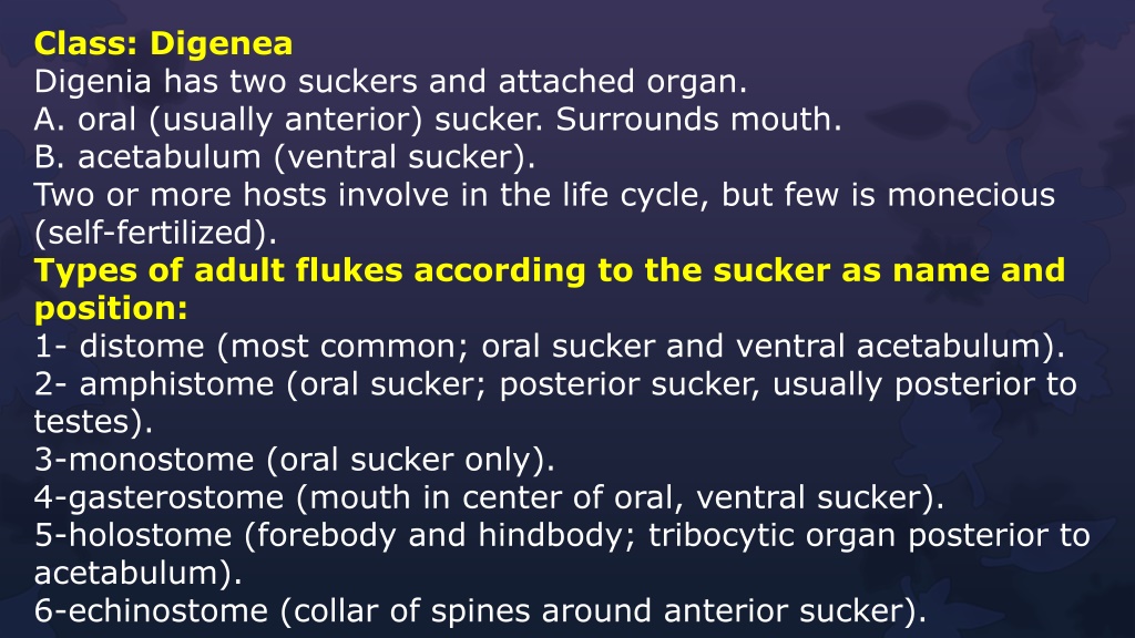
Class: Digenea Digenia has two suckers and attached organ. A. oral (usually anterior) sucker. Surrounds mouth. B. acetabulum (ventral sucker). Two or more hosts involve in the life cycle, but few is monecious (self-fertilized). Types of adult flukes according to the sucker as name and position: 1- distome (most common; oral sucker and ventral acetabulum). 2- amphistome (oral sucker; posterior sucker, usually posterior to testes). 3-monostome (oral sucker only). 4-gasterostome (mouth in center of oral, ventral sucker). 5-holostome (forebody and hindbody; tribocytic organ posterior to acetabulum). 6-echinostome (collar of spines around anterior sucker).
Tegument consist of two zones: 1. outer cytoplasmic syncytium provided with microvilli [mitochondria, ER, vacuoles, lipids, etc.] [pinocytosis; spines] 2. inner area of nucleated cell bodies cytons. 3. zones separated by basal lamina. 4. circular & longitudinal muscle under basal lamina. Numerous chemicals in the tegument: mucopolysaccharides to inhibit host digestive enzymes. acid & alkaline phosphatases, esterases, and aminopeptidases for digestion.
Alimentary tract consists of: incomplete gut. pharynx, when present, masticates food. esophagus leads to 2 blind cecae. entire gut secretion by cells along gut; proteases, lipases. in cecae, absorption process. * Belongings species usually feed on blood, mucus, and epithelium cells. Reproductive system consists of: Male reproductive system consists of: usually 2 testes; taxonomic importance, vas efferens - vas deferens - cirrus pouch, cirrus pouch encloses seminal vesicle, prostate glands, and cirrus; some with external seminal vesicle outside of pouch, sperm stored in seminal vesicle and prostate secretes fluid to keep sperm alive.
Female reproductive system consists of: usually single ovary, 2ndry oocytes released, through short oviduct, into ootype, 3 organs enter into ootype: Mehlis gland, cluster of unicellular glands, enhance egg tanning by maintaining correct pH. Different cell types in gland, secretions cause release of shell globules from vitelline glands, secretes first membrane around egg into lubricates uterus and activates sperm, which are passed down ootype. Common vitelline duct (passage of vitelline gland secretions for eggshell formation), Duct from seminal receptacle (absent in some species), Occasionally a fourth duct, vitelline reservoir, as diverticulum of vitelline duct.
Excretory system consists of: protonephridia (flame cells). excretory bladder in posterior, with excretory pore. Development: Egg: usually operculate, lid pops off during hatching (in Schistosomes, no lid; shell splits). Miricidium: hatches from egg and then penetrates mollusc (rarely annelids), and develop as asexual stages from miricidium, with morphologic characteristics: apical stylet, apical papilla [where ducts from glands open; also nerve endings for chemoreception], apical gland [histolytic enzymes], cephalic glands [lytic enzymes], photoreceptors [eyespots], germinal mass [initiate asexual stages], cilia [locomotion], excretory pore, actively swim, chemoreception to snail mucus, attaches to snail with apical papillae; lytic enzymes dissolve tissues. About 30 minutes for penetration
- Sporocyst: it is asexual stage with various shapes but does not have a mouth or digestive system, absorbs nutrients through tegument, may produce rediae, daughter sporocysts, or cercaria. Redia: can develop directly from miricidium, or from embryos generated from sporocysts, elongate; crawl actively, muscular pharynx, mouth, blind-ended sac caecum, 1-more ambulatory buds for movement often, because ingestion, many species cause extensive damage to host, will form daughter rediae or cercaria. Cercaria: emerged from redia or sporocyst, free-swimming, leaves snail and seeks host, miniature immature fluke with tail, penetrate skin of definitive host, encyst on vegetation as metacercaria, encyst as metacercaria in intermediate host or eaten by intermediate or definitive host or mesocercaria (unencysted juvenile) in tissues - -
- Classification based on sucker placement of cercaria monostome a. 1 sucker only, anterior b. 2 eyespots c. long, simple tails d. develop from redia e. give rise to monostome adults amphistome f. posterior sucker, often anterior too g. eyespots h. develop from redia i. give rise to amphistome adults j. all in superfamily: Paramphistomoidea
gasterostome k. mouth ventral l. develop into gasterostome adults m. all in family: Bucephalidae distome n. 2 suckers, one oral and one ventral o. most common type Additional classification, based on cercarial tail shape and other structures: pleurolophocercous furcocercous echinostome xiphidiocercariae ophthalmocercariae (with eyespots)
Trematoda is divided according to the site of infection in the definitive host: 1- Liver Flukes 2- Intestine Flukes 3- Lung Flukes 4- Blood Flukes
